3.2
Impact Factor
ISSN: 1449-1907
Int J Med Sci 2016; 13(6):412-417. doi:10.7150/ijms.14880 This issue Cite
Research Paper
The Expression of Gli3 and Teashirt3 in the Stenotic Tissue of Congenital Pelvi-Ureteric Junction Obstruction in Children
Department of Pediatric Surgery, Shengjing Hospital, China Medical University, Shenyang 110004, P.R. China
Received 2016-1-4; Accepted 2016-4-13; Published 2016-5-12
Abstract
Background: The aim of this study was to determine the expression pattern of Gli3 and Teashirt3 in stenotic segments in children with congenital hydronephrosis due to pelvi-ureteric junction obstruction (PUJO) versus in normal control subjects.
Materials and methods: 60 patients and 10 controls were included in this study. Immunohistochemistry, Western blot and real-time PCR were used to investigate into the expression of Gli3 and Teashirt3.
Results: Immunohistochemistry identified that Gli3 and Teashirt3 located in the cytoplasm of smooth muscle in normal ureter. However, the expression of Gli3 and Teashirt3 was negative in the PUJO group. Gli3 and Teashirt3 protein and mRNA expression was significantly decreased in PUJO group compared with control group on Western blot and real time PCR.
Conclusions: The expression of protein and mRNA of Gli3 and Teashirt3 was significantly decreased in the PUJO group. Gli3 and Teashirt3 protein was mainly located in the cytoplasm of smooth muscle in normal ureter. Gli3 and Teashirt3 might play an important role in the normal development of the ureter. The down-regulated Gli3 and Teashirt3 perhaps participated in the pathogenesis of the congenital hydronephrosis due to PUJO.
Keywords: Congenital hydronephrosis, Pelvi-ureteric junction obstruction, Gli3, Teashirt3, children
Introduction
Congenital hydronephrosis caused by pelvi-ureteric junction obstruction (PUJO) is a common pediatric congenital urinary malformation detrimental to children's health. Quite a few studies have shown that pelvi-ureteric junction obstruction results from the abnormal development of the ureteral smooth muscle at the junction[1], yet its molecular mechanisms are not clear. Recent researches have found that Shh signaling pathway is involved in embryonic development and morphogenesis of the kidney and ureter[2-4]. Animal experiments have identified that Shh downstream transcription factor Gli3 and Teashirt3 plays a major role in the differentiation of proximal ureteric smooth muscle [2,5-7]. However, there have been few studies on the expression of Gli3 and Teashirt3 in congenital unilateral hydronephrosis due to PUJO in the human. This study investigated the expression of Gli3 and Teashirt3 in stenotic segments in children with congenital hydronephrosis due to PUJO versus in normal control subjects using immunohistochemistry, Western blot and real-time PCR methods, aiming to probe into possible pathogenic mechanisms in congenital hydronephrosis due to PUJO.
Materials and methods
Patients and control samples
This study was approved by the Ethics Committee of China Medical University (Ethical Number:2012 PS81K). Stenotic segments of ureter tissues were obtained from 60 patients with congenital hydronephrosis during the operation in the department of pediatric urology, Shengjing hospital of China Medical University. The patients ranged from 1 month to 13 years old, with a mean age of 4.09 years. The diagnosis was based on the results of IVP, ECT, delayed three-dimensional contrast-enhanced CT and the PUJ morphology during operations. 10 control ureters were obtained from patients with Wilm's tumor, and the tissues were confirmed histologically to be unaffected. All tissue samples were storaged at -80℃ immediately after surgical removal.
Immunohistochemical labeling of Gli3 and Teashirt3
Endogenous peroxidase activity was blocked by incubation of the sections in 3% H2O2 for 20 min. Antigen retrieval was performed by heating the slides in 10 mmol/l citrate buffer (pH 6.0) at 98℃ for 10 min. Sections were incubated with primary anti-Gli3 (1:50 dilution, rabbit polyclonal, Santa Cruz Biotechnology, Santa Cruz, Calif., USA) or primary anti-Teashirt3(1:50 dilution, rabbit polyclonal, Santa Cruz Biotechnology, Santa Cruz, Calif., USA) and horseradish peroxidase (HRP)-conjugated secondary antibody (Santa Cruz Biotechnology). Antibody incubations were performed in phosphate-buffered saline (PBS) supplemented with 10% goat serum. Primary antibody was incubated on the sections at 4℃ for 16 h. Incubation with the secondary antibody was performed for 30 min at room temperature, and signals were visualized by using 3'3 P-diaminobenzidine (DAB; Sigma, UK). Sections were counterstained with hematoxylin. Negative controls were performed by either omitting the primary or secondary antibodies or incubating with the equivalent concentrations of nonimmune rabbit antiserum. Two pathologists independently reviewed immunohistochemically stained slides and agreed on results by consensus.
Protein preparation and Western blot
Protein extract (50µg) was denatured, separated by sodium dodecyl sulfate/polyacrylamide gel electrophoresis, and transferred to polyvinylidene fluoride membranes (Millipore, Billerica, Mass., USA), blocked with 5% fat-free milk in Tris-buffered saline (1h, room temperature) and incubated overnight at 4℃ in primary antibodies against Gli3 (1:2000; rabbit polyclonal, Santa Cruz Biotechnology), Teashirt3 (1:2000; rabbit polyclonal, Santa Cruz Biotechnology) and β-actin (1:2000; Santa Cruz Biotechnology). After washing, the membranes were incubated in secondary antibodies at room temperature for 1h. The membranes were washed and developed using a chemiluminescent substrate kit (SuperSignal West Pico, Pierce, Rockford, IL). For Western blot analysis, densitometric values were analyzed using the ECL Plus Western blot detection system.
RNA isolation and real-time RT-PCR
Total RNA was extracted from patients by use of TRIzol reagent (Invitrogen) according to the manufacturer's protocol. RNA (1µg) was reverse-transcribed by using the PrimeScript RT reagent Kit (TaKaRa) following the manufacturer' s instructions. Quantitative real-time PCR was accomplished with SYBR Premix Ex Taq (TaKaRa) on LightCycler-GmbH D-68298 (Roche Molecular Biochemicals) under the following conditions: 95℃ for 10 s, 45 cycles of 95℃ for 5 s, 58℃ for 20 s; 65℃ for 15 s. A dissociation procedure was performed to generate a melting curve for confirmation of amplification specificity. GAPDH was used as the reference gene. The relative levels of gene expression were determined as ΔCt=Ct gene-Ct reference, and the fold change in gene expression was calculated with the 2-ΔΔCt method. Experiments were repeated in triplicate. Primer sequences were as follows: Gli3 forward, 5'-GAGGGCCGTTACCATTAC-3', reverse, 5'-AGGGAGACTCGGAAGCAG-3'; Teashirt3 forward, 5'-GGAGCTGGTGAAAAAGGTCA-3', reverse, 5'-ACATGAATGATACGACGGCA-3'; GAPDH forward, 5'-GAGCCTGAGGCCGACTACTA-3', reverse, 5'-CTCAGTGTAGCCCAGGATGC-3'.
Statistical analysis
All data were presented as mean±SD. Significance of differences was evaluated by using two-sample t test. P value < 0.05 was considered to be statistically significant.
Results
1. HE stain
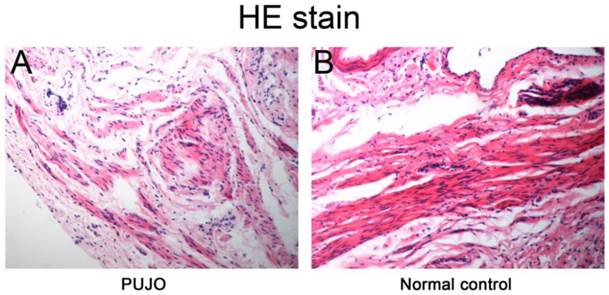
Microscopic findings: smooth muscle hypertrophy and mesenchyme hyperplasia with myofiber disarrangement in stenotic segments in PUJO group compared with no abnormality or tumor cell infiltration in controls (Figure 1).
2. Immunohistochemistry
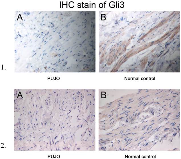
In PUJO group, no expression of Gli3 and Teashirt3 was found in PUJ stenotic segments (Figure 2-1A; 2-2A).
In normal control, positive expression of Gli3 and Teashirt3 was detected in ureteric smooth muscle cytoplasm (Figure 2-1B; 2-2B).
HE stain of stenotic segments in PUJO patients and ureter in normal controls (×200). A: PUJO group: smooth muscle hypertrophy and mesenchyme hyperplasia with myofiber disarrangement in stenotic segments; B: Normal control: no abnormality or tumor cell infiltration found.

1- Gli3 immunohistochemistry stain (×400). A: PUJO group: Gli3 not specifically stained in smooth muscle cytoplasm of the muscular layer in stenotic segments; B: normal control: Gli3 stained dark yellowish brown in smooth muscle cytoplasm of the muscular layer. 2- Teashirt3 immunohistochemistry stain (×400). A: PUJO group: Teashirt3 not specifically stained in smooth muscle cytoplasm of the muscular layer in stenotic segments; B: normal control group: Teashirt3 stained yellowish brown in smooth muscle cytoplasm of the muscular layer.

3. Western blot
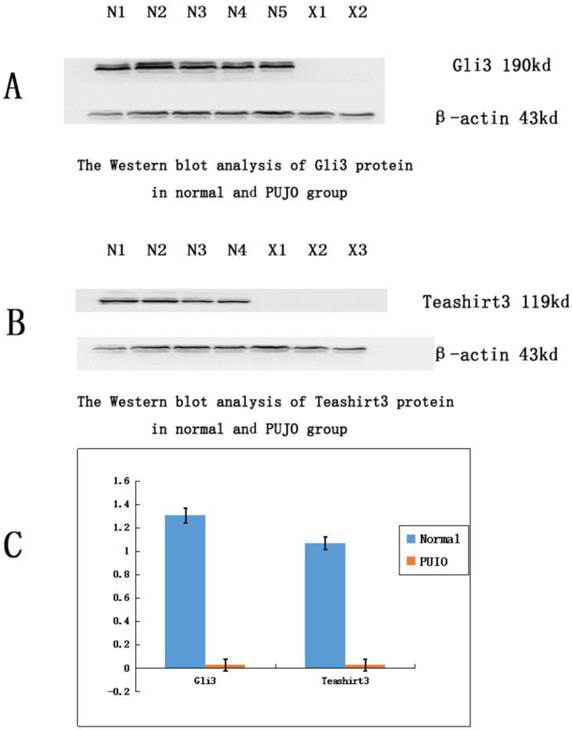
To further clarify the expression of Gli3 and Teashirt3 in PUJ, Western blot method was used to examine the expression level of Gli3 and Teashirt3 in PUJ and control subjects (Figure 3). Quantity One software was used to calculate the level of protein expression of Gli3 and Teashirt3, followed by the calculation of the relative intensity through standardization using the expression of β-actin as the internal control. Gli3 was 1.3068±0.0289 in normal controls and 0.0243±0.0819 in PUJO patients; Teashirt3 was 1.0682±0.0837 in normal controls and 0.0241±0.0719 in PUJO patients. The difference of Gli3 and Teashirt3 expression between the two group was statistically significant (P<0.05).
4. Real-time PCR
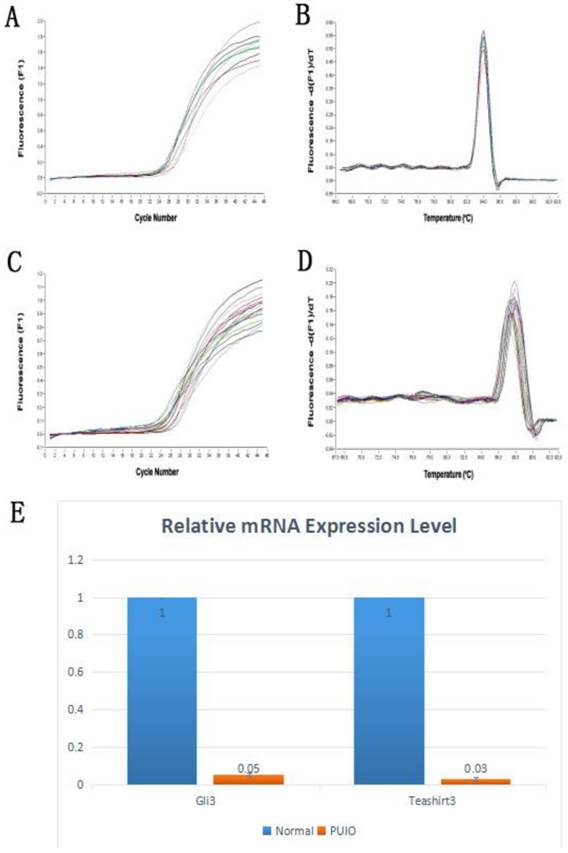
During the course of real-time PCR, amplification and melting curves were drawn according to the mRNA fluorescence values and cycle numbers of Gli3 and Teashirt3 (Figure 4). Using GAPDH as the internal control, samples exhibited satisfactory repeatability and consistent efficiency. Each sample was repeated three times in real-time PCR test and averaged as the Ct value. The differences in mRNA expression level of each gene were compared using 2-ΔΔCt relative quantification method (Table 1, Figure 4). The differences in mRNA expression of both Gli3 and Teashirt3 in the two groups were statistically significant (*P<0.05).
The relative ratio of Gli3 and Teashirt3 mRNA expression in each group (* P<0.05)
| Gli3 mRNA | Teashirt3 mRNA | |
|---|---|---|
| Control group | 1 | 1 |
| PUJO group | 0.05* | 0.03* |
Western blot analysis of Gli3 and Teashirt3 protein expression in PUJ of patients with congenital hydronephrosis and in normal controls. X indicates PUJO group and N normal group. The protein expression of Gli3 and Tshz3 in PUJ tissue in patients with PUJO is down-regulated significantly compared with that in control group.

Real-time RT-PCR analysis of Gli3 and Teashirt3 mRNA expression in PUJ of patients with congenital hydronephrosis and in normal controls. A, amplification curves for Gli3; B, melting curves for Gli3; C, amplification curves for Teashirt3; D, melting curves for Teashirt3; E, bar graphs of Gli3 and Teashirt3 mRNA expression. Gli3 and Teashirt3 mRNA expression in PUJ tissue was more significantly down-regulated in PUJO group than in control group.

Discussion
The congenital hydronephrosis caused by congenital pelvi-ureteric junction obstruction is a common urologic condition in children. It may result in complications such as pediatric renal dysfunction, hypertension or even renal failure, etc. and seriously affect children's quality of life. Quite a few studies have suggested that the cause of congenital hydronephrosis is the abnormal development of ureteric smooth muscle at the pelvi-ureteric junction [8], yet its molecular mechanism remains unclear. Recent studies have shown that Shh signaling pathway plays a key role in the embryonic development through its regulation in cell proliferation and differentiation. Shh signaling pathway is closely linked with the onset and development of ureteric abnormality in the human.
Shh signaling pathway consists of ligands, membrane spanning receptors, nuclear factor κB, downstream target genes as well as pathway regulation proteins. Gli3 and Teashirt3 act jointly as downstream transcriptors in the pathway and play a major role in the normal differentiation of proximal ureteric smooth muscle [2,5-7].
Xavier Caubit carried out a comparative study on mouse ureter with normal expression of Tshz3 versus Tshz3-null mouse ureter and found no abnormality of ureteric peristalsis in ureters with normally expressed Tshz3 whereas Tshz3-null ureters which was deficient in smooth muscle showed no normal peristalsis, suggesting Tsh/Tshz gene family was critical in the normal development of urinary tract in mammals [6]. They found in their experiments that Tshz3 was expressed in precursors of ureteric smooth muscle cells in the mouse. By contrast, congenital hydronephrosis was found in Tshz3-null mouse resulting not from anatomic obstruction, but from abnormal or absent ureteric peristalsis secondary to abnormal ureteric smooth muscle which caused ineffective contraction. In these mice, urine cannot be expelled normally and hydronephrosis ensues. Lye CM found in Tshz3-mutant proximal ureter, all Tshz3 upstream genes had normal expression with all downstream myocardial protein and SMC proteins absent [9].
As a zinc finger transcription factor in the vertebrate Shh signaling pathway, glioma-associated oncogene homolog is a core factor in the regulation of Shh signaling pathway. The change in its molecular function directly results in change in downstream gene transcription in Shh signaling pathway. Using Polymerase Chain Reaction, Jason E.Cain, etc. discovered in patients with Pallister-Hall Syndrome a mutant Gli3 segment and found Gli3 played a major role in the ureteric development of PHS mouse models and affected ureteric peristalsis [2]. Cain JE, etc. found in a mouse model study that blocking Gli3 signal could result in abnormality in ureteric peristalsis and subsequently lead to malformations like megaloureter and hydronephrosis.
The abovementioned animal studies have shown in the ureteric development of the mouse, Gli3 and Teashirt3 play an important role and they are related to congenital hydronephrosis due to pelvi-ureteric junction obstruction. Dagan Jenkins, etc., however drew a contrary conclusion from their comparative study on TSHZ2 and TSHZ3 expression in blood samples between 67 UPJO patients and controls using in situ hybridization method and they believed mutant TSHZ3 gene was not a major cause of PUJO [1]. As yet, studies based on Gli3 and Teashirt3 in UPJO patients have been scarce. In this study, we investigated the expression of Teashirt3 and Gli3 in Shh signaling pathway in ureteric tissue among UPJO patients. Under HE stain, smooth muscle hypertrophy and mesenchyme hyperplasia with myofiber disarrangement were found in PUJO patients. As for Immunohistochemistry stain, it showed positive expression of Gli3 and Teashirt3 in normal ureteric smooth muscle cytoplasm. And using the Western blot and real-time PCR, down- or absent regulation of Gli3 and Teashirt3 protein and mRNA were detected in stenotic PUJ tissue in congenital hydronephrosis, respectively. So we speculate that down-regulation of Gli3 and Teashirt3 in stenotic PUJ in congenital hydronephrosis may have a negative effect on the development of ureteric tissue in that site, which subsequently leads to ureteric dyskinesia and stenosis in PUJ and congenital hydronephrosis finally ensues. It stands to reason that there is a probable correlation between Gli3 and Teashirt3 down-regulation in stenotic PUJ in congenital hydronephrosis and the pathogenesis of PUJO.
Acknowledgements
This work was supported by the National Nature Science Foundation of China (No 81370772) and the Science and Technology Project of Liaoning Province, China (No 2012225076).
Competing Interests
The authors have declared that no competing interest exists.
References
1. Jenkins D, Caubit X, Dimovski A. et al. Analysis of TSHZ2 and TSHZ3 genes in congenital pelvi-ureteric junction obstruction. Nephrol Dial Transplant. 2010;25(1):54-60
2. Cain JE, Islam E, Haxho F. et al. GLI3 repressor controls nephron number via regulation of Wnt11 and Ret in ureteric tip cells. PLoS One. 2009;4(10):e7313
3. Hu MC, Mo R, Bhella S. et al. GLI3-dependent transcriptional repression of Gli1,Gli2 and kidney patterning genes disrupts renal morphogenesis. Development. 2006;133(3):569-578
4. Johnston JJ, Olivos-Glander I, Killoran C. et al. Molecular and clinical analyses of Greig cephalopolysyndactyly and Pallister-Hall syndromes:robust phenotype prediction from the type and position of GLI3 mutations. Am J Hum Genet. 2005;76(4):609-622
5. Cain JE, Islam E, Haxho F. et al. GLI3 repressor controls functional development of the mouse ureter. J Clin Invest. 2011;121(3):1199-1206
6. Caubit X, Lye CM, Martin E. et al. Teashirt 3 is necessary for ureteral smooth muscle differentiation downstream of SHH and BMP4. Development. 2008;135(19):3301-3310
7. Buttitta L, Mo R, Hui CC. et al. Interplays of Gli2 and Gli3 and their requirement in mediating Shh2-dependent sclerotome induction. Development. 2003;130:6233-6243
8. Karmarkar SJ. Long-term results of surgery for posterior urethral valves: a review. Pediatr Surg Int. 2001;17(1):8-10
9. Lye CM, Fasano L, Woolf AS. Ureter Myogenesis: Putting Teashirt into Context. J Am Soc Nephrol. 2010;21(1):24-30
Author contact
![]() Corresponding author: Dr. Yi Yang, yangy2org
Corresponding author: Dr. Yi Yang, yangy2org

 Global reach, higher impact