Impact Factor ISSN: 1449-1907
- Issue 9; 2026
- Issue 8; 2026
- Issue 7; 2026
- Issue 6; 2026
- Issue 5; 2026
- Volume 23; 2026
- Past Issues
- Editorial Board
- Cover Images
- Index & Coverage
- Cover Suggestion
- Special Issues
1. Introduction
2. Artificial intelligence...
3. Effectiveness of AI in...
4. Challenges
5. Future prospects of AI in...
6. Conclusion
Abbreviations
Supplementary Material
Acknowledgements
References

 Global reach, higher impact
Int J Med Sci 2026; 23(8):2548-2567. doi:10.7150/ijms.134301 This issue Cite
Review
Advances in Artificial Intelligence for Wrist Joint Injury Diagnosis
Hong Li1, Xilin Liu2 ![]()
1. Department of Nursing, China-Japan Union Hospital of Jilin University, Changchun, 130033, China.
2. Department of Hand and Foot surgery, China-Japan Union Hospital of Jilin University, Changchun, 130033, China.
Received 2026-3-12; Accepted 2026-4-8; Published 2026-7-1
Abstract

This study provides a comprehensive analysis of the application of artificial intelligence (AI) in diagnosing acute traumatic wrist joint injuries (WJIs), including fractures and ligament damage. AI has demonstrated significant potential in identifying fractures and ligament damage. The study highlights the use of various AI technologies and algorithms, including Convolutional Neural Networks (CNNs), Gradient Class Activation Mapping (Grad-CAM), deep learning models, object detection models, automated assessment algorithms, traditional machine learning techniques, data augmentation and preprocessing, Natural Language Processing (NLP), and integration with other imaging modalities. Compared with traditional diagnostic methods, AI offers substantial benefits, such as efficient processing of large datasets, minimizing diagnostic errors and missed cases, aiding in interpreting complex fracture patterns, optimizing workflow, enhancing diagnostic efficiency, and providing comprehensive diagnoses through multimodal integration. AI has significantly improved the precision and efficiency of fracture detection, reduced unnecessary imaging procedures, expedited the diagnostic and reporting process, optimized resource allocation, and improved patient outcomes. However, clinical application of AI faces challenges, including ethical considerations, regulatory hurdles, data privacy and security issues, algorithm transparency and interpretability problems, and unclear liability definitions. The future of AI in diagnosing WJIs is promising, with potential advancements in fracture detection, treatment planning, and rehabilitation strategies. However, challenges such as model validation and training of healthcare professionals must be addressed to fully integrate AI into orthopedic practice and advance the management of WJIs.
Keywords: artificial intelligence, wrist joint injuries, diagnosis, ligament damage, convolutional neural networks
1. Introduction
Artificial intelligence (AI) is transforming the diagnosis of wrist joint injuries (WJIs), improving diagnostic accuracy and operational efficiency for detecting fractures and ligament injuries. Recent studies have systematically evaluated AI-based models' applicability and diagnostic performance in identifying wrist fractures, representing some of the most prevalent injuries encountered in clinical practice [1-3]. AI is increasingly applied in medical imaging, with growing evidence supporting its use in fracture detection. Three clinically prevalent fracture types, including distal radius, ulnar styloid, and scaphoid fractures, are common in emergency settings and are often missed on initial radiographs. AI has shown potential to improve diagnostic accuracy and reduce missed injuries. This review provides a comprehensive analysis of AI technologies for WJIs, synthesizes available evidence on diagnostic performance, and discusses clinical benefits, challenges, and future directions.
Advancements in AI highlight its potential to play a crucial role in the early diagnosis and management of ligament injuries, thereby reducing the risk of progression to degenerative conditions [4]. Moreover, the application of AI in ultrasound imaging has been investigated as a novel approach for detecting wrist fractures. Another study evaluated the feasibility of combining three-dimensional ultrasound (3DUS) with AI for automated fracture identification. The findings demonstrated that AI-based analysis performed on par with human interpretation, achieving a sensitivity of 1.0 in fracture identification. This remarkable sensitivity suggests that AI-based ultrasound could provide a non-invasive and radiation-free alternative option to conventional radiography, particularly benefiting pediatric patients by minimizing unnecessary exposure to ionizing radiation [5,6]. Collectively, AI offers remarkable potential to transform the diagnosis and management of wrist injuries by improving the detection of fractures and ligament injuries. This technology not only supports clinical decision-making but also reduces the workload on healthcare providers and enhances patient outcomes. As research in this field advances, the use of AI in routine orthopedic practice may become more promising, providing a valuable tool for enhancing diagnostic accuracy and efficiency.
2. Artificial intelligence technologies and algorithms in wrist injury diagnosis
The application of AI and algorithm-driven approaches in the medical field has significantly advanced diagnostic capabilities across various conditions (Table 1). In musculoskeletal injuries, particularly wrist injuries, AI has remarkable potential to enhance diagnostic precision and clinical outcomes. Studies have found AI integration in various imaging modalities and diagnostic strategies to improve the identification and assessment of wrist injuries. In a study by Ozkaya et al. [7] the diagnostic efficacy of AI, specifically CNNs, was assessed for identifying scaphoid fractures in anteroposterior wrist radiographs. The study's results demonstrated the potential of AI in accurately identifying fractures, highlighting the application of AI in improving diagnostic capabilities for wrist injuries. Furthermore, Zhang et al. [5] focused on the diagnostic accuracy of 3DUS and AI in detecting pediatric wrist injuries. The study found that AI interpretation was similar to human interpretation, with a sensitivity of 1.0 in detecting fractures. This suggests that AI can play a crucial role in promoting the accuracy and reliability of diagnosing wrist injuries in pediatric patients. Similarly, Hendrix et al. conducted a study to compare the diagnostic performance of an AI algorithm with that of experienced musculoskeletal radiologists in identifying scaphoid fractures using conventional multi-view radiographs of the hand and wrist [8]. His research aimed to assess whether AI could support radiologists in making more accurate diagnoses. Their findings highlighted AI's potential to perform at a level comparable to musculoskeletal radiologists in detecting scaphoid fractures, emphasizing its value as a supportive tool for healthcare professionals in diagnosing wrist injuries.
The application of artificial intelligence technology in wrist joint injuries
| Year | Artificial Intelligence Techniques | Application Scenarios | Achievements/Contributions | Refs. |
|---|---|---|---|---|
| 2024 | DL, CNN | Detection of wrist fractures from plain wrist radiographs, comparing DL algorithms to healthcare experts' consensus | Conducted a systematic review and meta-analysis of DL algorithms for detecting wrist fractures. Found that CNNs achieved a summary sensitivity of 92% (95% CI: 80%-97%) and a summary specificity of 93% (95% CI: 76%-98%) compared to healthcare experts. Highlighted the need for robust reference standards, external dataset validation, and investigation of diagnostic performance of healthcare experts aided by CNNs. | [9] |
| 2021 | DL, CNN, Grad-CAM | Detection of wrist fractures (distal radius fractures) from X-ray images, comparing DL models to human experts on general and challenging cases. | Developed an open-source DL pipeline called DeepWrist for wrist fracture detection. Evaluated the pipeline on two test sets: a general population test set and a challenging test set requiring CT confirmation. While DeepWrist achieved near-perfect performance on the general test set (AUROC of 0.99), its performance dropped significantly on the challenging test set (AUROC of 0.84). Highlighted the limitations of current DL models in handling difficult cases and emphasized the need for robustness and safety assessments before clinical deployment. Investigated the use of Deep Ensembles to detect out-of-distribution cases but found limited success in distinguishing between general and challenging cases. | [10] |
| 2021 | DCNN | Automated Detection and Classification of Distal Radius Fractures. | Evaluated the performance of a DCNN for detecting and classifying distal radius fractures, metal, and cast on radiographs using labels from radiology reports. The models achieved high AUC values for detecting metal (0.99) and cast (1.00). Fracture detection performance approximated that of radiology residents (AUC 0.98). Demonstrated a positive correlation between training set size and model performance in most categories. Highlighted the potential of using low-effort data labeling from radiology reports for training AI models. | [11] |
| 2019 | CNN | Automated Detection and Localization of Fractures in Wrist Radiographs. Emergency Department Radiographs, Assisting Emergency Medicine Clinicians. | Demonstrated the feasibility of using an object detection CNN to detect and localize fractures on wrist radiographs with high sensitivity and specificity. Showed per-fracture sensitivity of 91.2% (frontal view) and 96.3% (lateral view). Per-image sensitivity of 95.7% (frontal view) and 96.7% (lateral view), with AUC values of 0.918 and 0.933, respectively. Per-study sensitivity of 98.1% and AUC of 0.895. Lower sensitivity for minimally displaced fractures compared to displaced fractures. Provided location information of fractures, enhancing trust and verifiability for clinicians. | [12] |
| 2021 | CNN | Automated Diagnosis of Distal Radius Fractures. Use of Biplane Plain X-rays | Developed an AI system capable of diagnosing distal radius fractures with high accuracy using relatively small datasets. Achieved diagnostic accuracy of 98.0% ± 1.6% for distal radius fractures and 91.1% ± 2.5% for ulnar styloid fractures. Obtained AUC values of 0.991 for distal radius fractures and 0.956 for ulnar styloid fractures. Demonstrated the effectiveness of using a pre-trained VGG16 model and image augmentation to enhance diagnostic performance with limited data. Proposed a two-stage diagnostic approach using both anteroposterior and lateral X-ray images to improve accuracy. | [1] |
| 2018 | DCNN | Automated Detection and Localization of Fractures in Radiographs. Emergency Department Radiographs. Assisting Emergency Medicine Clinicians | Developed a deep learning model to detect and localize fractures in radiographs with high accuracy. Trained the model using annotations from 18 senior subspecialized orthopedic surgeons on 135,409 radiographs. Significant improvements in diagnostic accuracy when clinicians used the model's assistance: Sensitivity increased from 80.8% to 91.5%. Specificity increased from 87.5% to 93.9%. Relative reduction in misinterpretation rate by 47.0%. The model's performance is comparable to that of senior orthopedic surgeons. | [13] |
| 2023 | DL, YOLOv8 algorithm | Fracture detection in pediatric wrist trauma X-ray images. Development of a GUI application ("Fracture Detection Using YOLOv8 App") to assist pediatric surgeons in analyzing X-ray images. | Achieved state-of-the-art mean average precision of 0.638 on the GRAZPEDWRI-DX dataset, outperforming YOLOv7 and other models. Improved detection accuracy for bone anomalies through data augmentation techniques. Developed a user-friendly application to assist pediatric surgeons in interpreting X-ray images, reducing the probability of misclassification. Provided a practical solution for hospitals with limited access to radiologists, especially in underdeveloped regions. | [14] |
| 2020 | DL, CNN | Detection and localization of distal radius fractures in wrist radiographs. | Achieved high performance comparable to radiologists using a relatively small dataset (AUC up to 0.96 on internal test set, sensitivity up to 0.90, specificity up to 1.0). Demonstrated the ability to localize fractures with over 90% alignment with radiologists' annotations Highlighted the potential of using deep learning systems as a computer-aided diagnostic tool in clinical workflows. | [15] |
| 2021 | CNN, Grad-CAM | Detection of wrist fractures in the emergency room using plain radiographs. Assisting emergency physicians in diagnosing fractures quickly and accurately. | Demonstrated that DenseNet-161 and ResNet-152 models achieved high performance in detecting wrist fractures with AUROCs of 0.962 and 0.947, respectively. Sensitivity, specificity, positive predictive value, negative predictive value, and accuracy for both models. The importance of using Grad-CAM to visualize and interpret fracture detection regions. Developed a model representative of real-world emergency room settings, addressing limitations of previous heterogeneous datasets. Provided insights into the potential use of AI models in clinical practice, especially in scenarios where radiologists are unavailable | [16] |
| 2022 | CNN, TL | Detection of hand fractures from plain radiographs in emergency departments. Assisting physicians in interpreting hand radiographs, especially in small hospitals or during night shifts/weekends | Developed a Computer-Aided Diagnosis method using deep learning to assist in the diagnosis of hand fractures. The effectiveness of transfer learning and data augmentation in improving model performance with limited data. Provided a potential solution for emergency departments where radiologists or orthopedic specialists may not be readily available. | [17] |
| 2022 | CNN, TL | DRF from wrist radiographs. Assisting hand orthopedic surgeons in interpreting radiographs. Improving diagnostic accuracy in emergency or outpatient settings | Achieved high diagnostic accuracy (99.3%) for DRF detection using combined AP and lateral views. Matched or exceeded the diagnostic performance of three experienced hand orthopedic surgeons. Demonstrated the effectiveness of using multiple views in improving diagnostic capability. Highlighted the potential of CNNs in fracture detection with limited training data through fine-tuning and data augmentation. Provided a practical solution for improving fracture detection in clinical settings, especially for non-specialists | [18] |
| 2024 | GAN | Reducing cast shadow artifacts in wrist radiographs. Improving diagnostic confidence and efficiency for radiologists. Potential for reducing patient radiation doses and improving clinical workflows. | Extended CycleGAN model with a perceptual loss function and self-attention layer to improve cast shadow suppression. The enhanced CycleGAN model produced high-quality cast-suppressed images comparable to original images. Radiologists could not distinguish between AI-enhanced and unenhanced images, indicating high realism of generated images. Improved diagnostic confidence in radiologists, leading to more decisive reports in some cases. Validated the model's clinical potential by comparing AI-enhanced image reports with follow-up imaging results. | [19] |
| 2022 | DL, CNN | Fracture detection in wrist X-ray images. Supporting physicians in diagnosing fractures, particularly in emergency services. Potential for real-time applications on portable devices. | Developed a unique ensemble model that achieved the highest average precision (AP50) of 0.8639 for fracture detection in wrist X-ray images. Identified and applied the most effective data augmentation methods for improving detection performance. First use of LRP metrics for evaluating fracture detection in biomedical images. Improved detection results by approximately 10% compared to individual models using ensemble techniques. | [20] |
| 2017 | CNN, TL | Automated fracture detection on plain radiographs. Improving radiology workflow efficiency and reducing diagnostic delays | Demonstrated the feasibility of using transfer learning from pre-trained CNNs for fracture detection on plain radiographs. Achieved an AUC score of 0.954, with sensitivity of 0.9 and specificity of 0.88. Provided proof of concept for using transfer learning in medical imaging with a moderate sample size. Contributed to the growing body of literature on AI applications in radiology, particularly in the context of fracture detection | [21] |
| 2022 | DL, CNN, GAN | Fracture Detection in Hand X-rays. Assisting Radiologists and Surgeons in Diagnosis. Improving Detection Accuracy and Workflow Efficiency. | Development of a Custom Dataset. 4346 hand X-rays (anteroposterior, lateral, and oblique views) with a significant number of hairline fractures. An attention mechanism-based GAN for automatic windowing enhancement, achieving 93% SSIM with manual windowing. A novel detection network with a feature extraction module (ResNeXt-TA) and a detection module (Soft-NMS) for improved accuracy. AP improvement of up to 7% compared to mainstream frameworks. Automated preprocessing and detection reduce the need for manual parameter tuning and increase clinical applicability. | [22] |
| 2023 | DL, CNN | Detecting scaphoid fractures in conventional multi-view hand and wrist radiographs. Assisting musculoskeletal radiologists in reducing misdiagnosis rates and improving diagnostic efficiency. | Diagnostic Performance: The AI algorithm achieved an AUC of 0.88, comparable to five experienced musculoskeletal radiologists (average AUC of 0.87). Diagnostic Efficiency: AI assistance significantly reduced radiologists' reading time (average reduction of 49.4%) and improved specificity and PPV for some radiologists. Value of Multi-views: Demonstrated that oblique and ulnar-deviated views enhance diagnostic performance compared to single PA views in AI detection. Clinical Potential: Preliminary evidence suggests that AI assistance can improve diagnostic efficiency but has limited impact on diagnostic accuracy. | [8] |
| 2023 | DL, CNN | Medical Imaging: Detecting pediatric wrist fractures, including subtle buckle fractures of the distal radius. Enhancing the diagnostic accuracy of trainee radiologists by providing AI-generated bounding boxes for suspected fractures. | Model Performance: Achieved an AUC of 0.92, accuracy of 88%, sensitivity of 88%, and specificity of 89% in identifying fractures. Identified 90% of buckle fractures with a sensitivity of 88% and specificity of 89%. Improved Resident Accuracy: Access to AI predictions increased resident accuracy from 80% to 93% for detecting any fracture (P < 0.001) and from 69% to 92% for detecting buckle fractures (P < 0.001). Human-AI Collaboration: Residents outperformed AI in cases of disagreement after accessing AI predictions (73% vs. 27%, P = 0.002). | [23] |
| 2022 | DL, TL | Fracture detection in wrist X-ray images to support physicians in diagnosing fractures. | Developed 20 different fracture detection procedures using various deep learning models. Achieved the highest average precision of 0.8639 with the WFD-C ensemble model. Used LRP as an evaluation metric for the first time in biomedical fracture detection. Demonstrated the potential for real-time fracture detection in clinical settings. | [20] |
| 2022 | DL, CNN, TL | Fracture Detection and Segmentation in Wrist X-rays. Automated Identification of Fractures in Resource-Limited Settings | Development of a deep neural network for detecting, localizing, and segmenting wrist fractures in X-ray images. Utilization of transfer learning with a surface crack dataset to improve model convergence. Modification of the backbone network by replacing the max-pool layer with Adaptive Concat Pool, Adaptive Avg Pool, and Adaptive Max Pool layers. Implementation of a two-phase training approach: inductive transfer learning (surface crack dataset) and transductive transfer learning (wrist fracture dataset). Achievement of an average precision of 92.278% for fracture detection and 79.003% (AP75), and 77.445% for fracture segmentation (AP50) and 52.156% (AP75). | [24] |
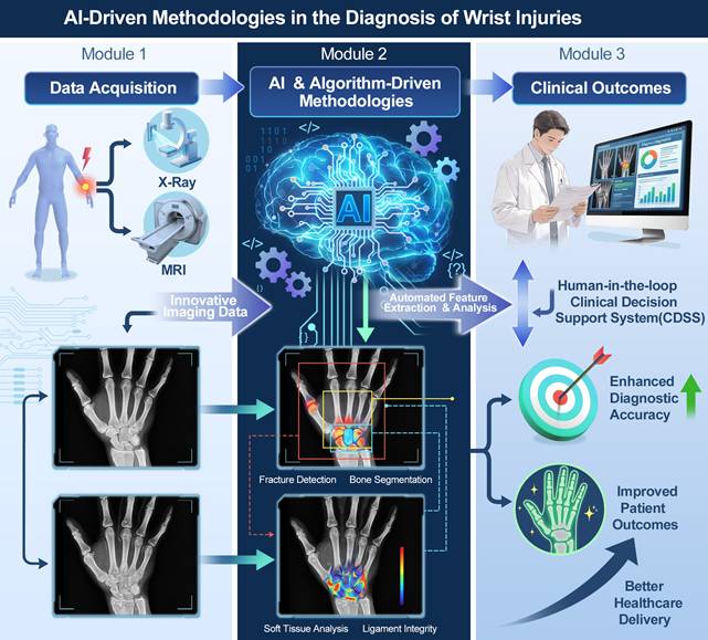
A comprehensive review of the literature underscores the increasing prominence of artificial intelligence and algorithm-driven methodologies in the diagnosis of wrist injuries. Collectively, these studies highlight the potential of AI to enhance diagnostic accuracy, improve outcomes, and aid healthcare professionals in effectively identifying and managing wrist injuries through innovative imaging and diagnostic approaches (Fig. 1).
AI Technologies in Wrist Injury Diagnosis. The process begins with data acquisition from X-ray and MRI, followed by AI-based feature extraction and analysis, ultimately leading to improved diagnostic accuracy, patient outcomes, and healthcare delivery.
CNNs are among the most extensively used AI algorithms for image classification, including detecting wrist fractures. These networks are highly effective in extracting features from images, enabling the recognition of intricate patterns indicative of fractures. Studies have shown that CNNs can attain high levels of sensitivity and specificity in identifying wrist injuries, often outperforming conventional diagnostic techniques [3,25].
CNNs have emerged as a widely used approach for image classification across various domains, including medical imaging. Within medical image analysis, CNN architectures are extensively employed for several tasks, pattern recognition and disease diagnosis [26]. Particularly, CNNs have been used to identify and classify fractures in medical images, including X-rays of extremity bones [27]. Recent studies have shown that AI-driven algorithms, particularly CNNs, can effectively detect and classify fractures of the wrist and long bones in X-ray imaging with high accuracy [25]. The use of deep learning, including CNNs, in medical image analysis has enabled the development of diagnostic models for various medical conditions, including fractures [27]. Deep learning models, once trained, can be used for tasks such as image classification, making them valuable tools for healthcare professionals [28].
The advancement of deep learning (DL) methodologies, particularly CNNs, has significantly contributed to the development of sophisticated diagnostic models for various medical conditions, including fractures. Once trained, these models exhibit a high degree of accuracy in image classification, rendering them indispensable tools for healthcare professionals [28]. Furthermore, advancements in AI, particularly the optimization of CNNs using bio-inspired algorithms, have led to significant improvements in medical imaging applications such as breast cancer detection in infrared scans [29]. In orthopedics, machine learning is increasingly utilized for diagnosing fractures and predicting their outcomes, providing valuable decision-making support for clinicians [30]. A promising development is the integration of multimodal AI, which combines imaging data with clinical information to improve diagnostic accuracy [31]. Further, deep learning-based object detection algorithms have enabled automated recognition of various medical conditions, including fractures, by leveraging neural network architectures [32]. The role of AI in medical image analysis, mainly through CNNs, is crucial in improving diagnostic efficiency and accuracy, especially for wrist fracture detection. These advancements provide potent tools for medical professionals, facilitating more reliable disease detection and classification [33]. As research progresses, the continued refinement of deep learning models is expected to enhance diagnostic capabilities further, thereby improving patient outcomes in orthopedics and other medical fields.
2.1. Deep learning models
Deep learning architectures, particularly those integrating CNNs with long short-term memory (LSTM) networks, have been developed to improve fracture detection accuracy. For instance, a fused model combining a dilated CNN and LSTM was proposed for wrist X-ray analysis, leading to enhanced diagnostic performance [34,35]. This approach enables both deep feature extraction and temporal data analysis, which is crucial for understanding wrist injury dynamics. Deep learning methods have been widely explored across various medical imaging domains, including fracture detection. For example, a diagnostic support system for distal humerus fractures was developed using deep learning and X-ray images, further illustrating its value in musculoskeletal diagnostics [36]. Moreover, CNN-based object detection was applied to security cameras and smart devices, emphasizing the role of pre-trained models in improving accuracy [37]. Similarly, a residual-based multi-stage deep learning framework was introduced for Alzheimer's disease detection, reinforcing the effectiveness of deep neural networks in medical diagnostics [38]. The reviewed literature demonstrates the increasing adoption of deep learning across multiple fields, particularly in medical imaging, security, and agriculture. These studies collectively highlight its potential to enhance diagnostic accuracy, predictive analytics, and object detection.
2.2. Gradient class activation mapping (Grad-CAM)
The Grad-CAM technique, a widely used Explainable Artificial Intelligence (XAI) method, visualizes image regions that significantly influence an AI model's classification process. By providing insights into the specific features that contribute to wrist fracture detection, Grad-CAM facilitates a deeper understanding of the model's decision-making [39,40]. This level of transparency is essential for clinical applications, as it enhances confidence in AI-assisted diagnostic systems and supports their integration into medical practice. The use of AI algorithms for wrist fracture detection has been a topic of interest in recent research. Till et al. [40] focused on the development and optimization of AI algorithms specifically for wrist fracture detection in children. The application of heat maps, particularly Grad-CAM, has proven to be an effective explainable AI technique for detecting wrist fractures in X-ray images. Studies utilizing emergency department datasets have demonstrated their effectiveness in highlighting crucial image features relevant to fracture identification [41]. This method allows for the visualization of the most relevant features for fracture detection in X-ray images. For instance, research on AI-based analysis of distal radius fractures has emphasized the significance of detecting key fracture-related characteristics to ensure accurate classification [42], with Grad-CAM emerging as a key tool for identifying critical image regions. In another study, deep learning models successfully learned to detect wrist fractures by generating activation maps that localized fracture-specific features. In another study, deep learning models were successfully trained to detect wrist fractures by generating activation maps that localized fracture-specific features [43]. Furthermore, transfer learning has been explored to enhance fracture detection, where pre-trained features from other tasks were adapted for wrist and humerus classification [44]. Integrating transfer learning with Grad-CAM provides valuable insights into the most relevant features across different bone structures. Overall, methods like CAM and Grad-CAM enhance the interpretability of deep learning models by visualizing significant activation regions linked to fracture detection [45]. Advancing these AI-driven algorithms could lead to more accurate and efficient diagnostic tools for wrist fractures.
2.3. Object detection models
The application of advanced object detection frameworks, such as You Only Look Once (YOLO), to wrist radiograph analysis has shown potential for significant improvements in fracture detection. These models trained using large annotated image datasets, can accurately and quickly locate fractures, potentially reducing the time radiologists spend on image review. The increasing adoption of object detection frameworks in medical imaging has led to significant advancements in fracture detection, with YOLO emerging as a prominent tool for analyzing radiographs. As a CNN-based detection system, YOLO has been particularly effective in identifying and localizing fractures, especially in pediatric wrist radiographs [33]. The development of dedicated datasets for fracture classification, localization, and detection further supports its growing role in this domain [46]. Recent studies have demonstrated the capabilities of YOLOv10 in automating pediatric wrist fracture detection, reflecting an ongoing shift toward automation in radiological analysis [47]. Furthermore, YOLO's utility extends beyond fracture detection to object localization and orientation estimation in medical imaging. Using oriented bounding boxes (OBBs) within the YOLO framework has facilitated improved localization and orientation assessment, highlighting its adaptability to complex medical imaging challenges [48]. The application of YOLOv3 in medical imaging has been explored extensively, with studies demonstrating its efficacy in detecting rib fractures on chest radiographs [49]. The integration of YOLO-based object detection with EfficientNet's classification network has also been utilized to automatically classify distal radius fractures, reinforcing the role of deep learning in radiographic fracture detection [50]. Moreover, cross-center validation studies have assessed YOLO-based deep learning algorithms for musculoskeletal radiography, providing further evidence of their diagnostic utility [51]. These findings suggest that object detection frameworks such as YOLO provide a robust approach for fracture detection and localization in radiographic imaging. The efficiency and accuracy of YOLO in identifying fractures across various radiograph types enhance its utility in automating and optimizing diagnostic workflows [52]. Furthermore, object detection methodologies have demonstrated superior performance to traditional segmentation-based fracture detection techniques, highlighting YOLO's role in advancing medical imaging technologies [53]. With the continuous expansion of YOLO's applications in medical imaging, its impact on automated fracture detection and localization is expected to grow further [52].
2.4. Automated assessment algorithms
Several studies have developed automated algorithms to assess the integrity of wrist ligaments, particularly the scapholunate ligament. These algorithms use deep learning methods to analyze radiographic images and correlate findings with clinical outcomes, aiding early decision-making in patient management [4]. Evaluating the integrity of wrist ligaments, particularly the scapholunate ligament (SL), is vital for diagnosing injuries and preventing long-term complications such as degenerative arthritis. Researchers have increasingly explored deep learning-based automation for ligament assessment, Keller et al. [4] designed an AI-driven algorithm to measure SL distance on radiographs, showing a significant correlation with arthroscopic results. Assessing wrist ligament integrity may be improved through automated methods. Radke et al. showed the potential of this approach by using CNNs to post-process real-time MRI images for quantifying wrist movement [54]. The development of reliable automated assessments is increasingly recognized as a valuable tool for diagnosing scapholunate ligament injuries [4], addressing a key need in current clinical practice. Deep learning algorithms offer a promising approach to evaluating wrist ligament integrity, especially the scapholunate ligament, potentially improving clinical decisions for wrist injury management.
2.5. Machine learning techniques
In addition to deep learning, traditional machine learning techniques, such as support vector machines (SVMs) and random forests, have been used for fracture detection. Their effectiveness is enhanced when paired with preprocessing methods that improve image quality and facilitate feature extraction [25,34]. Integrating machine learning algorithms into medical research has significantly advanced diagnostic accuracy and predictive modeling. In medical applications, including the detection of wrist fractures, machine learning algorithms such as SVM and Random Forests have considerable potential to improve diagnostic accuracy and decision-making processes [55,56]. The role of SVM in neurology was further exemplified by Xu et al. [57] who used wrist signal data to recognize tonic-clonic seizures, indicating its utility in seizure monitoring and prediction. Meanwhile, Mohanty et al. recommended SVM for detecting fractures from X-ray images, reinforcing its potential in radiological diagnostics [56]. These findings demonstrate the need to effectively utilize advanced technologies to improve patient care and healthcare results.
2.6. Data augmentation and preprocessing
Enhancing the performance of AI models often necessitates using data augmentation techniques. These methods generate additional training samples by modifying existing images through rotation, scaling, and flipping transformations. This process not only mitigates class imbalances but also contributes to the development of more generalized and resilient models capable of performing well across diverse datasets [34,35].
Data augmentation plays a significant role in improving AI model accuracy, particularly in wrist fracture detection. Ahmed and Manaf optimized the YOLOv8 algorithm for pediatric wrist trauma X-ray analysis [47]. Another study by Zhang et al. [58] applied data augmentation to increase dataset variability, thereby enhancing the performance of transfer-learning models in detecting wrist fractures. Beyond orthopedic applications, the benefits of augmentation extend to dentistry, where it has improved deep-learning model accuracy. Addressing class imbalance through techniques such as data augmentation significantly improves AI model performance across various medical imaging applications. In the context of wrist fracture detection, data augmentation has been shown to enhance diagnostic accuracy by increasing dataset diversity and minimizing overfitting. These improvements contribute to AI-assisted diagnostic tools' overall reliability and clinical applicability.
2.7. Natural language processing (NLP)
NLP in radiology has advanced diagnostic capabilities, particularly in fracture detection. Jungmann et al. used NLP to evaluate the temporal patterns of fracture diagnoses, particularly during the COVID-19 pandemic, providing insights into shifts in clinical presentation and healthcare utilization [59]. Chekmeyan et al. [60] introduced Cross-Check QA, a quality assurance framework designed to identify inconsistencies between AI-driven and radiologist interpretations of high-acuity CT scans, underscoring the role of AI in enhancing diagnostic reliability. Zech et al. [61] focused on the use of artificial intelligence to identify fractures in pediatric and young adult upper extremity radiographs. The study emphasized the challenges in detecting pediatric fractures due to the unique response of the pediatric skeleton to injuries. This suggests the need for specialized approaches when applying AI in fracture diagnosis for different age groups. The NLP has been explored for its role in analyzing radiology reports to aid fracture detection. One study highlighted its potential in extracting critical diagnostic information from these reports [62]. NLP applications extend beyond textual radiology reports, as machine learning-based approaches have been utilized to analyze Dutch radiology records of extremity injuries and chest radiographs, enabling the identification of fractures and pneumothorax [63]. This demonstrates NLP's adaptability in processing diverse medical datasets for diagnostic use. A review analyzed its role in orthopedic traumatology, particularly in fracture detection [64] This review emphasizes the critical role of NLP in advancing the precision of orthopedic fracture diagnosis. The integration of NLP methodologies into fracture diagnostic workflows offers a promising avenue for improving the accuracy and efficiency of these processes. By utilizing NLP algorithms to analyze radiology reports and other medical data, healthcare professionals can refine their ability to identify and classify fractures accurately. The studies indicated that NLP contributes substantially to improving fracture diagnosis by augmenting traditional diagnostic methods and AI-based systems.
2.8. Integration with other imaging modalities
AI algorithms are increasingly integrating with other imaging techniques, such as computed tomography (CT) and magnetic resonance imaging (MRI), to provide a more comprehensive assessment of wrist injuries. This multimodal approach can enhance diagnostic accuracy and facilitate better treatment planning [25]. AI in diagnosing wrist injuries uses various sophisticated algorithms and methodologies, particularly deep learning models such as CNNs, object detection frameworks, and automated assessment algorithms. These advanced tools increase diagnostic precision and streamline radiological evaluations, improving patient outcomes.
AI algorithms integrated with imaging techniques such as CT and MRI have shown potential in enhancing the comprehensive assessment of medical conditions. In orthopedics, AI has been used for fracture diagnosis in X-rays, improving accuracy and efficiency. It has also been combined with CT and MRI to provide detailed evaluations of wrist injuries [25]. The increasing use of medical imaging techniques, mainly CT and MRI, has resulted in a rise in incidental findings, underscoring the need for advanced technologies like AI to enhance interpretation and diagnosis [65]. AI can integrate data from multiple imaging modalities, including echocardiography, CT, MRI, and PET, enabling a comprehensive evaluation of conditions such as cardiovascular diseases [66]. Despite the advantages of traditional imaging methods, they have some limitations. For instance, MRI segmentation techniques for shoulder injuries may not fully capture the extent of tissue damage, limiting their diagnostic utility. AI-based analytical models can address these challenges by offering enhanced image interpretation, facilitating more precise injury characterization, and improving clinical outcomes [67]. The combination of different imaging methods, such as fMRI alongside EEG and MEG, provides a more detailed and holistic perspective on brain function, emphasizing the strengths of multimodal imaging [68]. In summary, incorporating AI-driven algorithms into imaging techniques like CT and MRI can significantly improve diagnostic precision and efficiency, facilitating more accurate evaluations of various medical conditions, including wrist injuries.
2.9. Quantitative synthesis of AI diagnostic performance for wrist fractures
Several recent meta-analyses have provided pooled estimates of AI diagnostic performance for wrist fracture detection (Table 2). A systematic review and meta-analysis by Suen et al. included 20 studies and reported that the pooled sensitivity and specificity for AI in detecting distal radius fractures were 0.92 (95% CI: 0.88-0.95) and 0.89 (95% CI: 0.84-0.92), respectively; for human readers, the corresponding values were 0.95 (95% CI: 0.91-0.97) and 0.94 (95% CI: 0.91-0.96) [69]. For scaphoid fractures, the pooled sensitivity and specificity for AI were 0.85 (95% CI: 0.73-0.92) and 0.83 (95% CI: 0.76-0.89), whereas human readers exhibited 0.71 (95% CI: 0.66-0.76) and 0.93 (95% CI: 0.90-0.95), respectively. These findings indicate that AI performance is comparable to human readers for distal radius fractures, while for scaphoid fractures, AI demonstrates higher sensitivity but lower specificity than human experts.
Diagnostic performance of AI models for wrist fracture detection
| Fracture type / Reader | Pooled sensitivity (95% CI) | Pooled specificity (95% CI) | AUC / LR | Refs. |
|---|---|---|---|---|
| Distal radius (AI) | 92% (88-95) | 89% (84-92) | — | [69] |
| Distal radius (human) | 95% (91-97) | 94% (91-96) | — | [69] |
| Scaphoid (AI) | 85% (73-92) | 83% (76-89) | — | [69] |
| Scaphoid (human) | 71% (66-76) | 93% (90-95) | — | [69] |
| CNNs (overall wrist) | 92% (80-97) | 93% (76-98) | — | [9] |
| Hand/wrist (overall) | — | — | AUC 0.946; LR+ 7.69 (6.40-9.19) | [71] |
A complementary meta-analysis by Hansen et al., which focused specifically on DL algorithms using CNNs, included six studies comprising 33,026 radiographs and found a summary sensitivity of 92% (95% CI: 80-97%) and a summary specificity of 93% (95% CI: 76-98%) for CNN-based models compared with a reference standard of healthcare expert consensus [70].
For hand and wrist fractures more broadly, a meta-analysis by Wong et al. of 36 studies reported that AI models achieved an area under the curve (AUC) of 0.946 (95% CI not reported), a positive likelihood ratio (LR+) of 7.69 (95% CI: 6.40-9.19), and a negative likelihood ratio (LR-) of 0.11 (95% CI: 0.08-0.15) [71].
A separate meta-analysis of 26 studies assessing AI assistance for clinicians found that AI support significantly improved pooled sensitivity from 77% (95% CI: 72-81) to 87% (95% CI: 83-90) and pooled specificity from 88% (95% CI: 85-90) to 92% (95% CI: 89-94), with the corresponding AUC increasing from 0.90 (95% CI: 0.87-0.92) to 0.95 (95% CI: 0.93-0.97) [72].
These pooled estimates confirm that AI, particularly CNN-based deep learning models, achieves high diagnostic accuracy for wrist fracture detection, with performance comparable to or exceeding that of human readers for certain fracture types, especially when used as an assistive tool.
2.10. Comparative analysis of AI architectures for wrist fracture detection
A critical comparative analysis of different AI architectures provides clinically actionable insights. The most widely applied architectures for wrist fracture detection fall into two broad categories: (1) CNNs optimized for image classification, and (2) object detection models that provide simultaneous fracture localization and classification.
CNN-based classification models treat fracture detection as a binary or multi-class classification problem, outputting a probability score without inherent spatial localization [3,70]. Their strengths include architectural maturity, availability of pre-trained models for transfer learning, and excellent performance on well aligned radiographs [73]. These models treat fracture detection as a binary or multi-class classification problem, outputting image-level probability scores without inherent spatial localization. While techniques such as Grad-CAM can provide approximate attention maps, these are less precise than bounding boxes for identifying multiple concurrent fractures.
Object detection models (single-stage architectures such as YOLO, and two-stage architectures such as Faster R-CNN) output bounding boxes around suspected fracture sites [10,74]. This localization capability reduces cognitive load, enables counting of multiple fractures, and supports automated measurement. However, they require larger annotated datasets and are computationally more intensive.
Quantitative comparisons on the GRAZPEDWRI-DX pediatric wrist dataset provide guidance for architecture selection. Ahmed et al. [75] evaluated YOLOv5, YOLOv6, YOLOv7, and YOLOv8 against Faster R-CNN, finding that YOLO models consistently outperformed Faster R-CNN. Specifically, YOLOv8m achieved a fracture detection sensitivity of 0.92 and a mean average precision (mAP) of 0.95 at IoU 0.5 for the fracture class, while YOLOv8x recorded the highest overall mAP of 0.77 across all abnormality classes [75]. These data confirm that single-stage object detectors offer superior performance for wrist fracture detection compared to two-stage architectures.
3. Effectiveness of AI in diagnosing wrist injuries: Advantages over traditional methods
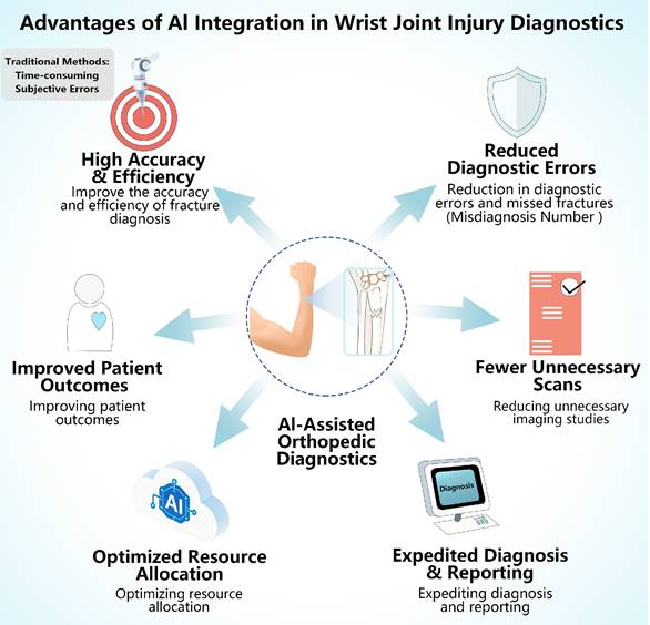
The application of AI in diagnosing wrist joint injuries has shown significant effectiveness, particularly in detecting common fractures such as those of the distal radius, ulnar styloid process, and scaphoid. A study demonstrated that an AI model trained on 4,432 medical images achieved high sensitivity and specificity in detecting fractures. The model yielded an AUC value of 0.903 for distal radius fractures, 0.925 for ulnar styloid fractures, and 0.808 for scaphoid fractures. When novice radiologists used the AI model as an assistive tool, their diagnostic accuracy for scaphoid fractures, frequently missed in clinical practice, improved significantly. This highlights the potential of AI to augment human performance in complex diagnostic settings [3]. One of the primary advantages of AI over traditional diagnostic methods is its ability to process large datasets quickly and consistently. The conventional approach to fracture diagnosis relies on radiologists, whose evaluations are inherently subjective and may be affected by fatigue, variability in interpretation, and cognitive biases. Recent advances in AI, especially deep learning methods such as CNNs, offer a promising alternative by recognizing patterns and features in medical images that might not be immediately obvious to the human eye. This capability not only reduces the likelihood of missed diagnoses but also improves the overall accuracy of fracture detection [6,25]. The application of AI in radiology enhances the interpretation of complex fracture patterns and subtle abnormalities that even expert radiologists may overlook. Scaphoid fractures, although uncommon, require rapid identification because of their potential for serious complications. Their detection on radiographs can be challenging, but AI's contribution to increasing the detection rate of such fractures is crucial. Failure to diagnose scaphoid fractures can result in severe outcomes, including non-union and avascular necrosis [3,25]. AI enhances diagnostic accuracy and optimizes operational workflows in radiology departments. By automating the preliminary evaluation of radiographs, AI reduces the burden on radiologists, enabling them to focus on more complex clinical cases and decreasing the time required for image interpretation. This improvement is particularly advantageous in busy clinical environments, where the ability to provide timely diagnoses is vital for effective patient management [25,76]. Integrating AI with other imaging modalities, such as CT and MRI, can provide a more comprehensive diagnostic approach. This multimodal approach enhances the precision of wrist injury classification and diagnosis, thereby contributing to improved patient outcomes [25]. Despite the advantages offered by AI in clinical practice, there are significant challenges to its implementation. These include ethical considerations, regulatory issues, and the need for high-quality annotated datasets for training AI models. Overcoming these obstacles is essential for AI's safe and effective use in medical diagnostics [25,76]. AI has demonstrated significant effectiveness in diagnosing wrist joint injuries, presenting clear advantages over traditional diagnostic methods (Fig. 2). These benefits encompass improved diagnostic accuracy, consistency, and operational efficiency. As AI progresses, its integration into clinical settings is poised to raise the standard of care in orthopedic diagnostics.
Advantages of AI in Wrist Injury Diagnosis. AI enhances diagnostic accuracy and efficiency, reduces errors and missed fractures, limits unnecessary imaging, accelerates diagnosis and reporting, optimizes resource use, and ultimately improves patient outcomes.
3.1. AI's Impact on the precision and effectiveness of diagnosing wrist injuries
Incorporating AI into the diagnosis of fractures has been demonstrated to significantly improve the accuracy and efficiency of diagnosing wrist injuries. AI holds the potential to improve diagnostic precision, optimize workflow, and enhance the reliability, convenience, and effectiveness of diagnostic procedures [77]. Advanced AI technology enables healthcare practitioners to achieve greater diagnostic accuracy and speed when identifying wrist injuries [25]. AI can also assist in minimizing missed or delayed diagnoses of hand and peripheral nerve injuries, which are often difficult to evaluate [78]. Commercially available AI tools have been demonstrated to enhance the diagnostic process, significantly boosting the performance of radiologists when used alongside traditional diagnostic techniques [79]. Studies have also explored the role of DL in medical image analysis, showing that these methods improve diagnostic accuracy and operational efficiency [28]. The integration of AI into healthcare has the potential to support healthcare professionals and patients across the care continuum, from diagnosis to treatment [80]. Although AI shows promise in diagnostic applications, there are still barriers to its integration into clinical treatment processes [81]. Incorporating AI into fracture diagnosis could significantly transform the process of diagnosing wrist fractures, increasing the precision and efficiency of detection and treatment.
3.2. Reduction in diagnostic errors and missed fractures
The integration of AI into fracture diagnostics has been extensively evaluated, with studies highlighting its role in reducing missed fractures and diagnostic errors. AI-assisted algorithms have demonstrated superior accuracy in detecting fractures on orthopedic X-rays [25]. Automated object detection algorithms have been proposed to enhance diagnostic reliability, particularly in high-pressure trauma settings where fractures may be overlooked [82]. AI-based systems for hip fracture diagnosis have shown rapid and precise identification capabilities, emphasizing their value in clinical settings [83]. Furthermore, AI models have significantly improved wrist trauma diagnostics, reducing reliance on additional diagnostic procedures [36]. AI-based models have also been shown to detect commonly missed fractures, such as distal radius fractures, one of the most frequent fracture types [84]. Similarly, across various anatomical regions, AI-supported fracture detection has demonstrated high diagnostic accuracy, particularly when combined with traditional methods [85]. Overall, the incorporation of AI into fracture diagnosis can substantially decrease diagnostic errors and overlooked fractures, thereby enhancing patient outcomes and optimizing the diagnostic workflow. AI-driven systems improve diagnostic accuracy and efficiency, facilitating timely and precise treatment strategies and contributing to superior patient care.
3.3. Reducing unnecessary imaging: AI's contribution to diagnostics
Integrating AI into medical diagnostics has demonstrated substantial potential in enhancing accuracy and efficiency. Within orthopedic practice, AI has been particularly effective in fracture detection, contributing to a reduction in unnecessary imaging studies [25]. The application of AI models for detecting fractures in radiographic images facilitates early diagnosis, thereby reducing redundant clinical evaluations [86]. This technological advancement streamlines diagnostic workflows and improves patient outcomes. A systematic review and meta-analysis by Wong et al. [71] evaluated AI's performance in diagnosing hand and wrist fractures and dislocations. The study demonstrated AI's potential to enhance diagnostic accuracy and reduce unnecessary imaging, reinforcing its value in clinical practice. The integration of AI into clinical practice has been a topic of interest in recent literature [77]. Advancements in deep learning for fracture detection, with a focus on scaphoid fractures, have led to improved diagnostic accuracy, potentially decreasing the incidence of unnecessary immobilization [87]. Integrating AI in fracture diagnosis enhances diagnostic speed and efficiency, improving patient outcomes. However, AI applications in this field remain constrained by certain limitations that warrant further exploration. For instance, some systematic reviews have excluded studies involving CT imaging, underscoring the necessity for more comprehensive research into AI-based fracture assessment [85]. To minimize risks associated with pediatric upper extremity imaging and to facilitate the effective integration of AI into global healthcare systems, further research is essential [88,89]. In summary, AI has the potential to significantly enhance fracture diagnosis by reducing redundant imaging, increasing diagnostic accuracy, and improving patient care. Future studies should address technological limitations, optimize AI algorithms for clinical application, and evaluate long-term impacts on orthopedic decision-making.
3.4. Expediting diagnosis and reporting
The application of AI in wrist fracture diagnosis represents a promising advancement in medical imaging, facilitating faster and more accurate diagnostic processes. While comprehensive frameworks for human-AI interaction have been established in general radiology, distinguishing integration models such as AI as triage tool and AI as secondary or safety-net reader-direct validation of these specific workflow models in the context of wrist fracture detection remains limited [90]. Most wrist fracture AI studies to date have evaluated AI as a concurrent decision support tool or as a second reader in retrospective simulation setting, rather than prospective implementation of triage or safety-net workflows. Future research should specifically assess these distinct integration strategies for wrist imaging to determine which model optimizes diagnostic accuracy, workflow efficiency, and cost-effectiveness in real-world clinical practice.
In the concurrent decision support model evaluated by Lee et al., AI assistance was provided to novice radiologists during image interpretation through heatmap visualizations and probability scores. This approach significantly improved diagnostic performance for scaphoid fractures, which are frequently overlooked in standard clinical assessments: the AUC for scaphoid fracture detection increased from 0.75 to 0.85 for one novice radiologist and from 0.71 to 0.80 for another. However, the study did not evaluate AI as an independent triage tool or as a sequential second reader; rather, AI served as a real-time assistive device during the interpretation process. The authors noted that interpretation time increased slightly with AI assistance, highlighting the importance of optimizing workflow integration [3].
Furthermore, the benefits of AI assistance extend beyond diagnostic accuracy improvements to include reductions in unnecessary diagnostic procedures and optimization of clinical decision-making. In a prospective study of emergency department clinical decision-making, Keller et al. [36] evaluated physicians using an AI tool for distal radius fracture detection. The AI-supported group was able to make a diagnosis without additional support in 75% of cases, compared to only 52% in the control group without AI assistance. The AI-supported group also required significantly fewer additional CT scans, thereby reducing unnecessary radiation exposure and potential healthcare costs. Notably, physicians using the AI tool reported significantly lower subjective stress levels, suggesting that AI assistance may alleviate cognitive burden in time-sensitive emergency settings. These findings indicate that reliable AI fracture detection models can not only enhance diagnostic confidence but also streamline clinical workflows by reducing reliance on confirmatory imaging and specialist consultations.
From a broader perspective, a systematic review by Kwee et al. [91] evaluating the commercially available AI tool BoneView across multiple anatomical regions found that AI assistance significantly improved sensitivity for fracture detection in the majority of readers, although specificity results were mixed. Importantly, reading speed was either unchanged or significantly improved in three out of four studies. This apparent improvement in reading speed contrasts with the slight increase in interpretation time reported by Lee et al. [3] for novice radiologists using concurrent AI assistance. This discrepancy may reflect differences in AI interface design, reader experience levels, the specific workflow integration model employed, or the complexity of the fracture types being evaluated. The systematic review also noted substantial heterogeneity across studies and highlighted methodological quality concerns, emphasizing the need for rigorous prospective validation before widespread clinical implementation.
In the specific context of pediatric wrist trauma, Ju et al. [14] demonstrated that a YOLOv8-based fracture detection model augmented with contrast and brightness adjustment achieved state-of-the-art mean average precision, outperforming YOLOv7-based models. The authors further developed a user-friendly graphical interface application designed to assist pediatric surgeons in regions with limited access to radiologists, illustrating how AI can democratize fracture diagnostic capabilities across diverse healthcare settings.
Collectively, these studies demonstrate promising tangible benefits of AI in wrist fracture diagnosis, including improved diagnostic accuracy for subtle fractures [3], reduced unnecessary CT scans and physician stress [36], and maintained or improved reading speed in most clinical contexts [91]. However, the optimal integration model for routine clinical practice-whether concurrent decision support, second reader, or triage tool-likely remains context-dependent and requires further prospective, multi-institutional validation to establish evidence-based guidelines for AI deployment. Improving AI-driven X-ray interpretation requires larger, higher-quality training datasets and incorporating advanced machine learning approaches, including deep reinforcement learning [25]. In a systematic review and meta-analysis conducted, the accuracy of AI models in identifying hand and wrist fractures and dislocations was determined, highlighting the potential of AI in fracture diagnosis [71]. A deep learning algorithm was developed and trained for detecting fractures in wrist X-rays, demonstrating high accuracy and sensitivity, as reported in a study on using artificial intelligence for fracture diagnosis in orthopedic X-rays [25]. Similarly, integrating AI models into clinical workflows has significantly improved diagnostic processes, as mentioned in Artificial Intelligence-Based Applications for Bone Fracture Detection [83]. Integrating AI into clinical practice offers significant advantages, including the ability to analyze large datasets efficiently and accurately. The research emphasizes AI's potential to accelerate wrist fracture diagnosis and reporting processes. The incorporation of AI into disease diagnosis and classification, highlighted by [92], can address various challenges and facilitate the smooth integration of AI into medical practice. In wrist fracture diagnosis, AI shows promise in accelerating diagnosis and enhancing the accuracy of reports. The application of cutting-edge machine learning models and the integration of AI systems into clinical workflows could significantly improve the practice of orthopedic X-ray analysis.
3.5. Workflow integration and cost-effectiveness
The application of AI in fracture diagnosis has demonstrated promising potential for optimizing resource allocation within healthcare settings [25]. Recent economic evaluations provide concrete evidence for the cost-effectiveness of AI-assisted fracture detection. A comprehensive budget impact analysis published in Value in Health evaluated the adoption of a commercially available AI fracture detection application across NHS emergency departments in England [93]. Over a one-year horizon, the analysis estimated that among 658,564 radiographs performed for suspected wrist, ankle, or hip fractures, AI integration would reduce the number of patients returning to the ED with a missed fracture by approximately 21,674 cases and reduce unnecessary referrals to fracture clinics by approximately 20,916 cases. The cost of current practice was estimated at £66,646,542, while the AI-integrated scenario reduced costs to £63,012,150, generating a net return on investment of £3,634,392 for the NHS.
Beyond economic considerations, successful integration of AI into clinical workflows requires a structured implementation framework. Shah et al. [94] proposed a four-phase framework for responsible AI deployment in radiology: (1) validation-retrospective or offline testing on institutional data to assess local performance; (2) deployment-progressing from limited trial to full deployment with emphasis on workflow integration and stakeholder feedback; (3) value assessment-longitudinal evaluation of financial and non-financial returns on investment; and (4) post-deployment surveillance-monitoring for data drift and maintaining AI safety. Similarly, Trivedi et al. [95] described a rubric-based approach for AI model evaluation and deployment at a large academic center, emphasizing holistic assessment beyond performance metrics.
A review on human-AI interaction in radiology outlines several workflow integration models, including AI as a triage tool (pre-screening normal cases), AI as a second reader (safety net for missed findings), and AI as a concurrent support tool (real-time decision aid) [90]. The choice of integration model depends on clinical context, AI performance characteristics, and local workflow requirements. Separately, the TRIAGE framework provides a clinically grounded evaluation structure for diagnostic AI testing aligned with real clinical use cases, including screening, triage, second reading, and confirmatory testing [96]. In addition to direct cost savings, AI integration offers non-monetary benefits including the potential to reduce physician burnout through workload redistribution, improve patient satisfaction through faster diagnostic turnaround, and enhance training opportunities for residents who can use AI-generated annotations as educational tools. By prioritizing the optimization of workflows and resource distribution, healthcare providers can elevate care quality while achieving measurable cost reductions. By streamlining workflows and improving cost efficiency, AI algorithms can significantly increase operational effectiveness [97]. This optimization supports better patient outcomes and personalized treatment planning [98]. Moreover, the integration of Internet of Things (IoT) applications, including wearable devices and medical technologies, strengthens the role of technology in patient care [99]. The real-time monitoring and data collection enabled by these tools are essential for improving resource management and healthcare delivery. As healthcare systems evolve, the need for efficient resource management grows in importance [88]. By utilizing AI and IoT to predict patient flow and streamline inventory management, healthcare systems can ensure better resource allocation and improved patient care [100]. By prioritizing the optimization of workflows and resource distribution, healthcare providers can elevate care quality and help shape the future of healthcare.
3.6. Improving patient outcomes
Incorporating AI into wrist fracture diagnosis has demonstrated the potential to improve patient outcomes. AI algorithms contribute to more precise treatment decisions, which enhance patient classification and overall results [25]. Medical imaging, such as X-rays, is integral to diagnosing and managing fractures. Incorporating AI into this imaging process could significantly elevate patient outcomes in orthopedic care [25]. One strategy to enhance AI interpretation of X-ray images is to expand both the size and quality of the datasets used for training while also adopting more advanced machine learning methods, including deep reinforcement learning [25]. AI integration in medical imaging can streamline clinical workflows, reduce diagnostic errors, and raise the standard of medical care, leading to better patient outcomes [101]. Studies have shown that incorporating AI into clinical practice can enhance medical care and improve health outcomes for patients with conditions such as burns, wounds, and diabetic retinopathy [77,98]. By incorporating AI along with pharmacogenomic data, healthcare providers can achieve better clinical results and provide more effective patient care [92]. The overall integration of AI into wrist fracture diagnosis holds the potential to revolutionize clinical care, ensuring accurate diagnoses, timely treatment decisions, and improved patient outcomes and quality of care [102].
4. Challenges
The incorporation of AI into the process of diagnosing fractures through orthopedic X-rays introduces significant ethical and legal challenges that require thorough consideration [25]. A critical concern raised in various studies is the potential influence of AI on the emotional experiences of patients [25]. In the healthcare setting, the application of AI raises four primary ethical issues, including the necessity for informed decision-making, that must be resolved to achieve its full potential [103]. The implementation of large language models in healthcare raises significant privacy and security risks, which must be managed thoughtfully to prevent harm to patients and avoid legal implications [104]. The emergence of mental health chatbots has brought critical ethical challenges, as these tools incorporate varying levels of artificial intelligence [105]. The broader integration of AI into healthcare raises fundamental ethical questions, particularly regarding fairness and transparency [106]. The potential biases inherent in AI models require careful evaluation, emphasizing the importance of addressing ethical concerns and biases in AI and machine learning systems [107]. As AI continues to reshape healthcare, the accompanying ethical, legal, and human resource challenges are becoming more prominent [108]. The increase in research on AI ethics reflects a growing awareness of the ethical issues surrounding AI adoption in healthcare [109]. Consequently, the application of AI in healthcare, including in fracture diagnosis, must be approached with a detailed assessment of both ethical and legal implications to guarantee responsible and effective use.
The integration of artificial intelligence into clinical practice raises concerns about transparency, accountability, and potential bias within algorithms [77]. Within the field of radiology, a critical ethical issue is whether AI can achieve diagnostic accuracy equivalent to that of human professionals in identifying fractures [110]. A systematic review of AI applications in orthopedic conditions highlights the necessity for transparent AI algorithms to promote ethical standards in healthcare [111]. Moreover, various initiatives led by government bodies are evaluating how federal agencies can effectively implement AI technologies [112]. AI and machine learning research in healthcare further emphasizes the importance of ensuring transparency, replicability, and ethical considerations in the development and adoption of these algorithms [113]. A primary ethical concern in AI adoption is the need for transparent and interpretable algorithms, particularly in applications such as wrist fracture diagnosis [25].
Integrating AI into medical imaging, with a particular focus on fracture diagnosis, has led to significant ethical challenges, primarily surrounding patient privacy and data protection. The literature emphasizes the transparency and explainability of AI algorithms as critical factors for the successful adoption of AI in medical settings [25]. However, patient data privacy and security concerns are significant limiting factors in applying AI technology within healthcare systems [92]. As the demand for medical imaging grows, radiologists face increased pressure, positioning AI as a valuable tool for assisting in diagnosis [110]. However, protecting patient privacy and concerns over liability associated with AI usage remain critical challenges in the field [108]. AI has demonstrated promising potential in improving diagnostic accuracy in orthopedic disease diagnosis, particularly in studies involving patients with wrist fractures [111]. However, ensuring patient privacy and securing data remain essential, necessitating robust ethical guidelines and legal frameworks to govern AI usage [77]. Concerns regarding data security and privacy in healthcare settings have been highlighted in the literature, emphasizing the need for stringent safeguards to protect sensitive medical data [88,99]. Addressing these ethical concerns as AI advances will be crucial for the responsible integration of AI technologies in patient care.
Beyond the general ethical and regulatory challenges discussed above, several barriers specifically impede the clinical deployment of AI for wrist fracture detection. Integration with existing PACS and RIS systems remains technically challenging, as many AI solutions require custom middleware or cloud-based processing that may not comply with institutional data governance policies [90]. Workflow disruption during the transition period can temporarily reduce radiologist efficiency, necessitating careful change management and parallel testing phases [94]. Algorithm performance drift over time—due to changes in imaging equipment, patient demographics, or referral patterns—requires ongoing post-deployment surveillance and model retraining protocols [94,96]. Liability and accountability questions remain unresolved: under current medical malpractice frameworks, there is no transfer of liability to AI systems, and the allocation of legal responsibility for errors arising from joint human-AI decisions remains unclear [90]. Automation bias, the tendency to over-rely on AI recommendations, and algorithmic aversion, excessive distrust of AI outputs, represent cognitive risks that require explicit mitigation strategies, especially for trainees. Addressing these implementation-specific barriers will be essential for successful and sustainable AI adoption in orthopedic radiology.
AI fracture detection software is regulated as Software as a Medical Device (SaMD). In the U.S., the FDA has authorized 950 AI/ML-enabled medical devices as of June 2024, with 97% cleared via the 510(k) pathway [114]. The FDA has issued two complementary guidances for AI-enabled medical devices in 2025. A draft guidance on Artificial Intelligence-Enabled Device Software Functions: Lifecycle Management and Marketing Submission Recommendations emphasizes a total product lifecycle approach, covering design, development, post-market monitoring, transparency, and bias mitigation throughout the device's lifecycle. A final guidance on Marketing Submission Recommendations for a Predetermined Change Control Plan for Artificial Intelligence-Enabled Device Software Functions establishes Predetermined Change Control Plans, which allow manufacturers to pre-specify algorithm updates—including modifications to improve sensitivity or specificity—that can be implemented without additional marketing submissions once authorized by the FDA as part of an initial 510(k), De Novo, or PMA application. These frameworks support iterative improvement of AI fracture detection software while maintaining regulatory oversight (Supplemental file 2). In the EU, AI-enabled medical devices must comply with both the Medical Device Regulation (MDR 2017/745) and the AI Act (2024/1689), creating overlapping requirements for conformity assessment, risk classification, clinical evaluation, and post-market surveillance. The AI Act envisages that there shall be no duplications, and notified bodies competent to assess medical devices under the MDR may also be designated under the AI Act to enable joint CE marking conformity assessment. However, the article notes that such combined conformity assessment may not always be straightforward, as there is no unified European regulatory institute to harmonize implementation across member states [115]. In the UK, NICE's early value assessment conditionally recommends four AI fracture detection technologies—BoneView, RBfracture, Rayvolve, and TechCare Alert—for use in NHS urgent care settings during a 2-year evidence generation period, subject to ongoing data collection and regulatory approval including NHS England's Digital Technology Assessment Criteria (DTAC) (Supplemental file 1). Manufacturers should align their development and quality management processes with internationally recognized standards. ISO 13485:2016 establishes requirements for medical device quality management systems and is recognized by the FDA, MHRA, and IMDRF. ISO 14971:2019 provides the framework for risk management throughout the device lifecycle, which IMDRF's software risk characterization guidance explicitly references. Additionally, IEC 62304 defines the software lifecycle processes required for medical device software development and maintenance, and is currently being revised to incorporate AI-specific requirements for AI-enabled medical devices (https://pureclinical.eu/news/mhra-imdrfs-latest-guidance-on-ai-and-medical-device-software;https://www.itu.int/en/ITU-T/studygroups/2025-2028/21/Pages/rm/dhealth.aspx). Understanding these pathways facilitates smoother market access and responsible clinical adoption.
While the diagnostic performance metrics reported in Section 3 demonstrate the potential of AI for wrist fracture detection, the generalizability of these findings across diverse populations and clinical settings remains a critical concern. A systematic review by Oliveira e Carmo et al. examined 36 studies developing CNNs for fracture detection and found that only four reported any form of external validation [116]. This scarcity of external validation greatly limits the potential for transferring these models from their development institutions to other hospitals with comparable diagnostic performance [116].
The impact of cross-population domain shift-distributional differences arising from demographic variability, variations in imaging protocols, scanner hardware, and differences in disease prevalence, can degrade model performance by 10-25% when models are tested on unseen populations [117]. For wrist fracture detection specifically, Raisuddin et al. reported that a deep learning model achieving near-perfect performance (AUROC 0.99) on a general population test set dropped substantially to an AUROC of 0.84 on a challenging test set comprising cases requiring CT confirmation, highlighting the vulnerability of AI models to shifts in case mix and disease severity [10].
Most AI fracture detection studies have been developed using datasets from single centers or geographically limited populations, raising concerns about their applicability to broader patient groups. A cross-population validation study by Ruitenbeek et al. evaluated an AI tool trained on over 1.5 million radiographs from Indian hospitals on a Dutch cohort of 14,311 radiographs, finding that classification performance remained robust, but fracture localization accuracy varied substantially across anatomical regions, from 90% for clavicle fractures to only 7% for rib fractures [118]. This demonstrates that while classification performance may generalize across populations, localization accuracy-critical for surgical planning-remains highly context-dependent and requires careful validation in target clinical settings.
Wrist fracture detection in older adults poses particular challenges, as age-related bone density loss, osteopenia, and osteoporosis alter radiographic bone texture, potentially affecting AI model performance. Although analogous systematic studies for wrist fracture AI are lacking, the principle that AI models can underperform in demographic subgroups underrepresented in training data is well established in radiology [110]. Given that fracture patterns and radiographic characteristics differ substantially between children and adults, wrist fracture AI models trained predominantly on adult or mixed-age populations may not generalize reliably to pediatric or geriatric populations without dedicated validation. For example, both Zech et al. and Ju et al. developed pediatric specific models trained exclusively on pediatric datasets, highlighting the importance of population matched training data [14,23].
To address these generalizability concerns, several strategies are recommended. Prospective external validation on geographically and demographically diverse, consecutively enrolled patient cohorts is essential before clinical deployment. Subgroup performance analyses by age, sex, and ethnicity should be routinely reported to identify potential biases. The use of reporting guidelines such as CONSORT-AI, SPIRIT-AI, and TRIPOD-ML can improve methodological rigor and facilitate critical appraisal of model performance [116]. Ongoing real-world surveillance, as recommended by Shah et al., should monitor for performance drift due to changes in patient demographics, imaging equipment, or referral patterns [94]. Trivedi et al. similarly advocate for continuous post deployment monitoring to ensure AI models remain safe and effective in real-world clinical settings [95]. While AI shows substantial promise for wrist fracture detection, current evidence is limited by a lack of rigorous external validation and insufficient evaluation across diverse populations. Researchers and clinicians should interpret reported performance metrics with caution, prioritize external validation on local, representative data before adoption, and actively work to ensure that training datasets reflect the demographic diversity of target patient populations.
5. Future prospects of AI in wrist injury diagnosis and treatment
The application of AI in the field of wrist injuries offers substantial potential for enhancing diagnostic accuracy, treatment planning, and patient outcomes. Various studies have emphasized the role of AI technologies across various aspects of wrist injury care, with a particular focus on fracture detection and rehabilitation. A significant advancement in this field is the development of AI algorithms for detecting wrist fractures in radiographs. Research suggests that AI models surpass non-specialized radiologists in identifying wrist fractures, with an observed sensitivity of 83%, compared to 76% for initial radiology reports made by non-specialized radiologists. When AI is used in conjunction with radiologist interpretation, sensitivity increases to 88%, although specificity slightly decreases to 92% [119]. This indicates that AI integration into clinical practice could enable more accurate and timely diagnoses, potentially reducing missed fractures, especially in complex cases such as scaphoid fractures, which are challenging to detect [3,119]. Research has explored the feasibility of using advanced imaging modalities, such as 3DUS coupled with AI, for evaluating pediatric wrist fractures. A study involving children with wrist injuries demonstrated that AI-based interpretation of 3DUS scans achieved a sensitivity of 100%, suggesting its effectiveness in ruling out fractures before traditional radiographic imaging [5]. This approach could significantly decrease unnecessary radiation exposure in pediatric populations, highlighting AI's potential to enhance patient safety while preserving diagnostic precision. In addition to fracture detection, AI is being leveraged to create predictive models for scaphoid fractures based on clinical factors such as pain location, age, and injury mechanism. Machine learning models trained on these parameters can assist clinicians in determining when advanced imaging is needed, thereby reducing unnecessary imaging procedures while ensuring appropriate care for high-risk patients [120]. The application of AI in rehabilitation medicine represents a promising field of development. Machine learning and AI tools are being used to develop individualized treatment protocols for patients with motor impairments, including those recovering from wrist injuries. By analyzing data collected from wearable sensors and robotic assistive devices, AI can guide adaptive neurorehabilitation approaches customized to each patient's needs [121]. This can potentially improve rehabilitation outcomes by optimizing recovery processes and enhancing patient satisfaction. Furthermore, developing XAI methods, such as Grad-CAM, enhances the interpretability of AI models in clinical settings. These methods allow clinicians to visually interpret the areas of an image that the AI model prioritizes when making diagnostic assessments, thereby strengthening the reliability of AI-driven clinical decisions [40]. As AI technologies progress, their transparency and reliability will be critical to their integration into clinical practice. However, challenges persist in the broad adoption of AI for managing wrist injuries. Robust validation of AI models across diverse clinical settings is necessary to ensure both their efficacy and safety. Moreover, healthcare professionals need proper training in interpreting AI-generated insights and seamlessly integrating them into routine clinical practice [121,122].
6. Conclusion
In conclusion, the potential of artificial intelligence in managing wrist injuries is vast, with significant advantages in areas such as fracture detection, treatment planning, and rehabilitation. Ongoing advancements and research are expected to produce more refined AI tools to improve patient care and treatment outcomes significantly. As these technologies evolve, they could become a foundational component of orthopedic practice, reshaping the management of wrist injuries.
Abbreviations
AI: artificial intelligence
AP: average precision
AUC: area under the curve
CNN: convolutional neural networks
CT: computed tomography
DCNN: deep convolutional neural networks
DL: deep learning
DRF: detection of distal radius fractures
GAN: generative adversarial network
Grad-CAM: gradient-weighted class activation mapping
IOT: internet of things
LRP: localization recall precision
LSTM: long short-term memory
Map: mean average precision
MRI: magnetic resonance imaging
MSK: musculoskeletal
NLP: natural language processing
SaMD: software as a medical device
SL: scapholunate ligament
SSIM: structural similarity index
SVMs: support vector machines
TL: transfer learning
WJI: wrist joint injuries
XAI: explainable artificial intelligence
3DUS: three-dimensional ultrasound
Supplementary Material
Supplementary file 1.
Supplementary file 2.
Acknowledgements
The authors would like to thank all the reviewers who participated in the review and MJEditor (www.mjeditor.com) for its linguistic assistance during the preparation of this manuscript.
Funding
This research was funded by the Department of Science and Technology of Jilin Province, with grant number (20250203164SF).
Data availability
The datasets generated and analysed during this study are available from the corresponding author on reasonable request.
Authors' contributions
H.L., and X.L., conceptualized the study, acquired the data, analysed the data, drafted and wrote the manuscript. They have all read and approved the final manuscript.
Declaration of AI use
During the preparation of this manuscript, the author(s) used [AI tool name and version, e.g., ChatGPT-4] for language polishing and reference format checking. After using this tool, the author(s) reviewed and edited the content as needed and take full responsibility for the content of the publication.
Competing Interests
The authors have declared that no competing interest exists.
References
1. Oka K, Shiode R, Yoshii Y, Tanaka H, Iwahashi T, Murase T. Artificial intelligence to diagnosis distal radius fracture using biplane plain X-rays. J Orthop Surg Res. 2021;16:694
2. Janisch M, Apfaltrer G, Hržić F. et al. Pediatric radius torus fractures in x-rays-how computer vision could render lateral projections obsolete. Front Pediatr. 2022;10:1005099
3. Lee K-C, Choi IC, Kang CH. et al. Clinical Validation of an Artificial Intelligence Model for Detecting Distal Radius, Ulnar Styloid, and Scaphoid Fractures on Conventional Wrist Radiographs. Diagnostics. 2023;13:1657
4. Keller G, Rachunek K, Springer F, Kraus M. Evaluation of a newly designed deep learning-based algorithm for automated assessment of scapholunate distance in wrist radiography as a surrogate parameter for scapholunate ligament rupture and the correlation with arthroscopy. Radiol med. 2023;128:1535-41
5. Zhang J, Boora N, Melendez S, Rakkunedeth Hareendranathan A, Jaremko J. Diagnostic Accuracy of 3D Ultrasound and Artificial Intelligence for Detection of Pediatric Wrist Injuries. Children. 2021;8:431
6. Ali M, Brogren E, Atroshi I. Assessment of a novel computer software in diagnosing radiocarpal osteoarthritis on plain radiographs of patients with previous distal radius fracture. Osteoarthritis and Cartilage Open. 2020;2:100112
7. Ozkaya E, Topal FE, Bulut T, Gursoy M, Ozuysal M, Karakaya Z. Evaluation of an artificial intelligence system for diagnosing scaphoid fracture on direct radiography. Eur J Trauma Emerg Surg. 2022;48:585-92
8. Hendrix N, Hendrix W, Van Dijke K. et al. Musculoskeletal radiologist-level performance by using deep learning for detection of scaphoid fractures on conventional multi-view radiographs of hand and wrist. Eur Radiol. 2022;33:1575-88
9. Hansen V, Jensen J, Kusk MW, Gerke O, Tromborg HB, Lysdahlgaard S. Deep learning performance compared to healthcare experts in detecting wrist fractures from radiographs: A systematic review and meta-analysis. European Journal of Radiology. 2024;174:111399
10. Raisuddin AM, Vaattovaara E, Nevalainen M. et al. Critical evaluation of deep neural networks for wrist fracture detection. Sci Rep. 2021;11:6006
11. Tobler P, Cyriac J, Kovacs BK. et al. AI-based detection and classification of distal radius fractures using low-effort data labeling: evaluation of applicability and effect of training set size. Eur Radiol. 2021;31:6816-24
12. Thian YL, Li Y, Jagmohan P, Sia D, Chan VEY, Tan RT. Convolutional Neural Networks for Automated Fracture Detection and Localization on Wrist Radiographs. Radiology: Artificial Intelligence. 2019;1:e180001
13. Lindsey R, Daluiski A, Chopra S. et al. Deep neural network improves fracture detection by clinicians. Proc Natl Acad Sci USA. 2018;115:11591-6
14. Ju R-Y, Cai W. Fracture detection in pediatric wrist trauma X-ray images using YOLOv8 algorithm. Sci Rep. 2023;13:20077
15. Blüthgen C, Becker AS, Vittoria De Martini I, Meier A, Martini K, Frauenfelder T. Detection and localization of distal radius fractures: Deep learning system versus radiologists. European Journal of Radiology. 2020;126:108925
16. Kim MW, Jung J, Park SJ. et al. Application of convolutional neural networks for distal radio-ulnar fracture detection on plain radiographs in the emergency room. Clin Exp Emerg Med. 2021;8:120-7
17. Üreten K. Use of deep learning methods for hand fractures detection from plain hand radiographs. Ulus Travma Acil Cerrahi Derg [Internet]. 2020 [cited 8 March 2025]; Available at: https://jag.journalagent.com/travma/pdfs/UTD-06944-CLINICAL_ARTICLE-URETEN.pdf
18. Suzuki T, Maki S, Yamazaki T. et al. Detecting Distal Radial Fractures from Wrist Radiographs Using a Deep Convolutional Neural Network with an Accuracy Comparable to Hand Orthopedic Surgeons. J Digit Imaging. 2022;35:39-46
19. Norris SA, Carrion D, Ditchfield M, Gubser M, Seah J, Badawy MK. Enhancing Radiographic Diagnosis: CycleGAN-Based Methods for Reducing Cast Shadow Artifacts in Wrist Radiographs. J Digit Imaging Inform med [Internet]. 2025 [cited 8 March 2025]; Available at: https://link.springer.com/10.1007/s10278-024-01385-3
20. Hardalaç F, Uysal F, Peker O. et al. Fracture Detection in Wrist X-ray Images Using Deep Learning-Based Object Detection Models. Sensors. 2022;22:1285
21. Kim DH, MacKinnon T. Artificial intelligence in fracture detection: transfer learning from deep convolutional neural networks. Clinical Radiology. 2018;73:439-45
22. Wang W, Huang W, Lu Q. et al. Attention mechanism-based deep learning method for hairline fracture detection in hand X-rays. Neural Comput & Applic. 2022;34:18773-85
23. Zech JR, Carotenuto G, Igbinoba Z. et al. Detecting pediatric wrist fractures using deep-learning-based object detection. Pediatr Radiol. 2023;53:1125-34
24. Joshi D, Singh TP, Joshi AK. Deep learning-based localization and segmentation of wrist fractures on X-ray radiographs. Neural Comput & Applic. 2022;34:19061-77
25. Sharma S. Artificial intelligence for fracture diagnosis in orthopedic X-rays: current developments and future potential. SICOT-J. 2023;9:21
26. Shen D, Wu G, Suk H-I. Deep Learning in Medical Image Analysis. Annu Rev Biomed Eng. 2017;19:221-48
27. Xie Y, Li X, Chen F. et al. Artificial intelligence diagnostic model for multi-site fracture X-ray images of extremities based on deep convolutional neural networks. Quant Imaging Med Surg. 2024;14:1930-43
28. Mall PK, Singh PK, Srivastav S. et al. A comprehensive review of deep neural networks for medical image processing: Recent developments and future opportunities. Healthcare Analytics. 2023;4:100216
29. Gonçalves CB, Souza JR, Fernandes H. CNN architecture optimization using bio-inspired algorithms for breast cancer detection in infrared images. Computers in Biology and Medicine. 2022;142:105205
30. Smets J, Shevroja E, Hügle T, Leslie WD, Hans D. Machine Learning Solutions for Osteoporosis-A Review. Journal of Bone and Mineral Research. 2020;36:833-51
31. Bozzo A, Tsui JMG, Bhatnagar S, Forsberg J. Deep Learning and Multimodal Artificial Intelligence in Orthopaedic Surgery. J Am Acad Orthop Surg. 2024;32:e523-32
32. Albuquerque C, Henriques R, Castelli M. Deep learning-based object detection algorithms in medical imaging: Systematic review. Heliyon. 2025;11:e41137
33. Ferdi A. Lightweight G-YOLOv11: Advancing Efficient Fracture Detection in Pediatric Wrist X-rays [Internet]. arXiv. 2025 [cited 24 January 2025]. Available at: https://arxiv.org/abs/2501.00647
34. Berke Erdaş Ç. Automated fracture detection in the ulna and radius using deep learning on upper extremity radiographs. Jt Dis Relat Surg. 2023;34:598-604
35. Rashid T, Zia MS, Najam-ur-Rehman, Meraj T, Rauf HT, Kadry S. A Minority Class Balanced Approach Using the DCNN-LSTM Method to Detect Human Wrist Fracture. Life. 2023;13:133
36. Keller M, Rohner M, Honigmann P. The potential benefit of artificial intelligence regarding clinical decision-making in the treatment of wrist trauma patients. J Orthop Surg Res. 2024;19:579
37. Naidu PB, Anusha P, Naveena G, Anusha T, Balaji C. Object Detection Using CNN. International Journal of Innovative Science and Research Technology (IJISRT). 2024:632-7
38. Hassan N, Musa Miah AS, Shin J. Residual-Based Multi-Stage Deep Learning Framework for Computer-Aided Alzheimer's Disease Detection. J Imaging. 2024;10:141
39. Pérez-González A, Roda-Casanova V, Sabater-Gazulla J. Predicting Wrist Joint Angles from the Kinematics of the Arm: Application to the Control of Upper Limb Prostheses. Biomimetics. 2023;8:219
40. Till T, Tschauner S, Singer G, Lichtenegger K, Till H. Development and optimization of AI algorithms for wrist fracture detection in children using a freely available dataset. Front Pediatr. 2023;11:1291804
41. Lysdahlgaard S. Utilizing heat maps as explainable artificial intelligence for detecting abnormalities on wrist and elbow radiographs. Radiography. 2023;29:1132-8
42. Russe MF, Rebmann P, Tran PH. et al. AI-based X-ray fracture analysis of the distal radius: accuracy between representative classification, detection and segmentation deep learning models for clinical practice. BMJ Open. 2024;14:e076954
43. Shen SC, Peña Fernández M, Tozzi G, Buehler MJ. Deep learning approach to assess damage mechanics of bone tissue. Journal of the Mechanical Behavior of Biomedical Materials. 2021;123:104761
44. Alammar Z, Alzubaidi L, Zhang J, Li Y, Lafta W, Gu Y. Deep Transfer Learning with Enhanced Feature Fusion for Detection of Abnormalities in X-ray Images. Cancers. 2023;15:4007
45. Ananda A, Ngan KH, Karabağ C, Ter-Sarkisov A, Alonso E, Reyes-Aldasoro CC. Classification and Visualisation of Normal and Abnormal Radiographs; A Comparison between Eleven Convolutional Neural Network Architectures. Sensors. 2021;21:5381
46. Abedeen I, Rahman MdA, Prottyasha FZ, Ahmed T, Chowdhury TM, Shatabda S. FracAtlas: A Dataset for Fracture Classification, Localization and Segmentation of Musculoskeletal Radiographs. Sci Data. 2023;10:521
47. Ahmed A, Manaf A. Pediatric Wrist Fracture Detection in X-rays via YOLOv10 Algorithm and Dual Label Assignment System [Internet]. arXiv. 2024 [cited 24 January 2025]. Available at: https://arxiv.org/abs/2407.15689
48. Kim KH, Koo H-W, Lee B-J. Deep Learning-Based Localization and Orientation Estimation of Pedicle Screws in Spinal Fusion Surgery. Korean J Neurotrauma. 2024;20:90
49. Wu J, Liu N, Li X. et al. Convolutional neural network for detecting rib fractures on chest radiographs: a feasibility study. BMC Med Imaging. 2023;23:18
50. Min H, Rabi Y, Wadhawan A. et al. Automatic classification of distal radius fracture using a two-stage ensemble deep learning framework. Phys Eng Sci Med. 2023;46:877-86
51. Hrubý R, Kvak D, Dandár J, Atakhanova A, Misař M, Dufek D. Cross-Center Validation of Deep Learning Model for Musculoskeletal Fracture Detection in Radiographic Imaging: A Feasibility Study [Internet]. Radiology and Imaging. 2024 [cited 24 January 2025]. Available at: http://medrxiv.org/lookup/doi/10.1101/2024.01.17.24301244
52. Ragab MG, Abdulkadir SJ, Muneer A. et al. A Comprehensive Systematic Review of YOLO for Medical Object Detection (2018 to 2023). IEEE Access. 2024;12:57815-36
53. Hržić F, Tschauner S, Sorantin E, Štajduhar I. Fracture Recognition in Paediatric Wrist Radiographs: An Object Detection Approach. Mathematics. 2022;10:2939
54. Radke KL, Wollschläger LM, Nebelung S. et al. Deep Learning-Based Post-Processing of Real-Time MRI to Assess and Quantify Dynamic Wrist Movement in Health and Disease. Diagnostics. 2021;11:1077
55. Mishra A. Machine Learning Classification Models for Detection of the Fracture Location in Dissimilar Friction Stir Welded Joint. Applied Engineering Letters. 2020;5:87-93
56. Mohanty S, Senapati MR. Fracture detection from X-ray images using different Machine Learning Techniques. In: 2023 1st International Conference on Circuits, Power and Intelligent Systems (CCPIS) [Internet]. Bhubaneswar, India: IEEE. 2023 [cited 24 January 2025]. Available at: https://ieeexplore.ieee.org/document/10291652/
57. Xu G, Chen C, Wang J, Zhou Y, Liang T. Total tonic clonic seizure recognition of wrist signals. In: 2022 3rd International Conference on Information Science, Parallel and Distributed Systems (ISPDS) [Internet]. Guangzhou, China: IEEE. 2022 [cited 24 January 2025]. Available at: https://ieeexplore.ieee.org/document/9874082/
58. Zhang X, Yang Y, Shen Y-W. et al. Diagnostic accuracy and potential covariates of artificial intelligence for diagnosing orthopedic fractures: a systematic literature review and meta-analysis. Eur Radiol. 2022;32:7196-216
59. Jungmann F, Kämpgen B, Hahn F. et al. Natural language processing of radiology reports to investigate the effects of the COVID-19 pandemic on the incidence and age distribution of fractures. Skeletal Radiol. 2022;51:375-80
60. Chekmeyan M, Baccei SJ, Garwood ER. Cross-Check QA: A Quality Assurance Workflow to Prevent Missed Diagnoses by Alerting Inadvertent Discordance Between the Radiologist and Artificial Intelligence in the Interpretation of High-Acuity CT Scans. Journal of the American College of Radiology. 2023;20:1225-30
61. Zech JR, Jaramillo D, Altosaar J, Popkin CA, Wong TT. Artificial intelligence to identify fractures on pediatric and young adult upper extremity radiographs. Pediatr Radiol. 2023;53:2386-97
62. Wang Y, Mehrabi S, Sohn S, Atkinson EJ, Amin S, Liu H. Natural language processing of radiology reports for identification of skeletal site-specific fractures. BMC Med Inform Decis Mak. 2019;19:73
63. Olthof AW, Shouche P, Fennema EM. et al. Machine learning based natural language processing of radiology reports in orthopaedic trauma. Computer Methods and Programs in Biomedicine. 2021;208:106304
64. Bhatnagar A, Kekatpure AL, Velagala VR, Kekatpure A. A Review on the Use of Artificial Intelligence in Fracture Detection. Cureus [Internet]. 2024 [cited 24 January 2025]; Available at: https://www.cureus.com/articles/204744-a-review-on-the-use-of-artificial-intelligence-in-fracture-detection
65. Gore JC. Artificial intelligence in medical imaging. Magnetic Resonance Imaging. 2020;68:A1-4
66. Chandra Sekar PK, Veerabathiran R. Emerging technologies and applications in multimodality imaging for ischemic heart disease: current state and future of artificial intelligence. Explor Cardiol. 2024;2:253-64
67. Mu X, Cui Y, Bian R. et al. In-depth learning of automatic segmentation of shoulder joint magnetic resonance images based on convolutional neural networks. Computer Methods and Programs in Biomedicine. 2021;211:106325
68. Yen C, Lin C-L, Chiang M-C. Exploring the Frontiers of Neuroimaging: A Review of Recent Advances in Understanding Brain Functioning and Disorders. Life. 2023;13:1472
69. Suen K, Zhang R, Kutaiba N. Accuracy of wrist fracture detection on radiographs by artificial intelligence compared to human clinicians. A systematic review and meta-analysis. European Journal of Radiology. 2024;178:111593
70. Briano S, May MC, Demontis G, Pachera G, Mazzola V, Vitali F, Galuppi A, Dapelo E, Zanirato A, Formica M. AI-driven Technologies for Wrist Fracture Prediction: A Narrative Review of Emerging Approaches. J Wrist Surg. 2025;14(6):500-508 doi: 10.1055/a-2674-3914
71. Wong CR, Zhu A, Baltzer HL. The Accuracy of Artificial Intelligence Models in Hand/Wrist Fracture and Dislocation Diagnosis: A Systematic Review and Meta-Analysis. JBJS Reviews [Internet]. 2024 [cited 24 January 2025]; 12. Available at: https://journals.lww.com/10.2106/JBJS.RVW.24.00106
72. Qin H, Ding Y, Ju J, Qu Z, Peng L. Enhanced fracture detection on radiographs with AI assistance for clinicians: a systematic review and meta-analysis. Annals of Medicine. 2026;58:2610079
73. Su Z, Adam A, Nasrudin MF, Ayob M, Punganan G. Skeletal Fracture Detection with Deep Learning: A Comprehensive Review. Diagnostics. 2023;13(20):3245 https://doi.org/10.3390/diagnostics13203245
74. Yoon AP, Chung WT, Wang C-W, Kuo C-F, Lin C, Chung KC. Can a Deep Learning Algorithm Improve Detection of Occult Scaphoid Fractures in Plain Radiographs? A Clinical Validation Study. Clin Orthop Relat Res. 2023;481:1828-35
75. Ahmed A, Imran AS, Manaf A, Kastrati Z, Daudpota SM. Enhancing wrist abnormality detection with YOLO: Analysis of state-of-the-art single-stage detection models. Biomedical Signal Processing and Control. 2024;93:106144
76. Droppelmann G, Rodríguez C, Jorquera C, Feijoo F. Artificial intelligence in diagnosing upper limb musculoskeletal disorders: a systematic review and meta-analysis of diagnostic tests. EFORT Open Reviews. 2024;9:241-51
77. Karalis VD. The Integration of Artificial Intelligence into Clinical Practice. Applied Biosciences. 2024;3:14-44
78. AlShenaiber A, Datta S, Mosa AJ, Binhammer PA, Ing EB. Large Language Models in the Diagnosis of Hand and Peripheral Nerve Injuries: An Evaluation of ChatGPT and the Isabel Differential Diagnosis Generator. Journal of Hand Surgery Global Online. 2024;6:847-54
79. Pauling C, Kanber B, Arthurs OJ, Shelmerdine SC. Commercially available artificial intelligence tools for fracture detection: the evidence. BJR|Open. 2023;6:tzad005
80. Lainjo B. Integrating Artificial Intelligence into Healthcare Systems: Opportunities and challenges. Academia Medicine [Internet]. 2024 [cited 24 January 2025]; 1. Available at: https://www.academia.edu/125160362/Integrating_Artificial_Intelligence_into_Healthcare_Systems_Opportunities_and_challenges
81. Bhuyan SS, Sateesh V, Mukul N. et al. Generative Artificial Intelligence Use in Healthcare: Opportunities for Clinical Excellence and Administrative Efficiency. J Med Syst. 2025;49:10
82. Inoue T, Maki S, Furuya T. et al. Automated fracture screening using an object detection algorithm on whole-body trauma computed tomography. Sci Rep. 2022;12:16549
83. Kutbi M. Artificial Intelligence-Based Applications for Bone Fracture Detection Using Medical Images: A Systematic Review. Diagnostics. 2024;14:1879
84. Zhang J, Li Z, Lin H. et al. Deep learning assisted diagnosis system: improving the diagnostic accuracy of distal radius fractures. Front Med. 2023;10:1224489
85. Husarek J, Hess S, Razaeian S. et al. Artificial intelligence in commercial fracture detection products: a systematic review and meta-analysis of diagnostic test accuracy. Sci Rep. 2024;14:23053
86. Das RK, Islam MSU. Application of Artificial Intelligence and Machine Learning in Libraries: A Systematic Review [Internet]. arXiv. 2021 [cited 24 January 2025]. Available at: https://arxiv.org/abs/2112.04573
87. Wang L, Laurentiev J, Yang J. et al. Development and Validation of a Deep Learning Model for Earlier Detection of Cognitive Decline From Clinical Notes in Electronic Health Records. JAMA Netw Open. 2021;4:e2135174
88. Ahmed Z, Mohamed K, Zeeshan S, Dong X. Artificial intelligence with multi-functional machine learning platform development for better healthcare and precision medicine. Database. 2020;2020:baaa010
89. Dittman LE, Dutta A, Baffour F, Pulos N. Updates in pediatric upper extremity imaging. Journal of the Pediatric Orthopaedic Society of North America. 2024;7:100037
90. Kocak B, Cuocolo R. Human-AI interaction and collaboration in radiology: from conceptual frameworks to responsible implementation. dir [Internet]. 2026 [cited 5 April 2026]; Available at: https://www. dirjournal.org/articles/human-ai-interaction-and-collaboration-in-radiology-from-conceptual-frameworks-to-responsible-implementation/doi/dir. 2026 263780
91. Kwee RM, Kwee TC. Artificial intelligence-assisted detection of fractures on radiographs with BoneView: a systematic review. European Journal of Radiology. 2025;190:112230
92. Li Y-H, Li Y-L, Wei M-Y, Li G-Y. Innovation and challenges of artificial intelligence technology in personalized healthcare. Sci Rep. 2024;14:18994
93. Gregory L, Boodhna T, Storey M. et al. Early Budget Impact Analysis of Artificial Intelligence to Support the Review of Radiographic Examinations for Suspected Fractures in National Health Service Emergency Departments. Value in Health. 2025;28:1161-8
94. Shah C, Ghodasara S, Chen D, Chen P-H. Does It Work, Help, and Stay? A Framework for Implementing Artificial Intelligence Tools in Radiology. Journal of the American College of Radiology. 2026;23:378-88
95. Trivedi H, Khosravi B, Gichoya J. et al. AI in Action: A Road Map From the Radiology AI Council for Effective Model Evaluation and Deployment. Journal of the American College of Radiology. 2025;22:1041-9
96. Fazilati F, Rajabi MZ, Alihosseini N. et al. TRIAGE: Trustworthy Reporting and Assessment for Clinical Gain and Effectiveness of AI Models. Diagnostics. 2026;16:666
97. Bonny T, Al Nassan W, Obaideen K, Al Mallahi MN, Mohammad Y, El-damanhoury HM. Contemporary Role and Applications of Artificial Intelligence in Dentistry. F1000Res. 2023;12:1179
98. Al-Antari MA. Artificial Intelligence for Medical Diagnostics-Existing and Future AI Technology!. Diagnostics. 2023;13:688
99. Li C, Wang J, Wang S, Zhang Y. A review of IoT applications in healthcare. Neurocomputing. 2024;565:127017
100. Rana MdS, Shuford J. AI in Healthcare: Transforming Patient Care through Predictive Analytics and Decision Support Systems. JAIGS [Internet]. 2024 [cited 24 January 2025]; 1. Available at: https://ojs.boulibrary.com/index.php/JAIGS/article/view/30
101. Cai PJ. Artificial Intelligence in Medical Imaging: Diagnosis and Beyond. Journal of Medical Imaging and Radiation Sciences. 2024;55:101454
102. Zeb S, Fnu N, Abbasi N, Fahad M. AI in Healthcare: Revolutionizing Diagnosis and Therapy. ijmdsa. 2024;3:118-28
103. Naik N, Hameed BMZ, Shetty DK. et al. Legal and Ethical Consideration in Artificial Intelligence in Healthcare: Who Takes Responsibility? Front Surg. 2022;9:862322
104. Ullah E, Parwani A, Baig MM, Singh R. Challenges and barriers of using large language models (LLM) such as ChatGPT for diagnostic medicine with a focus on digital pathology - a recent scoping review. Diagn Pathol. 2024;19:43
105. Coghlan S, Leins K, Sheldrick S, Cheong M, Gooding P, D'Alfonso S. To chat or bot to chat: Ethical issues with using chatbots in mental health. DIGITAL HEALTH. 2023;9:20552076231183542
106. Maleki Varnosfaderani S, Forouzanfar M. The Role of AI in Hospitals and Clinics: Transforming Healthcare in the 21st Century. Bioengineering. 2024;11:337
107. Hanna M, Pantanowitz L, Jackson B. et al. Ethical and Bias Considerations in Artificial Intelligence (AI)/Machine Learning. Modern Pathology. 2024 100686
108. Alowais SA, Alghamdi SS, Alsuhebany N. et al. Revolutionizing healthcare: the role of artificial intelligence in clinical practice. BMC Med Educ. 2023;23:689
109. Al-Matari AS. Artificial Intelligence and the Future of Teaching and Learning. 2023 [cited 24 January 2025]; Available at: https://rgdoi.net/10.13140/RG.2.2.28132.76160.
110. Mello-Thoms C, Mello CAB. Clinical applications of artificial intelligence in radiology. The British Journal of Radiology. 2023;96:20221031
111. Mohammed TJ, Xinying C, Alnoor A. et al. A Systematic Review of Artificial Intelligence in Orthopaedic Disease Detection: A Taxonomy for Analysis and Trustworthiness Evaluation. Int J Comput Intell Syst. 2024;17:303
112. Khan MS, Shoaib A, Arledge E. How to promote AI in the US federal government: Insights from policy process frameworks. Government Information Quarterly. 2024;41:101908
113. Machine learning and artificial intelligence research for patient benefit. 20 critical questions on transparency, replicability, ethics, and effectiveness. BMJ. 2024 q2390
114. Sivakumar R, Lue B, Kundu S. FDA Approval of Artificial Intelligence and Machine Learning Devices in Radiology: A Systematic Review. JAMA Netw Open. 2025;8:e2542338
115. Vardas EP, Marketou M, Vardas PE. Medicine, healthcare and the AI act: gaps, challenges and future implications. European Heart Journal - Digital Health. 2025;6:833-9
116. Oliveira E Carmo L, Van Den Merkhof A, Olczak J. et al. An increasing number of convolutional neural networks for fracture recognition and classification in orthopaedics: are these externally validated and ready for clinical application? Bone & Joint Open. 2021;2:879-85
117. Musa A, Prasad R, Onwualu P, Hernandez M. A Systematic Review of Cross-Population Shifts in Medical Imaging Analysis with Deep Learning. BDCC. 2026;10:76
118. Ruitenbeek HC, Sahil S, Kumar A. et al. Cross-validation of an artificial intelligence tool for fracture classification and localization on conventional radiography in Dutch population. Insights Imaging. 2025;16:150
119. Cohen M, Puntonet J, Sanchez J. et al. Artificial intelligence vs. radiologist: accuracy of wrist fracture detection on radiographs. Eur Radiol. 2022;33:3974-83
120. Bulstra AEJ, Buijze GA, Bulstra AEJ. et al. A Machine Learning Algorithm to Estimate the Probability of a True Scaphoid Fracture After Wrist Trauma. The Journal of Hand Surgery. 2022;47:709-18
121. Swarnakar R, Yadav SL. Artificial intelligence and machine learning in motor recovery: A rehabilitation medicine perspective. World J Clin Cases. 2023;11:7258-60
122. Lisacek-Kiosoglous AB, Powling AS, Fontalis A, Gabr A, Mazomenos E, Haddad FS. Artificial intelligence in orthopaedic surgery: exploring its applications, limitations, and future direction. Bone Joint Res. 2023;12:447-54
Author contact
![]() Corresponding author: Xilin Liu, liuxilinedu.cn.
Corresponding author: Xilin Liu, liuxilinedu.cn.