Impact Factor ISSN: 1449-1907




Int J Med Sci 2026; 23(7):2328-2354. doi:10.7150/ijms.134122 This issue Cite
Review
Biological Applications of Functionalized Cerium Oxide Nanoparticles
Siyu Chen*, Yuqi Gong*, Fagang Jiang  , Xinghua Wang
Department of Ophthalmology, Union Hospital, Tongji Medical College, Huazhong University of Science and Technology, Wuhan, Hubei Province, China.
*Equal contribution.
Received 2026-3-9; Accepted 2026-5-8; Published 2026-5-29
Abstract
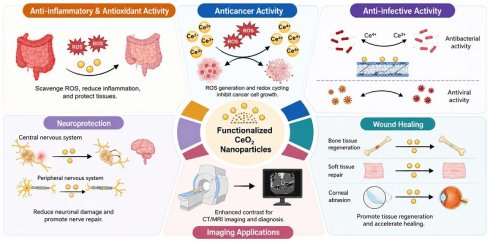
The rapid expansion and advancement of nanotechnology have presented potential applications in the diagnosis and treatment of various diseases. In recent years, cerium oxide nanoparticles (CeO2 NPs) have been extensively studied and applied in biological systems due to their unique physical and chemical properties, such as high antioxidant activity, enzyme-mimicking activity and good biocompatibility. This review summarizes the latest research progress of functionalized CeO2 NPs in anti-cancer activity, anti-infection, anti-inflammation, antioxidant activity, wound healing, neuroprotection and imaging applications. It explores their mechanisms of action and potential clinical application prospects, as well as the concerns about toxicity and safety.
Keywords: cerium oxide nanoparticles, biomedical applications, oxidative stress, toxicity
1. Introduction
1.1. Research Background: The Rise of CeO2 NPs
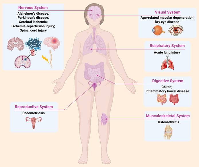
The rapid growth and development of nanotechnology have provided potential applications for the diagnosis and treatment of various diseases (1) (Figure 1). Among the numerous nanomaterials under intensive research, gold nanoparticles (Au NPs), silica nanoparticles (SiO2 NPs), iron oxide nanoparticles (IONPs), and polymer-based nanocarriers are the most commonly studied types, each with distinct advantages and inherent limitations. Specifically, Au NPs possess excellent optical properties and high biocompatibility, making them outstanding candidates for bioimaging and photothermal therapy, but their high synthesis cost and poor biodegradability restrict their large-scale clinical application (2). SiO2 NPs have a stable spherical structure, large specific surface area, and good biocompatibility, which make them ideal drug delivery carriers, yet their poor biodegradability may lead to long-term accumulation in the body and potential toxicity (3). IONPs exhibit unique superparamagnetic properties, enabling their application in magnetic resonance imaging (MRI)(4), but their antioxidant capacity is relatively weak, limiting their efficacy in treating oxidative stress-related diseases. Polymer-based nanocarriers, such as liposomes and polymeric micelles, have flexible structural design and good drug loading capacity, but they are prone to degradation and instability in the complex in vivo environment, affecting their delivery efficiency (5).
Applications of functionalized cerium oxide nanoparticles in disease treatment. Created with BioRender.com.
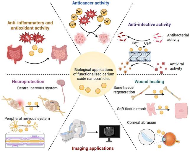
Unlike these commonly used nanomaterials, CeO2 NPs have attracted significant attention over the past decade due to their unique intrinsic biological effects, including high antioxidant activity (6), enzyme-mimicking activity (7,8), and biocompatibility (9). In recent years, increasing evidence suggests that CeO2 NPs have broad application prospects in the biomedical field (10) (Figure 2), and their unique properties have shown good potential in antioxidant, antitumor, anti-infection, and neuroprotective aspects.
Biological applications of functionalized cerium oxide nanoparticles. Created with BioRender.com.
The unique biological effects of CeO2 NPs are closely related to their three-core structural and chemical characteristics: reversible Ce3+/Ce4+ redox cycle, oxygen vacancies, and adjustable surface chemical states. The reversible conversion between Ce3+ and Ce4+ on the surface of CeO2 NPs allows them to continuously accept and donate electrons, thereby efficiently scavenging various reactive oxygen species (ROS) (such as superoxide anion, hydroxyl radical, and hydrogen peroxide) in biological systems and maintaining the redox balance of cells (11). Oxygen vacancies, as important surface defects, further provide active sites that synergistically enhance the enzyme-mimicking activity (e.g., superoxide dismutase (SOD) and catalase (CAT)) and overall antioxidant capacity of CeO2 NPs (12).
Notably, the modulation of these core structural and chemical characteristics is closely associated with the synthesis methods and functionalization strategies of CeO2 NPs, which can provide a more solid mechanistic basis for their unique biological effects. Specifically, functionalization strategies have been proven to significantly regulate the Ce3+/Ce4+ ratio and the catalytic performance of CeO2 NPs. Compositional modulation, such as transition metal doping (e.g., Fe, Cu), can alter the electronic cloud distribution around Ce atoms, promote the conversion of Ce4+ to Ce3+, and increase the Ce3+ content on the material surface. Meanwhile, structural defect engineering, such as the introduction of oxygen vacancies, can effectively increase oxygen vacancy density, which in turn synergistically enhances the redox activity of the Ce3+/Ce4+ cycle and optimizes the ROS scavenging capacity of CeO2 NPs (13).
1.2. The Necessity of Functional Modification: Addressing Issues of Aqueous Dispersibility, Targeting Capability, Biocompatibility, and Toxicity
CeO2 NPs exhibit a significant tendency to agglomerate, resulting in poor stability and dispersion in aqueous media (14). Agglomerated particles may impair their distribution and metabolism within the body, increasing potential toxicity risks (15). Functionalization modifications can improve their dispersion and stability (16). For example, surface functionalization with polyvinylpyrrolidone (PVP) can effectively improve the dispersion stability of CeO2 nanoparticles and prevent their aggregation (17).
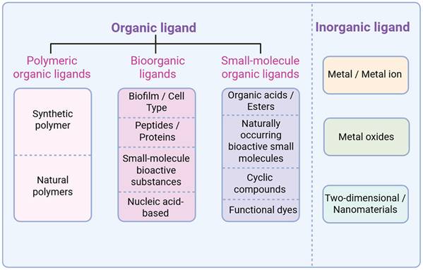
In the biomedical field, targeted functionalization of CeO2 NPs is essential to enable more precise delivery to diseased sites, enhance therapeutic efficacy, and minimize damage to healthy tissues. For instance, specific targeting molecules, including antibodies or ligands (Figure 3), can be anchored on the CeO2 NPs surface. This allows the CeO2 NPs to recognize and bind specifically to receptors on diseased cells or tissues, thereby achieving targeted delivery. Although related research remains relatively scarce, targeted modification represents one of the critical steps for realizing precision therapy with CeO2 NPs in biomedical applications.
Functionalized cerium oxide nanoparticle-modified ligands. Created with BioRender.com.
Uncoated CeO2 NPs exhibit limited biocompatibility (18), as their biopersistence and tendency to accumulate in mononuclear phagocytic system organs can lead to increased toxicity over time, including oxidative stress, inflammation, and tissue damage (19). Surface functionalization can improve the properties of CeO2 NPs, reducing adverse reactions with biological systems. For instance, by modifying CeO2 NPs with lipoic acid (LA) and polyethylene glycol (PEG), it is possible to prepare an LA-PEG-CeO2 nanocomposite system that exhibits good biocompatibility, low toxicity, and high free radical scavenging capacity (20).
High concentrations of CeO2 NPs have been shown to induce cytotoxicity and genotoxicity in vitro, raising safety concerns for potential in vivo applications (21). Functionalization modifications can reduce toxicity by altering CeO2 NPs' surface charge, particle size, and surface chemical composition. For example, in situ growth of CeO2 on negatively charged montmorillonite sheets optimizes surface properties and colloidal stability, effectively reducing systemic absorption and potential nanotoxicity for safer biomedical use (22).
1.3. Functional Modification Methods
The methods for functionally modifying CeO2 NPs using nanotechnology include surface chemical modification (including organic molecule modification, coupling agent modification, surfactant modification, as well as novel bioorganic ligand modification represented by elastin-based protein functionalization), surface physical modification (including polymer coating, metal nanoparticle coating, etc.), and in situ synthesis. Table 1 and Table 2 respectively present typical functionalization strategies for CeO2 NPs and their synthesis methods in different application scenarios. Functional modification is capable of improving their dispersion and stability, enhancing biocompatibility and targeting ability, boosting mechanical properties, increasing application efficiency, or endowing CeO2 NPs with novel physical and chemical characteristics. Figure 3 shows the ligands of functionalized CeO2 NPs.
Typical functionalization modification strategies for cerium oxide nanoparticles.
| Strategies | Type of modifier | Core modifier | Core role | Application scenarios | Reference |
|---|---|---|---|---|---|
| Polymeric organic ligands | Natural polymers | Dextran | Increase intracellular ROS levels to induce apoptosis in head and neck cancer cells | In vitro Treatment Research for Head and Neck Cancer | (34) |
| Bioorganic ligands | Amino acid | Histidine, glycine | Enhance antiviral activity by targeting and disrupting viral entry | Prevention and control of enveloped virus infections in vitro | (41) |
| Nucleic acid | miR146a | Targeted inhibition of inflammatory signaling pathways, efficient clearance of ROS | Treatment of chronic IBD | (44) | |
| ECM-derived structural proteins | mEl | Stabilizes dispersed CeO2, enhancing ROS scavenging and promoting angiogenesis | Microneedle Therapy for Oral Ulcers | (78) | |
| Small-molecule organic ligands | Organic acids | PA | Chelating excess intracellular Ca2+ and inhibiting P-gp expression to reverse tumor resistance | Chemotherapy Sensitization and Toxicity Mitigation in Doxorubicin-Resistant Hepatocellular Carcinoma | (39) |
| Inorganic nanocomposites | 2D MoS2 nanosheet | PEG-MoS2 nanosheets | Highly efficient photothermal antibacterial capability, enhanced antioxidant activity | Chronic wound treatment, particularly diabetic ulcer wounds | (28) |
| 2D graphene nanoplatelet | GNPs | Synergistically enhances antimicrobial activity | Prevention and control of multidrug-resistant bacterial infections | (40) |
Synthesis methods for functionalized modification of cerium oxide nanoparticles in different application scenarios.
| Synthesis method | Models | Main results | Applications | Reference |
|---|---|---|---|---|
| Precipitation Synthesis of Dex-CeO2 NPs | Cells: head and neck cancer cell lines A253, FaDu, SCC-25 | SD2 exhibits superior anticancer activity and stability, exerting its effects through the apoptosis pathway | Treatment of Head and Neck Cancer | (34) |
| Solvothermal synthesis of G/CeO2 nanotiles loaded with 12% and 25% GNPs | Bacteria:P. aeruginosa, S. aureus | G/CeO2-II (25% GNPs) exhibited the most optimal antibacterial properties, achieving inhibition rates of 82.67% and 89.48% against the two bacteria, outperforming pure CeO2 | As a novel nano-antibiotic for controlling multidrug-resistant bacteria | (40) |
| Conjugation of miR146a to CeO2 NPs via CDI chemistry | Animals:Establishing a Chronic Colitis Model Induced by T Cell Transplantation in Rag2-/- Mice | CNP-miR146a is orally stable and significantly alleviates colitis symptoms | Treatment of IBD | (44) |
| Hydrothermal synthesis of CeO2 nanospheres, reduced with hydrazine hydrate to load Cu and Pt, constructing PtCuOx/CeO2-x | Cells: SD rat primary chondrocytes (IL-1β-induced inflammation) Animals: SD rat ACLT osteoarthritis model | PtCuOx/CeO2-x exhibits high SOD/CAT activity, scavenging ROS/RNS, protecting mitochondria, and inhibiting the ROS/Rac-1/NF-κB pathway | Treatment of OA | (13) |
| Grow cerium oxide nanoclusters on BSA via biomineralization, then couple them with ICG. | Cells: J774 Macrophages Animals: Uterus-Specific Stat3 Knockout Mice (Pgrre/+Stat3f/f; Stat3d/d); Endometriosis Mouse Model | CeO2 can target ectopic lesions in mouse endometriosis, scavenge ROS, modulate immunity, inhibit pSTAT3, reduce lesions without affecting pregnancy, and enable non-invasive imaging when conjugated with ICG | Targeted Therapy for Endometriosis | (46) |
| Disperse CeO2 in water, add DA to adjust the pH to 9.0, and undergo oxidative polymerization to form Ce@P nanoenzymes coated with PDA | Cells: RAW264.7 macrophages (LPS-induced inflammation) Animals: SD rats administered LPS via tracheal instillation to establish an ALI model | Ce@P+NIR can efficiently scavenge ROS, reduce inflammatory factors, induce M2 polarization, and alleviate lung injury | Targeted Therapy for ALI | (47) |
| A one-step hydrothermal process enables α-CD to encapsulate cerium oxide via O-Ce bond formation, yielding CCNP. Subsequently, MOR is loaded through intermolecular interactions between α-CD and MOR | Cells: RAW264.7 macrophages (LPS-induced inflammation); Animals: C57BL/6 mice with laser-induced CNV model | M@ CCNP can eliminate ROS, inhibit M1 polarization, activate NRF2, reduce CNV area by 84.4%, and decrease leakage by 68.4%, demonstrating superior efficacy in suppressing leakage compared to anti-VEGF drugs | Treatment of AMD | (48) |
| Chemically synthesized CeO2 was ultrasonically mixed with HA solution, followed by ethanol precipitation and centrifugation to obtain HA-CeO2 | Cells:HCECs; Animals:C57BL/6 mice | HA-CeO2 effectively scavenges ROS, reduces IL-1β/MMP9, increases tear volume and tear film stability, and repairs the cornea | Treatment of DED | (49) |
| Aminated CeO2 was first prepared via silylation with APTES, followed by chemical coupling with ALG using EDC/NHS to construct Ce-ALG nanoparticles | Cells: Rabbit corneal epithelial cells (SIRC, LPS/H2O2-induced inflammation/oxidative stress); Animals: New Zealand white rabbits used to establish a CA model | Ce-ALG enhances adhesion and cellular uptake, enabling controlled release of β-1,3-glucan. It achieves 99% repair of abrasions, outperforming ketorolac and unmodified Ce | Treatment of CA | (50) |
| Solvothermal synthesis of CeO2 NPs fused with Pltm via ultrasonication and embedded in GelMa hydrogels yields Pltm@CeO2 NPs/Gel | Cells: HaCaT keratinocytes (H2O2-induced oxidative stress), RAW264.7 macrophages (LPS-induced inflammation); Animals: GK rats used to establish a diabetic wound model | Pltm@CeO2 NPs/Gel can scavenge ROS, suppress inflammation, promote angiogenesis and collagen alignment, significantly accelerating wound healing in diabetic rats | Diabetic Wound Care | (52) |
| CeO2 nanoparticles were mixed with a GelMA solution containing bone marrow-derived mesenchymal stem cells (BMSCs), and UV-cured to produce a GelMA-CeO2-BMSC composite hydrogel | Cells: BMSCs, RAW264.7 macrophages; Animals: SD rats used to construct a 5mm cranial critical defect model | GelMA-CeO2-BMSC promotes osteogenic differentiation of BMSCs, suppresses M1 polarization while promoting M2 polarization, and significantly repairs rat cranial defects | Repair and Regeneration of Bone Defects | (54) |
| PAA is modified on the surface of CeO2 to synthesize CeO2@PAA, which is then conjugated with biotinylated LXW7 via EDC reaction to construct CeO2@PAA-LXW7 | Cells: Mouse BV2 microglia, established as an inflammatory model by LPS induction | CeO2@PAA-LXW7 can scavenge ROS/NO, reduce TNF-α/IL-1β, and inhibit the phosphorylation of FAK and STAT3, exhibiting superior anti-inflammatory effects compared to CeO2@PAA or LXW7 used alone | Interventions for Neuroinflammation Associated with Central Nervous System Injury | (56) |
| After synthesizing CeO2 nanoclusters, the acidified ZnTPyP is self-assembled on their surface via hydroxyl interaction, and PLL is modified to obtain CZPN | Cells: Rat pheochromocytoma PC12 cells; Animals: ICR mice | CZPN can optically modulate neuronal activity, ensuring cellular safety, and induces TRP channel-mediated pain behavior in ICR mice | Treatment of Peripheral Nerve Disorders | (64) |
1.4. Heterostructure Engineering of CeO2-Based Nanozymes
Heterostructure engineering is a pivotal design strategy to elevate the catalytic and enzyme-mimetic performance of CeO2 nanozymes (23). By integrating CeO2 with transition metal oxides, rare-earth solids or noble metals to form well-defined interfaces, this strategy realizes multi-dimensional regulation of lattice strain, oxygen vacancy, component composition and interfacial electronic synergy, and optimizes the Ce3+/Ce4+ redox cycle at the atomic level to break the activity limit of pristine CeO2(24).
Lattice strain induced by heteroepitaxy can effectively boost oxygen vacancy generation (25). This mechanism, combined with the strategy of component complementation which broadens enzyme-mimetic adaptability (26), has been successfully applied to design high-performance ceria-based heterostructures for treating ROS-related diseases. For instance, epitaxially strained CeO2/Mn3O4 nanocrystals leverage enhanced oxygen vacancies and redox cycles to achieve superior radioprotection (27). Similarly, CeO2/MoS2 heterostructures optimize these properties to enhance ROS scavenging capacity, anti-inflammatory effects, antibacterial action, and wound healing (28). In a different direction, CeO2/Au@Pt nanospheres utilize dual enzyme-mimetic activities (peroxidase-like (POD-like) and CAT-like) to generate cytotoxic hydroxyl radicals and alleviate tumor hypoxia, enabling photothermal-catalytic synergistic therapy of tumors (29).
Compared with conventional modification, heterostructure engineering provides systematic lattice-electronic-compositional synergy, greatly expanding the biomedical applications of CeO2-based nanomedicines (23). Through precise interface regulation and electronic structure optimization, these representative heterostructure designs effectively tailor the catalytic behavior of CeO2 nanozymes. As summarized in Table 3, such structural engineering endows CeO2 nanozymes with enhanced performance and extended functional potential in biomedicine.
Synthesis methods, mechanism improvements, and biomedical applications of typical CeO2-based heterostructure nanoenzymes.
| Types of heterostructures | Synthesis Method | Key Mechanism | Representative biomedical applications | Reference |
|---|---|---|---|---|
| CeO2/Mn3O4 heterostructure | Epitaxial growth method | Lattice strain-induced formation of oxygen vacancies; Enhanced ROS scavenging capacity; Improved radiation protection efficacy | Radiation Protection | (27) |
| CeO2/MoS2 heterostructure | Solvothermal method + PEG modification | MoS2 provides photothermal antibacterial activity; CeO2 scavenges ROS for antioxidant and wound healing promotion | Healing of chronic wounds (such as diabetic ulcers) | (28) |
| CeO2/Au@Pt heterostructure | Seed-growth method + reduction deposition | CeO2 nanozymes with POD/CAT-like activities; Au@Pt provides photothermal effect; synergistic photothermal-catalytic tumor therapy | Photothermal-catalytic synergistic therapy for tumors | (29) |
| PtCuOx/CeO2-x heterostructure | Hydrothermal synthesis + reduction doping (Cu, Pt) | Cu doping increases the Ce3+/Ce4+ ratio and creates oxygen vacancies; Pt stabilizes Cu⁺ and enhances SOD/CAT-like activity; NIR response enhances catalytic efficiency | Treatment of OA | (13) |
As summarized in Table 4, different functionalization strategies exert distinct regulatory effects on the three major enzyme-mimetic activities of CeO2 NPs. Element doping (e.g., Cu, Pt) primarily enhances oxygen vacancy density and Ce3+/Ce4+ redox cycling, thereby boosting SOD/CAT-like activities (for ROS scavenging) or POD-like activity (for pro-oxidant therapy). Surface modification (e.g., with polymers, peptides, or polysaccharides) mainly improves colloidal stability, targeting, and biocompatibility while preserving or moderately enhancing intrinsic ROS-scavenging activities. Surface facet engineering further reveals that exposed crystal facets dictate catalytic preference, with {110} facets favoring SOD-like activity and {100} facets favoring POD-like activity (30). Collectively, these strategies allow tailored design of CeO2 nanozymes for either antioxidant therapy (SOD/CAT-dominant) or pro-oxidant therapy (POD-dominant), depending on the pathological context.
Effects of functionalization strategies on enzyme-mimetic activities of CeO2 NPs.
| Functionalization strategies | Typical Materials | SOD-like activity | CAT-like activity | POD-like activity | Key mechanisms | Key Use Cases | References |
|---|---|---|---|---|---|---|---|
| Element doping | PtCuOx/CeO2-x | Significantly enhanced | Significantly enhanced | — | Co-modification with a Cu-Pt bimetallic system increases the Ce3+/Ce4+ ratio, raises the oxygen vacancy concentration, and lowers the oxygen vacancy formation energy | Treatment of OA | (13) |
| Surface Modification | CeO2/Au@Pt-PEG | — | Significantly enhanced | Significantly enhanced | A stable core-shell Au@Pt structure effectively supports ultra-small CeO2 particles, addressing the issue of nanoenzyme aggregation; PEG modification enhances the material's water dispersibility, biocompatibility, and circulation time in vivo; the Au@Pt component provides highly efficient near-infrared photothermal conversion capabilities, enabling photothermal therapy | Photothermal-catalytic synergistic therapy for tumors | (29) |
| HA-CeO2 | — | Significantly enhanced | — | HA modification improves the dispersion stability and biocompatibility of CeO2, enhances its ability to adhere to and remain on the ocular surface, increases tear film thickness and stability, and exerts synergistic antioxidant and anti-inflammatory effects | Treatment of DED | (49) | |
| HA-5-HT@CeO2 | — | Enhanced | — | HA targets CD44+ macrophages; 5-HT acts as a substrate for MPO in inflamed colon, enabling targeted accumulation; CeO2 scavenges ROS (H2O2, •OH) and reduces pro-inflammatory cytokines (TNF-α, IL-6, IL-1β) while upregulating anti-inflammatory IL-10; repairs intestinal epithelial barrier (upregulates ZO-1, occludin, E-cadherin) | Treatment of UC | (43) | |
| M@CCNP | Significantly enhanced | Significantly enhanced | — | α-CD binds to the CeO2 surface via O-Ce bonds, enhancing the dispersibility, stability, and biocompatibility of the nanoparticles; MOR activates the NRF2 pathway and inhibits NF-κB, downregulating pro-inflammatory factors, suppressing M1 polarization of macrophages, and blocking the inflammatory cascade in AMD | Treatment of AMD | (48) | |
| Ce@P | Significantly enhanced | Significantly enhanced | — | PDA coating enhances the water dispersibility, colloidal stability, and biocompatibility of CeO2; PDA endows the material with highly efficient NIR photothermal conversion capabilities, and the synergistic photothermal effect enhances the ROS scavenging efficiency of CeO2; Ce@P efficiently scavenges ·OH, ·O2-, and H2O2 upon NIR activation, thereby breaking the oxidative stress-inflammation vicious cycle | Treatment of ALI | (47) | |
| Surface Facet Engineering | CeO2 nanocubes with {100} crystal faces exposed | lower | — | higher | {100} The {100} crystal plane has high surface energy and a high degree of reconstruction, resulting in stronger POD-like catalytic activity | Catalytic Applications | (30) |
| CeO2 nanorods with {110} crystal faces exposed | higher | — | lower | {110} The crystal surface is more conducive to ·O2- adsorption and protonation, resulting in superior SOD-like activity | Natural antioxidants | (30) |
Note: “—” indicates that the literature does not explicitly report or focus on the activity of this type of enzyme.
2. Biomedical Applications of Functional CeO2 NPs
2.1. Anticancer Activity
2.1.1. Inhibition of Tumor Growth
CeO2 NPs have the capability to suppress tumor growth by triggering apoptosis in tumor cells (31). Their core mechanisms of action include modulating the tumor microenvironment (TME) (e.g., scavenging excess ROS in normal tissues or generating ROS in acidic TME, enabling targeted delivery of therapeutics through surface functionalization to enhance selective killing of tumor cells and reduce damage to normal cells.
Engineering TME-responsive catalytic systems is critical for improving the selectivity and efficacy of nanocatalytic cancer therapy. A pH-responsive PN-CeO2-PSS system was prepared by electrostatically conjugating porous CeO2 nanorods with PSS, exerting catalytic activity only in acidic TME (32). Its nanoreactor structure traps oxygen and substrates, promotes charge transfer, and efficiently generates ·O2-, which induces oxidative stress (membrane damage, elevated malondialdehyde (MDA)), activates caspase-3-mediated tumor cell apoptosis and inhibits tumor growth. 70 kDa PSS modification was optimal: PN-CeO2-PSS with a hydrodynamic diameter of 306.5 nm balanced substrate capture and charge transfer efficiency, showing the highest oxidative and antitumor activity. It inhibited HepG2 tumors at 96.1% (vs. 51.1% for unmodified PN-CeO2), with no toxicity to normal cells/mouse organs, excellent biocompatibility and metabolic safety. This pH-responsive polymer-CeO2 hybrid provides a highly selective, effective and low-toxic nanocatalytic platform for tumor chemodynamic therapy (CDT), offering important insights for nanomedicine clinical translation.
For breast cancer nanotherapy, developing a smart-responsive, synergistic and low-toxic nanomedicine system is key to overcoming the drawbacks of traditional CeO2-based nanotherapeutics (weak POD-mimetic activity, poor monotherapy efficacy) and chemotherapeutics (severe toxic side effects). A study fabricated the smart-responsive CCCs nanosystem via hydrothermally doping Cu2+ into CeO2 lattice, electrostatically loading DOX on Cu-CeO2 surface and ultrasonicating MDA-MB-231 cell membrane coating (33). Cu-CeO2 NPs show enhanced POD-mimetic activity in acidic TME, generating toxic OH via Fenton-like reactions to induce tumor cell apoptosis. CCCs realize precise tumor targeting and pH-responsive DOX release, achieving synergistic tumor suppression with CDT; cell membrane coating endows it with targeting ability and biocompatibility. In vitro, 200 μg/mL CCCs kill 95.8% of MDA-MB-231 cells with low cytotoxicity to C2C12 cells. In vivo, it inhibits 98.5% of tumor growth without obvious organ damage or abnormal blood parameters, realizing highly effective and low-toxic breast cancer treatment. Moreover, the system exerts antioxidant activity under normal physiological conditions, scavenging excess ROS to alleviate DOX-induced normal tissue damage and ensure treatment safety and sustainability.
In head and neck cancer nanotherapy, surface modification to regulate nanoparticle bioactivity and targeting is key for optimizing efficacy, with modifier molecular weight drawing much attention. Tarakci et al. (34) synthesized two Dex-CeO2 NPs (SD1: 9-11 kDa dextran; SD2: 6 kDa dextran). SD2 markedly elevated intracellular ROS at high concentrations, inducing apoptosis. Cytotoxicity assays on A253, FaDu and SCC-25 cells showed SD2 exerted high cytotoxicity at 500/1000 µg/mL, significantly reducing viability. Gene expression analysis revealed that SD2 exerted anticancer effects by upregulating pro-apoptotic genes (TP53, CASP3, BAX), contributing to tumor cell apoptosis.
2.1.2. Enhancing Radiotherapy Effects
When used in combination with radiotherapy, CeO2 NPs can increase the sensitivity of tumor cells to radiotherapy while reducing damage to normal cells (35).
A core challenge in improving radiotherapy efficacy and safety is balancing precise tumor radiosensitization with normal tissue protection, where cell-type-selective redox-regulating nanotherapeutics are key. Zamyatina et al. (36) developed pyrroloquinoline quinone-modified CeO2 NPs (CeO2@PQQ NPs). These NPs enhance cancer cell (EMT6/P) radiosensitivity to X-rays by inducing oxidative stress: post 5 Gy irradiation, glutathione (GSH) levels (key antioxidant) decreased by 32%, impairing ROS scavenging and accumulating oxidative damage. They also disrupted mitochondrial function, reducing mitochondrial membrane potential (MMP) by 29% to trigger pro-apoptotic pathways. CMN assay showed a 2.5-fold increase in micronuclei (chromosomal damage marker), and clonogenic assay revealed a 2-fold reduced survival fraction, exacerbating DNA damage and inhibiting repair. In contrast, for normal L929 cells, CeO2@PQQ NPs post-radiotherapy maintained viability, elevated GSH and MMP, reduced micronuclei, and enhanced survival, exerting radioprotection. With cell-type-specific antioxidant/pro-oxidant properties, CeO2@PQQ NPs lay a theoretical foundation for novel radiosensitizers/antioxidants.
Radiotherapy resistance induced by tumor hypoxia, the lack of precision guidance during treatment, and inadequate control of radiotherapy toxicity are key issues limiting the improvement of radiotherapy efficacy for solid tumors. The construction of an integrated nanoplatform with TME responsiveness, hypoxia alleviation, efficacy enhancement, and imaging guidance functions is of great significance for addressing the aforementioned problems. Pi et al. (37) developed core-shell CeO2-MnO2 nanoparticles, a novel radiosensitizer with excellent biocompatibility and clinical translation potential, enhancing radiotherapy efficacy while reducing toxicity. In vitro, MnO2 catalyzes O2 generation from TME H2O2 (alleviating hypoxia) and depletes GSH (weakening antioxidant defenses), while CeO2 boosts X-ray absorption and synergistically generates ROS to increase radiosensitivity. Mn2+ from MnO2 degradation enhances T1-weighted MRI signal (peak at 4 h post-injection) for tumor localization and guidance. In vivo 21-day experiments showed CeO2-MnO2 + X-rays significantly reduced tumor volume, with no toxicity to normal MIHA cells or mouse major organs, and normal blood/biochemical parameters. Integrating TME responsiveness, hypoxia alleviation, and MRI guidance, CeO2-MnO2 provides a new strategy for effective, low-toxicity solid tumor radiotherapy.
2.1.3. Tumor Targeting Ability
The tumor targeting capacity of CeO2 NPs can be enhanced by modifying their surfaces, such as through conjugation with specific peptides. These modifiers are capable of specifically binding to certain receptors or molecules on tumor cell surfaces, which directs CeO2 NPs to reach tumor cells and promotes their accumulation at the tumor site.
The insufficient tumor targeting of nanophototherapeutic agents is a key bottleneck leading to low precision in photothermal therapy for breast cancer and a high risk of thermal damage to normal tissues. Constructing an integrated nanozyme with "active targeting-photothermal conversion-imaging guidance" functions has become a core direction for improving treatment accuracy and safety. Researchers successfully developed a targeted nanozyme (HCeO2@ICG-RGD) by loading indocyanine green (ICG) onto mesoporous cerium oxide nanocatalysts (HCeO2) and modifying the surface of HCeO2@ICG with arginine-glycine-aspartic acid (RGD) peptides, enabling specific recognition and efficient photothermal therapy for breast cancer (38). This peptide exhibits specific recognition and binding to αvβ3 integrins on tumor cell surfaces, thereby enabling targeted identification of tumor cells and enhancing their targeting efficiency. The targeting property of HCeO2@ICG-RGD in tumor tissue was studied by near-infrared-II (NIR II) fluorescence imaging, and results showed that HCeO2@ICG-RGD has good targeting property. The results from confocal laser scanning microscopy (CLSM) also showed that the amount of RGD modified HCeO2@ICG-RGD nanozymes absorbed by 4T1 breast cancer cells was greatly increased compared with HCeO2@ICG.It proved that the RGD peptide could enhance the targeting ability of the nanozymes for tumor cells. In photothermal therapy, CeO2 NPs can absorb light energy of a specific wavelength and convert it into heat. When they accumulate in the tumor site, they can increase the local temperature of the tumor through external light source irradiation, thereby killing tumor cells. Its targeting ability for tumor can improve the accuracy and effectiveness of photothermal therapy and reduce the thermal damage for the surrounding normal tissues, and it has certain application value in cancer treatment, and also provides new ideas and methods for cancer treatment.
2.1.4. Reversal of Tumor Drug Resistance
Abnormally elevated intracellular Ca2+ levels are intimately associated with tumor drug resistance. High Ca2+ concentrations can activate drug efflux proteins (such as P-glycoprotein (P-gp)), prompting the efflux of chemotherapeutic agents from tumor cells and consequently diminishing drug efficacy.
A study conducted by Tian et al. (39) engineered phytic acid (PA)-modified cerium dioxide nanoparticles (CeO2@PA). Leveraging PA's robust coordination capacity with Ca2+, the nanoparticles enable effective modulation of intracellular Ca2+ levels. This intervention reduces intracellular Ca2+ concentrations, inhibits P-gp expression, and markedly enhances drug accumulation in drug-resistant tumor cells, thereby achieving efficient reversal of tumor drug resistance. This study indicates that CeO2@PA is expected to be a new type of Ca2+ inhibitor for reversing tumor drug resistance, providing new strategies and methods for overcoming tumor drug resistance.
2.2. Anti-Infection
2.2.1. Antibacterial Activity
The spread of multidrug-resistant bacteria has greatly reduced the efficacy of traditional antibiotics, urgently calling for novel high-efficiency antimicrobial materials. CeO2 features reversible Ce3+/Ce4+ conversion, which enables in-situ ROS generation for bacterial inhibition without UV irradiation, yet pure CeO2 nanotiles show limited antimicrobial activity. Graphene's high conductivity and sharp-edge structure can synergistically boost the antibacterial properties of metal oxides. Accordingly, Rehman et al. (40) synthesized CeO2 nanotiles and graphene/cerium oxide (G/CeO2) nanocomposites via a solvothermal method to explore their synergistic antibacterial effects. The results show that the incorporation of graphene significantly enhances antimicrobial activity, with the 25% graphene-loaded G/CeO2-II performing optimally, achieving inhibition rates of 82.67% against P. aeruginosa and 89.48% against S. aureus. The excellent antibacterial activity of G/CeO2-II mainly comes from the unique nanostructures of graphene and CeO2 nanotiles and their synergy: graphene's sharp edges damage bacterial cell membranes and induce charge imbalance; CeO2's reversible Ce3+/Ce4+ conversion generates ROS to oxidize bacterial proteins and nucleic acids; the blade-like CeO2 nanotiles physically pierce bacterial cell membranes. This triple synergy is believed to contribute to bacterial death. G/CeO2 nanocomposites show superior inhibitory effects against Gram-positive S. aureus in comparison with Gram-negative P. aeruginosa, owing to structural differences in bacterial cell membranes. Gram-positive bacteria possess a thicker peptidoglycan layer, which renders them more vulnerable to physical damage. As a promising candidate for combating drug-resistant bacteria, G/CeO2 nanocomposites are expected to promote technological advances in the fields of nanomedicine and anti-infection therapy.
Studies have shown that MoS2-CeO2 nanocomposites formed by combining CeO2 NPs with polyethylene glycol-modified molybdenum disulfide (PEG-MoS2) exhibit excellent photothermal antibacterial properties under 808 nm laser irradiation. CeO2 NPs have intrinsic antibacterial activity, derived from surface oxygen vacancies and ROS generation via reversible Ce3+/Ce4+ redox reactions; PEG-MoS2 converts near-infrared (NIR) light energy into heat, disrupting bacterial cell membranes through photothermal effects to exert antibacterial action. The combination yields a distinct synergistic effect, where CeO2 NPs' antibacterial property and PEG-MoS2's photothermal antibacterial capacity reinforce each other, greatly boosting overall antimicrobial efficacy. This nanocomposite can alleviate two key issues in chronic wounds, namely infection and oxidative stress. It holds great application potential for diabetic ulcers and offers an efficient, convenient and innovative strategy for clinical chronic wound treatment (28).
2.2.2. Antiviral Activity
The invasion mechanisms of enveloped viruses (such as pH-dependent fusion in endocytic pathways and dual-pathway invasion) represent key targets for antiviral material design. CeO2 NPs, with good biocompatibility and antiviral potential, are a research focus, but bare CeO2 NPs suffer from insufficient stability and limited targeting, restricting therapeutic efficacy. To address this, Dupkalová et al. (41) functionalized CeO2 NPs with essential amino acids histidine (CH) and glycine (CG), both forming stable bonds with CeO2 via carboxyl groups. Notably, histidine's imidazole ring exhibits pH-responsive protonation, matching the acidic endosomal microenvironment (pH 5.8-6.2) during invasion of enveloped viruses like vesicular stomatitis virus (VSV). By introducing “histidine-mediated low-pH protonation” into surface design, the team triggered nanoparticle aggregation and charge reversal in the viral fusion acidic environment, achieving an “endosome-targeting-fusion-blocking” strategy. This simple amino acid functionalization enhanced CeO2 NPs' therapeutic index against VSV by 1-2 orders of magnitude without increasing toxicity. The study revealed that antiviral activity strongly depends on viral entry pathways: it is highest for the endosomal pathway of VSV, intermediate for the dual pathways of bovine beta coronavirus (BCoV-1), and minimal for the non-endosomal pathway of herpes simplex virus (HSV), laying a foundation for “pathway-selective” nanovirals. Functionalization (especially histidine) significantly boosts CeO2 NPs' ability to capture viruses and block fusion in acidic endosomes, showing high therapeutic potential against enveloped viruses (e.g., VSV) relying on acidic endosomal conditions (pH 5.8-6.2) for membrane fusion and entry.
2.3. Anti-inflammatory and antioxidant activity
2.3.1. Colitis and Inflammatory Bowel Disease (IBD)
ROS are key drivers in the pathological progression of colitis and IBD. Their excessive accumulation exacerbates the cycle of intestinal injury and inflammation, making them important therapeutic targets. Consequently, ROS scavengers offer a promising comprehensive strategy for effective treatment.
CeO2 NPs with their enzyme-mimicking activity (mimicking CAT, POD, and SOD), efficiently scavenge various ROS while exhibiting excellent biocompatibility, making them ideal candidates for inflammation treatment. To further optimize dispersion, stability, and in vivo targeting, Yang et al. (42) synthesized PEG-modified hollow CeO2 NPs (H-CeO2-PEG) via in-situ CeO2 growth on SiO2 NPs, subsequent SiO2 core removal, and PEG surface modification. Their study first revealed that H-CeO2-PEG alleviates colitis by inhibiting the MAPK signaling pathway (ERK1/2, JNK, p38, c-Jun). Possessing exceptional ROS-scavenging capacity, H-CeO2-PEG converts hydroxyl radicals (•OH), hydrogen peroxide (H2O2), and superoxide anions (•OOH) into water and oxygen. In a dextran sulfate sodium (DSS)-induced colitis mouse model, it significantly mitigates symptoms through multiple mechanisms: scavenging ROS, suppressing pro-inflammatory cytokines (IL-6, TNF-α, IL-1β, IL-18), and inhibiting MAPK pathway activation. Additionally, H-CeO2-PEG exhibits good in vivo biosafety, with no significant toxicity to mice's major organs at therapeutic doses, laying an important foundation for clinical applications.
Ulcerative colitis (UC) is a chronic non-specific IBD. Traditional drugs for UC are severely limited by their poor targeting, low bioavailability, and significant side effects, which prevent them from acting specifically on inflammatory sites and balancing excessive ROS accumulation and intestinal barrier damage. UC inflamed regions feature elevated myeloperoxidase (MPO) expression and enriched CD44+ receptors on macrophages, while excess ROS worsens epithelial barrier disruption and inflammatory cytokine release. Though CeO2 NPs efficiently scavenge ROS, their inadequate targeting causes systemic exposure. Hyaluronic acid (HA) specifically binds CD44+ receptors, and serotonin (5-HT) targets MPO, with their synergy compensating for individual limitations. Accordingly, researchers developed a novel targeted UC nanomedicine: hyaluronic acid/5-hydroxytryptamine-modified CeO2 NPs (HA-5-HT@CeO2). Studies show HA-5-HT@CeO2 excels at scavenging ROS and inflammatory mediators, repairing the intestinal epithelial barrier, and alleviating colitis symptoms. It exhibited good biocompatibility with no significant toxicity in both in vitro cell and in vivo animal experiments, holding high clinical application potential (43).
The treatment of IBD still faces numerous bottlenecks. Existing biologics are associated with high costs, immunogenicity, and side effect risks. JAK inhibitors are also limited by contraindications in specific populations. Approximately 30% of patients do not respond to current therapies, urgently requiring the development of novel treatment options with unique mechanisms and convenient delivery. As a key anti-inflammatory molecule, miR146a suppresses NF-κB activation by targeting IRAK1 and TRAF6, reducing pro-inflammatory factor release, but its RNA nature renders it vulnerable to gastrointestinal acid and enzymatic degradation, hindering oral delivery. CeO2 NPs exhibit excellent ROS-scavenging capacity, biocompatibility, and serve as stable biomolecule carriers. To address these challenges, Apte et al. (44) constructed an oral nanoparticle formulation (CNP-miR146a) by chemically conjugating miR146a with CeO2 NPs, resolving miR146a's gastrointestinal stability issues while leveraging synergistic antioxidant (CeO2 NPs) and anti-inflammatory (miR146a) effects. The study first validated CNP-miR146a's stability in simulated gastrointestinal environments, showing resistance to degradation and significant therapeutic efficacy in a mouse model of chronic colitis. Results demonstrated reduced colitis symptoms: diminished inflammatory cell infiltration, lower oxidative stress marker 8-OHdG levels, and decreased pro-inflammatory cytokines (IL-6, TNF). Though no clinical trials were conducted, CNP-miR146a's oral stability, notable in vivo therapeutic effects, and novel mechanism support its potential for clinical trials, offering a new IBD treatment strategy and insights for oral RNA drug delivery.
2.3.2. Osteoarthritis (OA)
OA, characterized by chronic inflammation and cartilage degeneration, severely impacts the lives of hundreds of millions of people worldwide. Current treatments primarily focus on symptom management rather than addressing the underlying disease mechanisms. Research on functionalized CeO2 NPs offers new avenues for developing nanotherapeutics targeting inflammatory joint diseases.
The core pathological mechanism of OA is closely associated with oxidative stress induced by excessive ROS and reactive nitrogen species (RNS), as well as abnormal activation of the ROS/Rac-1/NF-κB pathway. This leads to chondrocyte apoptosis, release of inflammatory mediators, and degradation of the extracellular matrix, while existing therapeutic approaches struggle to precisely regulate the inflammatory microenvironment. Traditional CeO2 NPs possess SOD/CAT-like enzymatic activity but suffer from limited oxygen vacancy density and insufficient catalytic efficiency, restricting their therapeutic potential. To address this, researchers employed a defect engineering strategy by co-doping copper (Cu) and platinum (Pt): Cu increases the Ce3+/Ce4+ ratio to boost oxygen vacancy density, while Pt stabilizes the more active Cu+ and forms strong metal-carrier interactions with CeO2. Their combined light absorption properties confer photothermal responsiveness to the material, with NIR irradiation further enhancing enzymatic catalytic activity. Based on this, Yang et al. (13) designed a novel oxygen vacancy-engineered ceria nanozyme (PtCuOx/CeO2-x) through co-doping copper (Cu) and platinum (Pt). Experimental results demonstrate that PtCuOx/CeO2-x efficiently scavenges IL-1β-induced ROS/RNS within chondrocytes, protects mitochondrial function (restoring membrane potential and adenosine triphosphate (ATP) production), inhibits chondrocyte apoptosis, downregulated proinflammatory factors and matrix degrading enzymes, and upregulated chondroprotective factors. In the ACLT rat OA model, PtCuOx/CeO2-x + NIR therapy significantly repaired cartilage damage and improved rat gait function. Immunohistochemistry confirmed its ability to inhibit Rac-1 and p-p65 expression, blocking the ROS/Rac-1/NF-κB pathway. Leveraging its core advantages of “high-efficiency SOD/CAT-like activity, photothermal responsiveness, and excellent biosafety”, PtCuOx/CeO2-x demonstrates clear clinical translation potential in treating ROS-mediated inflammatory diseases, particularly in the field of OA.
As a typical multi-stage degenerative disease, the pathological process of OA encompasses an initial phase of intense inflammatory response followed by a subsequent stage of cartilage repair and proliferation. Current monotherapies with single nanoparticles only target specific stages, limiting full-course intervention and therapeutic efficacy. Collagen hydrogels, biocompatible and injectable with cartilage extracellular matrix-mimicking properties, are ideal nanomedicine carriers. Researchers have developed a thiol-crosslinked collagen hydrogel (CSH-CeO2-pFe2O3) by leveraging the antioxidant and anti-inflammatory properties of CeO2 NPs and the chondrogenic effects of Fe2O3 nanoparticles (Fe2O3 NPs). CeO2 NPs are directly embedded in the hydrogel matrix, while Fe2O3 NPs are encapsulated in PLGA microspheres before loading, with a ROS-responsive release mechanism enabling "rapid anti-inflammation in the inflammatory phase + sustained regeneration promotion in the proliferative phase" to address OA's pathological progression comprehensively. Experimental results confirm CSH-CeO2-pFe2O3 has excellent mechanical strength, injectability, and biocompatibility, significantly enhancing cell adhesion, proliferation, and chondrogenic differentiation. It intelligently responds to high ROS levels by rapidly releasing CeO2 NPs to alleviate inflammation, followed by sustained Fe2O3 release for cartilage regeneration, improving treatment precision and effectiveness with remarkable therapeutic outcomes. This hydrogel provides a novel strategy for clinical OA treatment, promising more effective pain and inflammation relief via local sustained drug release (45).
2.3.3. Endometriosis
Endometriosis is characterized by ectopic implantation and growth of endometrial tissue, chronic inflammatory activation, and oxidative stress overload, severely impacting reproductive health and quality of life in women of childbearing age. Current treatments primarily rely on hormonal interventions or surgical excision, which can temporarily alleviate pain and disease progression but are limited by significant side effects and high recurrence rates, failing to target the fundamental mechanisms of the disease. The anti-inflammatory properties, ROS scavenging capabilities, and targeted delivery characteristics of functionalized CeO2 NPs open new avenues for developing precision nanotherapeutic drugs for endometriosis.
Rahman et al. (46) developed CeO2 NPs as a non-steroidal anti-inflammatory agent for endometriosis therapy. Synthesized via biomineralization on a bovine serum albumin (BSA) substrate, the CeO2 NPs possess antioxidant and enzymatic activities. Benefiting from albumin's properties, they passively target inflammatory sites (especially ectopic lesions) while sparing healthy tissues. Conjugated with near-infrared fluorescent dye ICG, CeO2 NPs enable non-invasive imaging for real-time tracking of drug distribution, aiding clinical applications. In vitro, CeO2 NPs reduced M1 macrophage marker CD80 expression and increased M2 marker ARG1 expression, demonstrating potent anti-inflammatory effects. In a mouse endometriosis model induced by autologous uterine tissue transplantation, intravenous administration of CeO2 NPs (120 μg/kg) was evaluated via fluorescent/photoacoustic imaging (for lesion accumulation) and histological analysis. Results showed significant reduction in ectopic lesion numbers without impairing early pregnancy (implantation/decidualization). As CeO2 NPs passively target ectopic lesions and allow real-time tracking via non-invasive imaging without disrupting normal pregnancy, this nonsteroidal agent integrates anti-inflammatory therapy with lesion imaging, offering a novel “precision treatment + real-time monitoring” theranostic approach for endometriosis with substantial clinical translation potential.
2.3.4. Acute Lung Injury (ALI)
ALI is characterized by acute inflammatory bursts in the lungs, disruption of the pulmonary epithelial/endothelial barrier, and excessive oxidative stress, posing a severe threat to the lives and prognosis of critically ill patients. Current treatments primarily focus on respiratory support and broad-spectrum anti-inflammatory therapies, emphasizing symptom relief while failing to target the fundamental pathophysiological mechanisms involving the interplay of inflammation and oxidative stress. The anti-inflammatory, antioxidant, and tissue-protective properties of functionalized CeO2 NPs open new avenues for developing precision nanotherapeutic agents specifically for acute lung injury.
Polydopamine (PDA) serves as a biocompatible material that combines NIR photothermal conversion capability with ROS scavenging activity. Its phenolic hydroxyl groups enhance ROS removal, while its photothermal effect can modulate nanozyme catalytic activity, thereby addressing the limitations of CeO2 in terms of limited catalytic efficiency and lack of synergistic enhancement mechanisms. Based on this, Yin et al. (47) developed a novel PDA-coated ceria nanoenzyme (Ce@P), whose anti-inflammatory efficacy arises from PDA-CeO2 synergy, further amplified by NIR irradiation. The reversible Ce3+/Ce4+ conversion in CeO2 cooperates with PDA's phenolic hydroxyl groups to scavenge excess pulmonary ROS (O2-, ·OH, H2O2) and suppress ROS-mediated inflammatory signaling (e.g., NF-κB activation). In LPS-induced RAW264.7 macrophages, PDA and CeO2 jointly downregulated pro-inflammatory factor transcription, induced M2 macrophage polarization, and reduced inflammatory cell infiltration. Such anti-inflammatory effects can be further enhanced by NIR irradiation. NIR not only boosts Ce@P's ROS scavenging efficiency but also promotes heat shock protein gene (HSP70) expression via thermal activation, strengthening anti-inflammatory and tissue repair capacities. Notably, Ce@P exhibits excellent biocompatibility, causing no inflammatory damage to non-pulmonary organs (heart, liver, spleen, kidney). After intravenous injection, it significantly enriches in the lungs and degrades gradually. Endowed with targeted enrichment, ROS scavenging, anti-inflammatory effects, photothermal synergy, and favorable biosafety, Ce@P holds promising prospects for ALI treatment.
2.3.5. Age-related Macular Degeneration (AMD)
AMD is characterized by chronic oxidative stress, inflammatory damage, and photoreceptor degeneration in the macular region. It poses a severe threat to the visual health of tens of millions of elderly individuals worldwide and can even lead to irreversible blindness. Current treatments lack effective interventions for dry AMD, while wet AMD relies on anti-vascular endothelial growth factor drugs to mitigate vascular proliferation. Neither therapeutic strategy targets the fundamental disease mechanisms driven by oxidative stress and inflammation. Functionalized CeO2 NPs, with their efficient ROS scavenging, targeted anti-inflammatory effects, and excellent biocompatibility, open new avenues for developing precision nanotherapeutics against AMD.
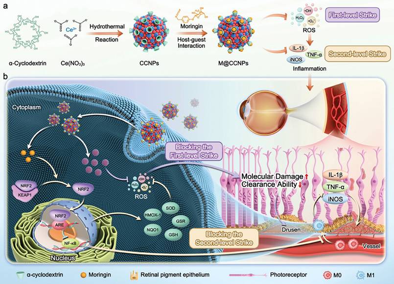
The pathogenesis of AMD follows a “two-stage” model: Stage I involves excessive oxidative stress damaging retinal pigment epithelial cells and forming drusen; Stage II triggers inflammatory responses. These stages mutually exacerbate, activate downstream vascular endothelial growth factor (VEGF) signaling, and ultimately lead to choroidal neovascularization (CNV). Single-target therapies cannot fully intervene, creating an urgent need for integrated platforms blocking both stages. CeO2 NPs, with reversible Ce3+/Ce4+ conversion for renewable ROS scavenging, inhibits the first stage. Moringin (MOR) exerts potent anti-inflammatory effects via NRF2 pathway activation but suffers from poor solubility due to hydrophobicity. As a biocompatible carrier, α-cyclodextrin (α-CD) enhances the biocompatibility of CeO2 NPs through O-Ce bond encapsulation and improves the solubility of MOR via host-guest interactions. As shown in Figure 4, Xu et al. (48) constructed MOR-loaded α-CD-coated CeO2 NPs (M@CCNP) via supramolecular engineering. This nanotechnology platform combines the self-regenerating antioxidant capacity of CeO2 NPs with the anti-inflammatory properties of MOR, enabling dual inhibition of oxidative stress and inflammatory pathways. In vitro, M@CCNP significantly inhibited LPS-induced ROS production and inflammation, with good biocompatibility and cellular uptake. In a mouse CNV model, it remarkably reduced CNV lesions, decreased leakage area, and restored fundus structure integrity. This treatment showed superior efficacy in reducing leakage compared with aflibercept in the experimental model, a traditional anti-VEGF agent. Surface modification endows CeO2 NPs with distinct advantages in drug loading and targeting, further emphasizing their great potential as versatile delivery platforms.
Schematic illustration of M@CCNP for management and amelioration of AMD via blocking the two-level strike. a) The synthesis of M@CCNP by one-pot hydrothermal method for CCNP preparation and host-guest interaction for MOR loading. b) The underlying therapeutic mechanism of M@CCNP via its unique properties of auto-regenerative ROS scavenging and inflammation suppression, thus blocking the two-level pathological response of AMD. Reprinted from Ref. (48). Open-access article. Permission not required.
2.3.6. Dry Eye Disease (DED)
Dry eye disease is a common ocular condition affecting a substantial proportion of the global population. Current treatments, such as artificial tears, have limitations including long-term side effects and slow onset of action, necessitating the development of novel therapeutics with antioxidant and anti-inflammatory properties.
HA, as an endogenous biocompatible biomolecule, improves the thickness and stability of the tear film while exerting a moisturizing effect on the ocular surface. Meanwhile, CeO2 acts as a highly efficient ROS scavenger by virtue of the reversible redox conversion between Ce3+ and Ce4+ valence states. Leveraging their complementary advantages, Wu et al. (49) developed a novel nanomaterial (HA-CeO2) based on HA-modified CeO2 for the treatment of DED. In vitro studies showed that HA-CeO2 exhibited remarkable antioxidant and anti-inflammatory effects, efficiently scavenging ROS, alleviating oxidative stress, and downregulating the expression of inflammatory factors. In a dry eye mouse model, HA-CeO2 significantly improved corneal epithelial damage, tear secretion, and tear film stability, indicating promising therapeutic potential.
2.4. Wound Healing
2.4.1. Corneal Abrasion (CA)
CA are common ocular surface injuries, with core pathologies involving corneal epithelial damage, oxidative stress, and inflammatory responses. Without timely and effective intervention, they may progress to severe corneal pathologies. Current treatments primarily rely on analgesics like ketorolac, which alleviate pain but fail to promote wound healing. Prolonged use may induce side effects such as tissue ulceration.
β-1,3-glucan, a natural wound-healing promoter that regulates cell migration and proliferation, is limited by low bioavailability and short corneal retention, with the latter attributable to the ocular blink reflex and tear drainage. CeO2 NPs have attracted attention for ocular surface disease treatment owing to their antioxidant activity (from reversible valence conversion) and porous structure enabling drug loading, but pure CeO2 suffers from poor mucoadhesion and insufficient cellular uptake, restricting therapeutic efficacy. Alginate (ALG), a biocompatible natural polysaccharide, enhances mucoadhesion and prolongs corneal retention via hydrogen bonding. It also has unique ion-responsive properties: forming stable "egg-crate structures" with Ca2+ for efficient drug encapsulation and undergoing ion exchange with tear Na+ for sustained drug release, while improving nanoparticle cellular uptake. Based on this, Ger et al. (50) developed an alginate-functionalized CeO2 NP (Ce-ALG) eye drop formulation for CA treatment. In vitro experiments confirmed Ce-ALG's good biocompatibility and enhanced antioxidant, anti-inflammatory, and anti-apoptotic activities. In a rabbit CA model, Ce-ALG significantly reduced corneal damage area, accelerated epithelial recovery, and inhibited inflammation. Thus, Ce-ALG holds promise as an effective drug carrier for corneal injury treatment.
2.4.2. Soft Tissue Repair
CeO2 NPs demonstrate remarkable efficacy and favorable biocompatibility in the wound healing process. Their multiple mechanisms of action include antioxidant properties, antibacterial effects, and the promotion of wound healing. It can not only effectively clear ROS in the wound, alleviate oxidative stress, but also exert antibacterial effects by catalyzing the production of hydroxyl radicals, and promote cell proliferation and migration by regulating the wound microenvironment, accelerating wound healing. In addition, CeO2 NPs can also be used in combination with other bioactive ingredients such as Bletilla striata polysaccharide (BSP) and epidermal growth factor (EGF) to get the synergistic effect and further improve the effect of wound healing (51).
A novel multifunctional hydrogel (Pltm@CeO2 NPs/Gel) was developed, which embeds platelet membrane-camouflaged CeO2 NPs in a gelatin methacrylate (GelMA) matrix to accelerate diabetic wound healing. CeO2 NPs possess the capability to scavenge ROS, which can mitigate oxidative stress and inflammatory responses at wound sites, thereby establishing a conducive microenvironment for wound healing. Using platelet membrane (PLTm) to encapsulate CeO2 NPs simulates the function of platelets, promotes platelet aggregation and activation at the wound site, and accelerates blood coagulation and angiogenesis. At the same time, the GelMA hydrogel provides a suitable microenvironment for wound healing, which can absorb exudate and maintain moisture. This study integrates antioxidant, anti-inflammatory, and pro-angiogenic functions into a hydrogel system, providing a comprehensive solution for diabetic wound healing (52).
2.4.3. Bone Tissue Regeneration
Bone tissue defect repair presents a significant challenge in clinical orthopedics. Bone defects caused by trauma, tumor resection, congenital deformities, and other factors often fail to achieve ideal repair due to limited regenerative capacity or issues with traditional treatments (such as autologous bone grafting and allogeneic bone grafting), including donor shortages, immune rejection, and infection risks. The rapid advancement of nanomaterials has brought new breakthroughs to bone tissue regeneration. Among these, CeO2 NPs have emerged as highly promising regenerative medical materials due to their unique redox activity, excellent biocompatibility, and low cytotoxicity.
Considering bioactive glass (BG) as a classic bone repair material, while it can stimulate bone matrix formation by releasing calcium and silicon ions, its inherent brittleness and lack of antioxidant and anti-inflammatory activity make it difficult to address the vicious cycle of oxidative stress and inflammatory responses in the early stages of bone defects. 3D printing enables precise fabrication of porous scaffolds matching defect morphology to support cell infiltration and new bone growth, while CeO2 NPs efficiently scavenge ROS, regulate inflammation, and promote osteoblast proliferation/differentiation. Zhang et al. (53) developed a multifunctional 3D-printed scaffold by integrating CeO2 NPs into BG via ball milling (CeO2-BG), combining CeO2's antioxidant/osteogenic properties with BG's structural support for sequential inflammation management and osteogenesis. This modification enhanced BG's mechanical strength (from 2.17 MPa for pure BG to 34.24 MPa for CeO2-BG-20%) and endowed it with antioxidant, anti-inflammatory, and osteogenic capabilities. In vitro, CeO2-BG promoted rat osteoblast proliferation and osteogenic differentiation by increasing mineral deposition, alkaline phosphatase (ALP) activity, and osteogenic gene expression, while efficiently scavenging excess ROS to reduce oxidative stress. In a rat tibial defect model, micro-CT and histological analysis showed significantly higher new bone formation in the CeO2-BG-20% group than in pure BG and blank groups, with superior bone volume/tissue volume (BV/TV), trabecular thickness (Tb.Th), and trabecular number (Tb.N). Thus, CeO2-functionalized BG scaffolds offer innovative potential for clinical bone defect repair as next-generation bone regeneration materials.
Bone defect repair faces key challenges: inflammatory microenvironment interference, low stem cell survival post-transplantation, and insufficient osteogenic induction. Inflammatory responses at defect sites drive macrophage M1 polarization, releasing excessive inflammatory mediators that inhibit bone marrow-derived mesenchymal stem cell (BMSC) proliferation and osteogenic differentiation. Traditional gelatin methacrylate (GelMA) hydrogels, while mimicking the extracellular matrix (ECM) and loading BMSCs, lack mechanical strength, anti-inflammatory activity, and osteogenic capacity, failing to meet complex bone repair demands. CeO2 NPs, with unique antioxidant properties, immunomodulatory functions (inhibiting M1 and promoting M2 polarization), and osteogenic effects, address GelMA's limitations. Wang et al. (54) constructed a GelMA-CeO2-BMSC composite hydrogel by combining CeO2 NPs, GelMA, and BMSCs. In vitro, it showed superior osteoinductive and immunomodulatory properties compared with traditional GelMA hydrogels by regulating macrophage polarization to alleviate inflammation and construct a favorable microenvironment for BMSC osteogenic differentiation, which is a critical mechanism for bone repair. Both in vitro and in vivo experiments confirmed its significant bone defect repair efficacy, highlighting promising application prospects.
Periodontal bone defects from periodontitis are a key challenge in periodontal regenerative therapy. Existing guided tissue/bone regeneration (GTR/GBR) membranes prevent fibrous tissue invasion but suffer from insufficient mechanical strength, inadequate osteogenic induction, and improper degradation rates, failing to meet complex regeneration demands. Electrospun fiber membranes mimic the natural ECM's porous structure, serving as ideal tissue engineering scaffolds. The PCL-gelatin composite system offers biocompatibility and degradability but lacks active osteogenic properties. CeO2 NPs possess excellent biocompatibility, stem cell osteogenic differentiation-promoting capacity, and antioxidant activity, which enables them to mitigate this deficiency. Nevertheless, their poor hydrophilicity limits their application in the biomedical field. To resolve this, Ren et al. (55) synthesized CeO2 NPs via chemical methods and modified them with citric acid to enhance hydrophilicity. They fabricated a PG-CeO2 composite fiber membrane by loading these NPs into PCL-gelatin via electrospinning. This membrane features an ECM-mimicking porous structure that facilitates cell migration, adhesion, proliferation, and differentiation, with good mechanical properties and sustained CeO2 NPs release for continuous bioactive support. Results confirmed CeO2 NPs' biocompatibility and ability to promote human periodontal ligament stem cell (hPDLSC) proliferation and osteogenic differentiation. PG-CeO2 fiber membranes exhibited excellent bone regeneration capacity both in vitro and in vivo, emerging as a promising novel biomaterial for periodontal bone regeneration.
2.5. Neuroprotection
2.5.1. Central Nervous System (CNS)
CNS-related neurodegenerative diseases (such as Alzheimer's disease and Parkinson's disease) are characterized by chronic inflammation, oxidative stress-mediated neuronal damage, and synaptic dysfunction. These conditions severely threaten the cognitive and motor functions of tens of millions of people worldwide, often leading to irreversible functional loss. Current treatments primarily alleviate symptoms symptomatically, failing to interrupt the fundamental disease mechanism driven synergistically by inflammation and oxidative stress. Functionalized CeO2 NPs, with their targeted antioxidant, anti-inflammatory, and neuroprotective properties, open new avenues for developing precision nanotherapeutic drugs targeting CNS inflammation-related diseases.
CNS inflammation is a core pathological mechanism in neurodegenerative diseases and post-brain injury conditions, driven primarily by excessive microglial activation. LPS and other stimuli trigger microglia to secrete proinflammatory factors (TNF-α, IL-1β), ROS, RNS and NO, which activate the FAK/STAT3 signaling pathway downstream of integrin αvβ3, forming a detrimental "inflammation-oxidative stress" cycle that aggravates neuronal damage. Conventional monotherapies targeting single mechanisms fail to comprehensively block this pathological process. Jia et al. (56) synthesized CeO2@PAA nanoparticles by modifying CeO2 with PAA, and further prepared CeO2@PAA-LXW7 composites via EDC-mediated covalent conjugation of biotinylated RGD cyclic peptide LXW7 to the carboxyl groups on PAA. LXW7, a peptide that specifically binds integrin αvβ3, synergizes with CeO2@PAA to strengthen the inhibition of integrin signaling. In vitro studies showed that CeO2@PAA-LXW7 potently suppressed LPS-induced BV2 microglial activation, reduced the production of TNF-α, IL-1β and other proinflammatory cytokines, and inhibited the release of NO and ROS. It also significantly downregulated integrin αvβ3 expression and the activation of its downstream FAK/STAT3 pathway (including the phosphorylation of FAK and STAT3) in LPS-stimulated BV2 cells. By alleviating neuroinflammation and oxidative stress, CeO2@PAA-LXW7 exerts robust neuroprotective effects, offering novel targets and mechanisms for neuroinflammation treatment. This composite thus holds promise as an inhibitor of CNS inflammation, warranting further in vivo validation in animal models of neurodegenerative diseases or brain injury.
Following spinal cord injury (SCI), the CNS struggles to regenerate spontaneously. A series of secondary injuries, including uncontrolled inflammation, oxidative stress and glial scar formation, aggravate neuronal death and axonal damage, which ultimately induces motor function loss and intractable neuropathic pain. Existing treatments struggle to simultaneously address the dual demands of structural repair and pathological microenvironment regulation. PCL composite nanofiber scaffolds, mimicking ECM architecture with excellent biocompatibility and degradability, emerge as ideal structural support carriers for SCI repair. However, standalone scaffolds lack antioxidant and anti-inflammatory activity, failing to interrupt the vicious cycle of secondary injury. CeO2 NPs combine potent ROS scavenging capacity with anti-inflammatory activity, suppressing microglial activation (reducing Iba-1 expression) and alleviating neuroinflammation while promoting neuronal survival and axonal stability. Rahimi et al. (57) compounded CeO2 NPs with gelatin and stably integrated them onto the surface of gelatin-PCL nanofiber scaffolds using electrospray technique. The incorporation of CeO2 NPs into the scaffold was aimed at leveraging their antioxidant and anti-inflammatory properties to promote nerve regeneration and alleviate pain. The results showed that the nanofiber scaffolds significantly improve motor function and pain symptoms after spinal cord injury, providing a new strategy for the treatment of spinal cord injury.
Alzheimer's disease (AD) is characterized by a vicious cycle of Aβ aggregation and oxidative stress, with excessive ROS exacerbating neuronal damage and the blood-brain barrier (BBB) hindering drug delivery. Current therapies fail to simultaneously address Aβ clearance, oxidative stress regulation, and BBB penetration. Resveratrol (RES) has anti-inflammatory, antioxidant, and Aβ aggregation-inhibiting properties but is limited by poor water solubility, low bioavailability, and inability to cross the BBB. CeO2 NPs scavenge ROS but lack sufficient catalytic activity and targeting. To overcome these drawbacks, Hu et al. (58) synthesized manganese-doped CeO2 hollow nanoparticles (LMC) and loaded them with RES to form LMC-RES, enhancing RES's solubility and bioavailability. Manganese doping and lactoferrin modification improved the nanoparticles' catalytic activity, biocompatibility, and BBB penetration, enabling LMC-RES to reach and act on brain neurons and microglia. In vitro, LMC-RES mitigated Aβ-induced oxidative stress via the Nrf-2/HO-1 pathway, reducing ROS and protecting neural cells. In AD model mice, it lowered ROS levels, inhibited Aβ aggregation, protected neurons, and significantly ameliorated cognitive dysfunction.
Aggregation of β-amyloid (Aβ1-42), abnormal aggregation of tau protein forming neurofibrillary tangles, and cholinergic dysfunction caused by abnormally elevated acetylcholinesterase (AChE) and butyrylcholinesterase (BuChE) activity make it difficult for single-target therapies to halt the progression of AD. Existing drugs like donepezil inhibit cholinesterases but show no significant effect on protein aggregation and carry notable side effects. Natural products geniposide and harpagoside exhibit potential for neuroprotection and anti-inflammatory effects, yet suffer from poor water solubility, low bioavailability, and limited BBB penetration. CeO2 NPs, with their excellent antioxidant properties and drug-loading capacity, can serve as delivery vehicles for natural compounds. However, when used alone, they have only a limited inhibitory effect on protein aggregation. Pérez Gutiérrez et al. (59) synthesized CeO2 NPs via the sol-gel method and combined them with geniposide and harpagoside to form GH/CeO2 NPs. Encapsulation of GH within CeO2 NPs enhanced nanoparticle stability, biocompatibility, and brain delivery. The fabrication of GH/CeO2 nanoparticles enhanced the water solubility and bioavailability of GH, rendering it more appropriates for neuroprotective applications. GH/CeO2 NPs not only suppressed the aggregation of Aβ1-42 and Tau proteins but also improved cognitive function by inhibiting AChE and BuChE, illustrating multi-target therapeutic potential. The study revealed the mechanism of neuroprotection by GH/CeO2 NPs through the inhibition of protein aggregation and cholinesterase activity, providing a theoretical basis for future drug development.
Acute cerebral ischemia-reperfusion injury is a major challenge post-thrombolytic therapy for ischemic stroke. Reperfusion triggers an oxidative stress surge in the ischemic penumbra, with excessive ROS directly inducing neuronal apoptosis and disrupting BBB integrity, exacerbating cerebral edema and brain damage. Conventional thrombolytics only restore vascular patency without blocking this pathological process. CeO2 NPs exhibit potent ROS-scavenging activity but lack brain targeting, leading to systemic distribution and insufficient brain accumulation that limits efficacy. Integrin αvβ3 is selectively overexpressed in ischemic brain regions during reperfusion, serving as an ideal target for site-specific delivery. LXW7, a high-affinity integrin αvβ3 inhibitor, binds specifically to this target and has a structure suitable for coupling with nanoparticles. Zhang et al. (60) synthesized bLXW7-CeNP by conjugating LXW7 with CeO2 NPs. Via LXW7-integrin αvβ3 binding, CeO2 NPs were efficiently targeted to the ischemic penumbra, enhancing local antioxidant capacity and alleviating ischemia-reperfusion injury. Experiments showed both bLXW7-CeNP and CeO2 NPs improved neurological deficit scores (no significant intergroup difference), but the bLXW7-CeNP group had significantly smaller infarct volumes than the CeO2 NPs and control groups, with enhanced neuroprotection. Additionally, bLXW7-CeNP better maintained BBB integrity (less disruption vs. CeO2 NPs and control groups), showed superior oxidative stress indicators (GSH/GSSG ratio and SOD activity), and reduced expression of apoptotic protein cleaved caspase-3, demonstrating stronger antioxidant and anti-apoptotic effects. This study provides experimental evidence for developing novel cerebral ischemia therapies, potentially overcoming limitations of existing thrombolytics and reducing post-ischemic neuronal damage.
CeO2 NPs hold promises as neuroprotective agents, yet their biological activity remains highly dependent on surface stabilizers. Stabilization with citric acid (CA) alone may induce pro-oxidative effects, while EDTA alone may compromise colloidal stability, making it difficult to simultaneously optimize enzyme activity, stability, and biocompatibility. To overcome this challenge, optimizing stabilizer combinations is essential to regulate the surface properties of CeO2 NPs, balancing their antioxidant activity with in vivo behavior. Based on this, Estevez et al. (61) focused on regulating the ratio of CA to EDTA, systematically investigating the effects of different ratios on the enzyme-mimetic activity, stability, and neuroprotective efficacy of CeO2 NPs. Their aim was to identify an optimal formulation combining high antioxidant efficiency with good biocompatibility, offering a novel approach for treating ischemic brain injury. They synthesized CeO2 NPs by varying the ratio of CA and EDTA, revealing that different CA/EDTA ratios significantly impact the antioxidant enzyme-mimicking activity (including CAT, SOD, and oxidase activity) and neuroprotective effects of CeO2 NPs. Specifically, CeO2 NPs with equal proportions of CA/EDTA (50/50) exhibited the best antioxidant and neuroprotective effects, significantly reducing cell death and oxidative stress induced by ischemia/reperfusion injury. These CeO2 NPs also demonstrated good bioavailability in vivo, accumulating and maintaining activity in brain tissue. Therefore, CeNPs exhibit solid potential for clinical translation in oxidative stress-related neurological diseases.
A composite carrier composed of PEG and poly(lactic-co-glycolic acid) (PLGA) exhibits excellent biocompatibility, prolonged circulation properties, and controlled release capabilities. PEG reduces clearance of nanoparticles by the reticuloendothelial system, while PLGA protects CeO2 and enables sustained release. Their synergistic action significantly enhances the BBB penetration efficiency and targeting of nanomaterials. Gao et al. (62) developed a nanocomposite by combining CeO2 with PEG and PLGA. This composite leverages the properties of PEG and PLGA to enhance the biocompatibility and targeting of the nanoparticles, enabling CeO2 to better penetrate the BBB and exert neuroprotective effects. In the middle cerebral artery occlusion (MCAO) model, the CeO2-PEG/PLGA nanocomposite significantly reduced infarct volume and brain edema, demonstrating promising neuroprotective effects and offering a novel strategy for cerebral ischemia treatment.
The BBB impedes targeted drug delivery in Alzheimer's disease therapy. Natural bioactive compounds like curcumin (Cur) require high concentrations to reverse the glial cell phenotype, yet suffer from poor water solubility and insufficient local concentrations. While CeO2 NPs can scavenge ROS and depolymerize Aβ, they lack precise targeting and controlled release mechanisms, hindering multi-target synergistic therapy. To overcome these challenges, a delivery system must be developed that combines BBB penetration capability, responsive release within the lesion microenvironment, and multi-effect synergistic functionality. Han et al. (63) selected a polymer with thermosensitive deformation properties (pNIPAAm-co-pAAm). At temperatures above the lower critical solution temperature (LCST), the polymer can be compressed to encapsulate hydrophobic drugs (such as Cur) and fixed on ultra-small CeO2 NPs through elastic compression. A dopamine self-polymerization shell was formed to further encapsulate the drug, and apolipoprotein (apoA-I) modification was used to enhance brain transport. The dopamine shell undergoes degradation triggered by ROS, leading to expansion of the internal polymer structure and restoration of hydrophilicity, which enables rapid release of Cur and complete exposure of CeO2. This polymer-based biomimetic assembly can respond to ROS in the brain by undergoing elastic expansion and drug release. The rapid release of Cur can concentration-dependently convert Aβ-activated microglia into normal microglia, reducing the secretion of inflammatory factors, increasing the secretion of anti-inflammatory factors, and promoting the uptake and degradation of Aβ. CeO2 NPs can depolymerise Aβ fibres, reduce Aβ deposition, and promote the uptake and degradation of Aβ by microglia. In AD mouse models, APPCeO2/Cur could significantly reduce Aβ deposition, inflammatory factor expression, and oxidative stress, and improve neuronal damage and cognitive ability.
2.5.2. Peripheral Nervous System (PNS)
Peripheral neuropathy (PN) is characterized by chronic inflammation, oxidative stress-mediated nerve fiber damage, and myelin degeneration. It severely impairs sensory and motor function in hundreds of millions of patients worldwide, often accompanied by pain, numbness, and even limb disability. Current treatments primarily focus on symptom relief (e.g., pain management, nerve nutrition) without addressing the fundamental disease mechanism driven by the synergistic interaction of inflammation and oxidative stress. Functionalized CeO2 NPs, with their potent anti-inflammatory properties, targeted ROS scavenging, and neuroprotective effects, open new avenues for developing precision nanotherapeutics targeting inflammatory disorders of the peripheral nervous system.
Transient receptor potential (TRP) channels serve as key regulators of neuronal excitability and are activated by intracellular redox changes. Exogenous ROS induce Ca2+ influx by modifying channel cysteine residues to depolarize neurons, while excessive ROS trigger oxidative stress and neuronal damage. This process forms the core “activation-damage” paradox in ROS-mediated neuromodulation. To address this, Liu et al. (64) developed a hybrid nanosystem (CZPN) by coating CeO2 nanocrystals (with inherent antioxidant activity) with metalloporphyrin ZnTPyP, enabling reversible TRP channel modulation and neuronal excitability regulation via photo-induced electrochemical reactions and antioxidant synergy. Upon light irradiation, ZnTPyP transfers electrons to CeO2 (converting Ce4+ to Ce3+) and activates ambient O2 to produce ROS. The altered redox state activates TRP channels and induces neuronal depolarization, while the nanosystem's high Ce3+/Ce4+ ratio scavenges excess ROS, protecting neurons from oxidative damage. This safe, effective, and universal photo-induced neuromodulation strategy offers a versatile photoelectrochemical approach for peripheral nervous system regulation and applications.
2.6. Imaging Applications
2.6.1. For CT-guided diagnosis and treatment of IBD
The clinical management of IBD faces a dual challenge of “limited therapeutic efficacy” and “insufficient monitoring methods.” Traditional medications (e.g., 5-aminosalicylic acid) merely alleviate symptoms without interrupting the vicious cycle of ROS-mediated intestinal mucosal damage. Among existing non-invasive diagnostics, iodinated CT contrast agents suffer from short intestinal retention time, poor targeting of inflammatory sites, and insufficient signal intensity, preventing dynamic monitoring of treatment response and leading to a disconnect between diagnosis and therapy.
Cerium (Ce) has a higher K-edge (40.4 keV) than iodine (33.2 keV), making it compatible with clinical CT energies and a promising theranostic carrier. However, pure CeO2 NPs aggregate and precipitate in the acidic, enzyme-rich gastrointestinal environment, limiting oral application. Inulin (IN) is a naturally degradable polysaccharide that resists gastric acid and undergoes colonic microbiota-specific degradation, a characteristic that confers colon-targeting capability. Additionally, it improves nanoparticle dispersion and biocompatibility via steric hindrance. Li et al. (65) developed orally administered inulin-modified CeO2 nanozymes (CeO2@IN NPs), integrating antioxidant therapy and CT imaging on a single platform. In vitro, CeO2@IN showed a linear correlation between CT values and concentration, outperforming iodine-based ioversol at 80-140 kV with high sensitivity/resolution even at 0.5-10 mg/mL. In healthy mice, oral CeO2@IN visualized the entire gastrointestinal tract within 5 minutes, with sustained imaging for 6 hours. In DSS-induced colitis mice, it significantly accumulated in inflamed colon regions (retaining signal for 12 hours), while ioversol was largely cleared within 6 hours. CT 3D reconstruction revealed diminished signal in inflamed areas with treatment progression, synchronized with tissue repair and inflammation resolution, achieving “treatment-monitoring integration.” With superior CT contrast efficiency, inflammation-targeted retention, and dynamic monitoring capabilities, CeO2@IN outperforms traditional iodine contrast agents, enabling precision imaging-guided therapy for IBD.
2.6.2. For MRI/CT dual-modality imaging-guided tumor therapy
The clinical translation of nanocatalytic chemotherapy (NCDT) is constrained by insufficient endogenous H2O2 levels, inefficient nanocatalyst performance, and the non-targeted depletion of H2O2 by CAT and GSH within the TME. Additionally, the lack of real-time visualization methods to precisely track treatment progression results in suboptimal efficacy and difficulty in therapeutic regulation. Dong S et al. (66) employed the Kirkendall effect to transform solid CeO2 into a hollow/ yolk-shell mesoporous structure. Co-doped with Mn2+/Zr4+, loaded with 3-amino-1,2,4-triazole (3-AT), and covalently modified with PEG-NH2, they constructed a CeO2 nanocatalyst platform (PHMZCO-AT) featuring precise multi-enzyme activity regulation, dual MRI/CT imaging, and TME responsiveness. Under TME conditions (pH 5.5), Mn2+ release increases, raising the r₁ value from 0.30 mM-1s-1 (pH 7.4) to 1.41 mM-1s-1 (pH 5.5), achieving tumor-specific signal enhancement. Leveraging Zr's high atomic number (Zr: 40) and strong X-ray attenuation properties enables high-resolution CT imaging. PHMZCO-AT achieves pH-responsive T1-MRI through Mn2+ and high-contrast CT imaging via Zr4+. Their synergy enables tumor-targeted localization and visual monitoring of therapeutic processes, providing a model for image-guided precision catalytic therapy.
2.6.3. For CT and photothermal dual-modality imaging-guided colorectal cancer theranostics
The early diagnosis and precision treatment of colorectal cancer face dual challenges: early symptoms are often subtle, leading to high rates of missed diagnoses, while traditional diagnostic methods (such as colonoscopy and conventional CT scans) suffer from high invasiveness or insufficient contrast. Single treatment approaches (surgery, radiotherapy, or chemotherapy) are prone to side effects and have limited efficacy, making it difficult to balance tumor targeting with therapeutic synergy. Nanozyme-mediated “diagnosis-treatment integration” strategies offer novel solutions to this dilemma, but require overcoming bottlenecks including poor stability, separation of imaging and therapeutic functions, and lack of multi-mechanism coordination. Mei et al. (67) synthesized spherical CeO2 via hydrothermal synthesis, reduced HAuCl4 with sodium citrate to grow Au nanoparticles in situ on the CeO2 surface, dispersed CeO2@Au in IP6 solution, and formed a negatively charged organic shell layer through phosphate group chelation with metal ions. This resulted in a metal nanoenzyme CeO2@Au@IP6 (CeAIP). CeAIP enables high-contrast CT localization and real-time photothermal imaging via Au NPs, providing dual imaging support for precise diagnosis and image-guided multimodal therapy of colorectal cancer.
2.6.4. For bimodal bioimaging of optical imaging and CT
Biomedical imaging demands deep tissue localization accuracy and cellular-level sensitivity, but CT (relying on high-atomic-number elements for high resolution) lacks cellular sensitivity, while traditional optical imaging (e.g., fluorescence) suffers from tissue autofluorescence, photobleaching, short lifetimes, and surface quenching. Quantum dots have stable luminescence but contain toxic metals (e.g., cadmium), and inorganic luminescent materials (e.g., rare-earth-doped aluminates) lose efficiency due to surface defects, limiting dual-modality imaging. An ideal platform also requires biocompatibility and dispersion stability to avoid in vivo aggregation. Calatayud et al. (68) fabricated a core-shell CeO2@Eu,Dy:SrAlO structure by in situ growing a ~10 nm CeO2 shell on Eu2+/Dy3+ co-doped strontium aluminate (SrAlO) luminescent nanosheets. The CeO2 shell—rich in high-atomic-number Ce (Z = 58)—exhibits strong X-ray attenuation (validated by X-ray diffraction) for CT imaging, enabling deep tissue localization when combined with the luminescent core. For optical imaging, the CeO2 shell suppresses surface quenching; in living CHO cells, the fluorescence lifetime second component (τ2) of CeO2@Eu,Dy:SrAlO prolonged from 2.34 ns (bare core) to 2.50 ns (core-shell), enhancing FLIM signal stability. Post-encapsulation, the material retains long-lived phosphorescence (>100 ns) for high signal-to-noise ratio lifetime imaging with low background interference. Under 800 nm two-photon excitation, the coated nanosheets generate detectable lifetime signals, suitable for deep tissue imaging and cell tracking. CeO2 in CeO2@Eu,Dy:SrAlO serves as a CT contrast agent for deep imaging and enhances the luminescent core's lifetime and stability via shell engineering, providing an integrated platform for nanoscale biological imaging.
2.6.5. For display devices and LEDs, fluorescent lamps, etc
Stable, efficient multiband emission is core for luminescent materials in displays (e.g., LEDs, fluorescent lamps) and bioimaging. With a cubic fluorite structure, excellent chemical stability and ionic conductivity, CeO2 serves as an ideal luminescent host. However, its 4f orbitals are electron-deficient, which leads to weak intrinsic luminescence and necessitates rare-earth ion doping to introduce active centers. Er3+ ions, with unique 4f energy levels, enable visible/near-infrared (NIR) luminescence via energy transitions and can be excited by low-cost commercial laser diodes, making them preferred dopants. Chandrakar et al. (69) enhanced CeO2 NPs' luminescence by Er3+ doping. Under 251 nm UV excitation, the NPs emitted intense blue light (main peak 413 nm) with additional peaks at 470 and 594 nm (visible spectrum), suitable for displays (LEDs, FL, CFL) and fluorescence imaging. Under 980 nm infrared excitation, they exhibited NIR emission (848, 870, 980 nm), applicable for NIR fluorescence imaging or tissue penetration imaging. Though no specific bioimaging experiments were conducted, their visible/NIR emission properties highlight significant potential for optical imaging, with proven practical value in displays and future extensibility to bioimaging.
2.6.6. For near-infrared fluorescence imaging-guided tumor therapy
To achieve integrated fluorescence imaging guidance and synergistic SDT/CDT combination, while enhancing treatment precision and enabling real-time drug distribution tracking, Xu et al. (70) constructed a multifunctional nanoplatform (CC@PP) by coating CeO2 and Ce6 with PEG-PLGA (polyethylene glycol-polylactic-polyhydroxyacetic acid copolymer) via the nanoprecipitation method. Functionalized CeO2 NPs, co-loaded with Ce6, acquired near-infrared fluorescence imaging capability, demonstrating excellent performance in tumor-targeted imaging, drug distribution monitoring, and treatment guidance. The fluorescence signal from Ce6 enabled dynamic monitoring of CC@PP distribution and metabolic processes in vivo. In vivo experiments revealed that after tail vein injection of CC@PP in mice, fluorescence signals progressively intensified in tumor regions at 6h, 12h, and 24h post-injection, peaking at 24h. This demonstrates effective tumor tissue accumulation of CC@PP and provides temporal guidance for subsequent ultrasound therapy. The imaging application of CC@PP not only enhances treatment visualization and precision but also lays the foundation for future multimodal imaging (e.g., fluorescence + ultrasound + photoacoustic) and expands its application scope.
3. Biodistribution and delivery strategies of functionalized CeO2 NPs
The therapeutic efficacy and biosafety of functionalized CeO2 NPs are highly dependent on their in vivo biodistribution, clearance rate, and administration route, which represent a critical conceptual gap in the current understanding of CeO2-based nanomedicines. A clear distinction between systemic administration and local delivery is essential to optimize targeted accumulation, minimize off-target effects, and promote clinical translation (71).
3.1. Systemic administration
Systemic infusion (e.g., intravenous injection) is the most common route for nanomedicine delivery, but it faces inherent bottlenecks for CeO2 NPs. Unmodified or poorly functionalized CeO2 NPs tend to rapidly accumulate in the reticuloendothelial system, particularly the liver and spleen, due to non-specific phagocytosis (72). Surface modification strategies such as PEGylation can prolong blood circulation time by reducing opsonization and immune clearance, but they rarely guarantee efficient delivery to pathological target tissues (e.g., solid tumors, inflamed intestinal mucosa, or injured neural tissues). This mismatched biodistribution leads to low local drug concentration, compromised therapeutic outcomes, and potential systemic toxicity caused by reticuloendothelial system accumulation (73).
3.2. Local delivery
Local delivery strategies outperform systemic administration by achieving high local concentration, reduced systemic exposure, improved therapeutic index, and lower dosage requirements, making them highly suitable for cutaneous (74), ocular (75), and local inflammatory lesions (76). Several representative local delivery platforms have demonstrated remarkable translational value for functionalized CeO2 NPs:
Gastrointestinal (GI) retention oral delivery systems are designed to address the shortcomings of oral nanomedicines for intestinal diseases (e.g., IBD), which are limited by rapid clearance and low mucosal retention. Specifically, the SHIELD system is fabricated by incorporating C18 ligand-functionalized CeO2 NPs into acid-treated sucralfate, forming a cohesive coacervate that uniformly coats the intestinal mucosa. As an oral nanoparticle aggregate, the SHIELD system forms a continuous protective coating over the full gastrointestinal tract, effectively covering widely distributed areas including inflamed regions. In colitis and radiation enteritis models, this system significantly enhances local therapeutic effects while effectively restricting systemic exposure, addressing the core contradiction between oral delivery and systemic toxicity (77).
Microneedle-based local delivery platforms Microneedle patches enable direct, non-invasive delivery of CeO2 NPs to mucosal or cutaneous lesions. For example, delivery of metabolized elastin-complexed CeO2 NPs to oral ulcer sites via microneedles markedly improves drug retention on accessible mucosal surfaces, enhances local ROS scavenging and anti-inflammatory effects, and validates the superiority of local administration for superficial lesions (78).
Hydrogel-based carriers, such as GelMA (79) and collagen (80), have been developed for the local delivery of CeO2 NPs. Notably, certain hydrogel systems, including zwitterionic hydrogels (81), have been shown to provide a sustained-release microenvironment for CeO2 NPs. Such hydrogel systems have demonstrated therapeutic efficacy in various applications, including diabetic wounds (82), bone defects (83), and OA (84), where they facilitate synchronized tissue repair.
3.3. Translational significance of delivery optimization
Tailoring biodistribution and delivery routes is a prerequisite for the clinical translation of functionalized CeO2 NPs. Local delivery strategies minimize reticuloendothelial system accumulation and systemic side effects (71), while targeted modification (e.g., ligand conjugation, microenvironment responsiveness) further improves the precision of systemic delivery (85). Clarifying the relationship between delivery strategies, biodistribution, and therapeutic outcomes will bridge the gap between laboratory research and clinical application of CeO2-based nanomedicines.
4. Toxicity and safety concerns of functionalized cerium oxide nanoparticles
The toxicity and safety of functionalized CeO2 NPs fundamentally depend on the type of functionalization modification (86), exposure dose (87,88), and the biological system involved (89).
4.1. Cases of reduced toxicity
4.1.1. Biocompatible carrier / Functional molecule loading modification: Reducing CeO2 exposure to mitigate toxicity
CeO2 is a highly efficient ROS scavenger, yet its positive charge, poor gastrointestinal stability and easy intestinal absorption cause systemic exposure risks when used alone. Montmorillonite (MMT), an intestinal mucosal protector with negative charge, strong gastrointestinal stability and non-absorbability by the human body, can target positively charged inflammatory sites via electrostatic interactions but has no anti-inflammatory activity. To integrate their merits and overcome their drawbacks for an IBD therapy with oral stability, inflammation targeting, and low toxicity plus high efficacy, Zhao et al. (22) prepared CeO2@MMT nanozymes by in-situ growth of CeO2 NPs on MMT sheets. MMT's negative charge modified CeO2's surface, which not only enhanced CeO2's stability and dispersion but also reduced its gastrointestinal systemic absorption, thus alleviating potential nanotoxicity. The nanozymes' structure and composition were characterized by TEM, XRD and XPS. Regulating the CeO2/MMT ratio optimized the nanozyme composition, improved its gastric acid stability and enhanced oral performance. CeO2@MMT (1:9) exhibited favorable therapeutic effects and low toxicity in both in vitro and in vivo models. This functional design offers a vital reference for developing novel nanomedicines, proving that material compounding and surface modification can effectively reduce nanomaterial toxicity and boost their biomedical application potential.
Bone marrow mesenchymal stem cell-derived exosomes (BMSC-exos) serve as natural delivery vehicles, exhibiting not only excellent biocompatibility and immune evasion capabilities but also being rich in polyunsaturated fatty acids (PUFAs). They can assist in blocking lipid peroxidation chain reactions and enhance tissue penetration and targeted accumulation through their biomembrane properties. To integrate the antioxidant activity of CeO2 with the delivery advantages of BMSC-exos, addressing the therapeutic limitations of single-component approaches. Meng et al. (90) created a composite nanostructure, BMSC-exos@CeO2, by loading CeO2 NPs into bone marrow mesenchymal stem cell-derived exosomes (BMSC-exos), achieving functional modification of CeO2 NPs via natural exosomes. Compared to direct use of CeO2 NPs, BMSC-exos@CeO2 exhibited significantly reduced cytotoxicity. In vitro experiments using the mouse hepatocellular carcinoma cell line Hepa 1-6 demonstrated that BMSC-exos@CeO2 markedly alleviated Deoxynivalenol (DON)-induced cellular damage, oxidative stress, and inflammatory responses, outperforming either BMSC-exos or CeO2 alone. In vivo experiments in C57BL/6J mice demonstrated that BMSC-exos@CeO2 stably persisted in the liver for 72 hours with gradual metabolism, reducing accumulation risks. Compared to CeO2 NPs, it exhibited higher and more stable concentrations in the liver, indicating superior targeting and retention properties. BMSC-exos@CeO2 significantly reduced alanine aminotransferase/aspartate aminotransferase (ALT/AST) levels, protected liver function, suppressed inflammatory responses, modulated AMPK and JAK1/STAT3 signaling pathways, improved lipid metabolism and energy homeostasis, without inducing additional toxicity. Concurrently, the negative surface charge (-21.57 mV) of BMSC-exos@CeO2 reduces non-specific adsorption, prolongs circulation time, and ensures excellent dispersion with uniform particle size after exosome encapsulation, enhancing stability and tissue permeability. The exosome membrane structure possesses immune evasion capabilities, lowering the risk of immune clearance. BMSC-exos@CeO2 maintains potent antioxidant and anti-inflammatory activity while significantly reducing cytotoxicity, immune clearance risk, and in vivo accumulation risk, achieving a “safer and more effective” nanomedicine delivery system. However, future studies should conduct long-term toxicity research and preclinical safety assessments to ensure safety at higher doses or during prolonged use.
4.1.2. Surface property modification: Mitigating the bioaccumulation toxicity of CeO2
Adding natural organic matter such as HA to the exposure medium of CeO2 NPs significantly reduces their toxicity. When the HA concentration is sufficiently high relative to CeO2 NPs, the bioaccumulation of cerium decreases (91). This is attributed to surface property modification: HA adsorbs onto positively charged CeO2 NPs, reversing their surface charge from positive to negative and promoting nanoparticle aggregation. These changes mitigate the electrostatic attraction between the nanoparticles and negatively charged biological membranes (e.g., the cuticle of Caenorhabditis elegans), thereby reducing cellular uptake and subsequent bioaccumulation and toxicity.
4.2. Cases of increased toxicity
4.2.1. Surface positive charge modification
Researchers synthesized CeO2 NPs with positively charged, negatively charged, and neutral coatings, finding that positively charged CeO2 NPs exhibited significantly higher toxicity and greater bioaccumulation in Caenorhabditis elegans (91). Another study exposed C. elegans to cerium oxide nanomaterials coated with cationic polymer (DEAE), anionic polymer (CM), and nonionic polymer (DEX). The mortality and reproductive toxicity of DEAE-CeO2 were approximately one order of magnitude higher than those of CM-CeO2 or DEX-CeO2(92).
4.2.2. Specific intracellular localization
CeO2 NPs with positively charged or neutral surfaces can enter most studied cell lines. When localized within the lysosomes of cancer cells, they exhibit significant cytotoxicity, whereas toxicity is minimal when localized in the cytoplasm or when the nanoparticles fail to enter the cells (93).
5. Challenges in the functionalization of CeO2 NPs
To address the various challenges facing the biomedical translation of functionalized CeO2 NPs, it is necessary to gain a deep understanding of how particle size, morphology, surface chemistry, protein crown formation, in vivo distribution, and the conflicting effects of functionalization collectively determine the safety and efficacy of CeO2 NPs.
5.1. Size, morphology, and facet effects
Particle size is a dominant challenge for functionalized CeO2 NPs. Clearance depends critically on diameter: particles < 6 nm undergo rapid renal excretion, while larger ones are retained in the liver and spleen via hepatobiliary clearance, raising risks of chronic accumulation. Although a 2-10 nm window with optimal coating appears relatively safe in short-term studies, small particles may cross biological barriers (e.g., the blood-brain barrier) unpredictably, whereas larger ones tend to aggregate and trigger persistent macrophage activation. Moreover, size and surface functionalization interact non-linearly: a suitable coating cannot compensate for an inappropriate core diameter. Thus, balancing rapid clearance with minimal off-target retention and stable functionalization remains a major hurdle, requiring case-by-case optimization rather than a universal solution (71).
Beyond size, the morphology and exposed crystal facets of CeO2 NPs profoundly influence their enzyme-mimicking activities. CeO2 nanocubes with exposed {100} facets exhibit higher POD-like but lower SOD-like activity than nanorods with exposed {110} facets, despite comparable Ce3+/Ce4+ ratios and oxygen vacancy contents (30). This indicates that the spatial arrangement of surface Ce3+ sites, rather than their total concentration, dictates catalytic preference. Consequently, a one-size-fits-all surface modification strategy cannot be assumed to yield predictable biological effects across different CeO2 morphologies.Furthermore, Pütz et al. showed that for mesoporous CeO2-x, the Ce3+/Ce4+ ratio scales inversely with ζ-potential, allowing selective tailoring of haloperoxidase- and POD-like activities (94).
5.2. Surface chemistry, protein corona, and cellular interactions
The surface chemistry of CeO2 NPs determines not only their colloidal stability but also their biological identity upon contact with physiological fluids, a phenomenon mediated by the protein corona. Mazzolini et al. demonstrated that CeO2 NPs in serum-containing media acquires a protein corona enriched in transferrin, which mediates cellular entry via clathrin-mediated endocytosis. Under these conditions, CeO2 NPs does not affect cell growth, viability, or metabolism even at high concentrations. Conversely, in serum-free conditions, the same nanoparticles induce plasma membrane disruption and cause significant changes in cellular metabolism (95). This finding carries profound implications for the interpretation of in vitro toxicity studies of functionalized CeO2 NPs: results obtained in serum-free media may not accurately reflect the biological response in vivo, where protein corona formation is inevitable. Functionalization strategies must therefore be evaluated in physiologically relevant media to account for corona-mediated alterations in cellular uptake and toxicity.
The surface charge of CeO2 NPs also dictates their subcellular localization and consequent cytotoxicity. Asati et al. found that positively charged CeO2 NPs are predominantly localized within lysosomes of cancer cells, where they exhibit significant cytotoxicity, whereas negatively charged or neutral NPs show minimal toxicity when localized in the cytoplasm or when failing to enter cells (93). This charge-dependent intracellular trafficking pathway suggests that the same functionalized CeO2 NPs may exhibit drastically different toxicological profiles depending on the surface charge imparted by the modification strategy. Such an outcome is difficult to predict a priori without detailed mechanistic studies.
5.3. Biodistribution, metabolism, and clearance
The in vivo behavior of CeO2 NPs is characterized by substantial inter-individual and inter-strain variability, largely attributable to differences in immune status and reticuloendothelial system activity (96). Yokel et al. demonstrated that intravenously administered 30 nm CeO2 NPs are primarily retained in organs of the mononuclear phagocyte system, including the liver, spleen, and bone marrow. Over 90 days, there was little decrease in tissue cerium content, and this retention was accompanied by granuloma formation in the liver (72). Ernst et al. further reported that 3 nm CeO2 NPs accumulate in the spleen and liver, with exponential decay such that approximately 50 % of the nanoparticles are excreted within 100 days, primarily via urinary (cerium ions) and hepatobiliary (nanoparticles) routes (97). The long-term persistence of CeO2 NPs in RES organs, lasting months after a single administration, poses a significant challenge for functionalization strategies aimed at reducing systemic accumulation. The rate and pathway of clearance are further modulated by particle size, coating, and surface chemistry, but systematic structure-clearance relationships remain to be established.
5.4. Paradoxical effects of functionalization: from protection to new sources of toxicity
Functionalization is widely employed to improve the dispersion, stability, and targeting of CeO2 NPs, yet emerging evidence indicates that such modifications can paradoxically introduce new toxicity mechanisms or exacerbate adverse effects relative to the unmodified nanoparticles. Fisichella et al. conducted a combined physicochemical and whole-genome expression analysis of CeO2 NPs before and after simulated environmental or gastrointestinal degradation (86). They found that citrate-coated CeO2 NPs, upon degradation by light or gastric acid, partially shed their protective surface coating, exposing a “bare core.” This exposed core induced significantly enhanced gene expression disruption and mitochondrial dysfunction, exhibiting toxicity patterns closer to those of uncoated CeO2 rather than the intact functionalized particles. This study suggests that the degradation products of functionalized CeO2 may exhibit greater toxicity than the parent nanoparticles. Notably, this observation is especially relevant for oral formulations, which inevitably come into contact with the acidic environment of the stomach.
The role of surface functionalization in altering the in vivo fate of CeO2 NPs has been further explored by Rahman et al., who demonstrated that biomineralization on a bovine serum albumin substrate produces CeO2 NPs that passively target inflammatory sites via albumin's properties, sparing healthy tissues. While this functionalization strategy confers targeting advantages, it simultaneously introduces new variables, including protein conformation, surface charge, and aggregation state. These variables are difficult to control across batches and may unpredictably influence immunogenicity and clearance (46).
Surface charge-mediated toxicity represents another paradoxical outcome of functionalization. Arndt et al. exposed C. elegans to CeO2 nanomaterials coated with cationic polymer (DEAE), anionic polymer (CM), and nonionic polymer (DEX) (92). The EC30 for reproductive toxicity of DEAE-CeO2 was approximately two orders of magnitude lower than that of CM-CeO2 or DEX-CeO2. These findings collectively indicate that the biological outcome of functionalized CeO2 NPs depends on a delicate balance of surface properties, where modifications intended to enhance biocompatibility may inadvertently increase toxicity if the surface charge is not carefully optimized.
The formation of a protein corona on functionalized CeO2 NPs further complicates this picture. While protein corona formation generally attenuates direct membrane damage by providing a biological interface, corona composition varies with surface functionalization. Certain proteins become enriched within the corona, which can redirect cellular uptake pathways and alter intracellular trafficking (95). For example, transferrin in the corona facilitates clathrin-mediated endocytosis, which may bypass certain degradation pathways and prolong nanoparticle retention. Functionalization strategies that unintentionally alter corona composition thus risk producing unanticipated long-term accumulation and chronic toxicity profiles.
5.5. Challenges in large-scale preparation and cost control
The production of high-quality, reproducible functionalized CeO2 NPs still relies on complex synthesis routes such as sol-gel and hydrothermal methods, which suffer from significant batch-to-batch variability, high costs, and substantial environmental pressures. Ahamed et al. highlighted that persistent challenges in CeO2-based nanomaterials include stabilizing oxygen vacancies under realistic conditions and achieving scalable application in environmental and biomedical technologies (23).
6. Conclusion
Functionalized CeO2 NPs hold broad application prospects in the biomedical field, demonstrating outstanding potential in areas such as anticancer, anti-infection, anti-inflammatory, antioxidant activity, wound healing, and neuroprotection.
Despite the tremendous potential of CeO2 NPs in biological applications, further research is still needed on their long-term effects in vivo, toxicity, and optimal delivery systems. Future approaches may involve reducing toxicity through improved synthesis methods and surface modifications, enabling the customization of CeO2 NPs with minimal or no toxicity to biological systems. Biocompatible polymer coatings and/or targeted CeO2 NPs could enhance therapeutic activity by minimizing side effects and improving pharmacokinetics. Furthermore, delving into its mechanism of action remains a key focus for future research.
Acknowledgements
Siyu Chen and Yuqi Gong contributed equally to this work. The authors acknowledge the Hubei Provincial Key Research and Development Program of Technological Innovation (No. 2023BCB147) for financial support.
Funding
This work was supported by Hubei Provincial Key Research and Development Program of Technological Innovation (No. 2023BCB147).
Author contributions
Investigation, S.C.; writing—original draft preparation, S.C. and Y.G.; writing—review and editing, X.W. and F.J.; supervision, X.W.; funding acquisition, F.J. All authors have read and agreed to the final version of this manuscript.
Competing Interests
The authors have declared that no competing interest exists.
References
1. Rezaei R, Safaei M, Mozaffari HR, Moradpoor H, Karami S, Golshah A. et al. The role of nanomaterials in the treatment of diseases and their effects on the immune system. Open Access Maced J Med Sci. 2019;7:1884-1890
2. Kumar Sarangi M, Padhi S, Rath G, Sekhar Nanda S, Kee Yi D. Advances in immunological and theranostic approaches of gold nanoparticles - a review. Inorg Chem Commun. 2023;153:110858
3. Zhang Y, Lin X, Chen X, Fang W, Yu K, Gu W. et al. Strategies to regulate the degradation and clearance of mesoporous silica nanoparticles: A review. Int J Nanomed. 2024;19:5859-5878
4. Geppert M, Himly M. Iron oxide nanoparticles in bioimaging - an immune perspective. Front Immunol. 2021;12:688927
5. Qian J, Guo Y, Xu Y, Wang X, Chen J, Wu X. Combination of micelles and liposomes as a promising drug delivery system: A review. Drug Deliv Transl Res. 2023;13:2767-2789
6. Singh KR, Nayak V, Sarkar T, Singh RP. Cerium oxide nanoparticles: Properties, biosynthesis and biomedical application. RSC Adv. 2020;10:27194-27214
7. Korsvik C, Patil S, Seal S, Self WT. Superoxide dismutase mimetic properties exhibited by vacancy engineered ceria nanoparticles. Chem Commun (Camb). 2007(10):1056-1058
8. Fisher TJ, Zhou Y, Wu T-S, Wang M, Soo Y-L, Cheung CL. Structure-activity relationship of nanostructured ceria for the catalytic generation of hydroxyl radicals. Nanoscale. 2019;11:4552-4561
9. Kalyanaraman V, Naveen SV, Mohana N, Balaje RM, Navaneethakrishnan KR, Brabu B. et al. Biocompatibility studies on cerium oxide nanoparticles - combined study for local effects, systemic toxicity and genotoxicity via implantation route. Toxicol Res (Camb). 2019;8:25-37
10. Lam TV, Tran GT, Nguyen NTT, Phan N-K-N, Nguyen DTC. Recent advances in biological, photocatalytic and adsorption applications of biosynthesized CeO2 nanoparticles. Nanoscale Adv. 2025;7:7459-7482
11. Nag S, Mitra O, P S, Bhattacharjee A, Mohanto S, Gowda BHJ. et al. Exploring the emerging trends in the synthesis and theranostic paradigms of cerium oxide nanoparticles (CeONPs): A comprehensive review. Materials Today Chemistry. 2024;35:101894
12. Bai Y, Li Y, Li Y, Tian L. Advanced biological applications of cerium oxide nanozymes in disease related to oxidative damage. ACS Omega. 2024;9:8601-8614
13. Yang J, Xiao S, Deng J, Li Y, Hu H, Wang J. et al. Oxygen vacancy-engineered cerium oxide mediated by copper-platinum exhibit enhanced SOD/CAT-mimicking activities to regulate the microenvironment for osteoarthritis therapy. J Nanobiotechnology. 2024;22:491
14. Caputo F, Mameli M, Sienkiewicz A, Licoccia S, Stellacci F, Ghibelli L. et al. A novel synthetic approach of cerium oxide nanoparticles with improved biomedical activity. Sci Rep. 2017;7:4636
15. Bruinink A, Wang J, Wick P. Effect of particle agglomeration in nanotoxicology. Arch Toxicol. 2015;89:659-675
16. Nigro L, Freitas R, Maggioni D, Hamza H, Coppola F, Protano G. et al. Coating with polysaccharides influences the surface charge of cerium oxide nanoparticles and their effects to Mytilus galloprovincialis. NanoImpact. 2021;24:100362
17. Titova SA, Kruglova MP, Stupin VA, Manturova NE, Achar RR, Deshpande G. et al. Excipients for cerium dioxide nanoparticle stabilization in the perspective of biomedical applications. Molecules. 2025;30:1210
18. Fu Z, Chen L, Mo L, Zhan Y, Ma L. Cerium oxide nanoparticles for enhanced healing of diabetic foot ulcers. Front Bioeng Biotechnol. 2025;13:1695821
19. Yokel RA, Hussain S, Garantziotis S, Demokritou P, Castranova V, Cassee FR. The yin: An adverse health perspective of nanoceria: uptake, distribution, accumulation, and mechanisms of its toxicity. Environ Sci Nano. 2014;1:406-428
20. Zhang D, Ji L, Yang Y, Weng J, Ma Y, Liu L. et al. Ceria nanoparticle systems alleviate degenerative changes in mouse postovulatory aging oocytes by reducing oxidative stress and improving mitochondrial functions. ACS Nano. 2024;18:13618-13634
21. De Marzi L, Monaco A, De Lapuente J, Ramos D, Borras M, Di Gioacchino M. et al. Cytotoxicity and genotoxicity of ceria nanoparticles on different cell lines in vitro. Int J Mol Sci. 2013;14:3065-3077
22. Zhao S, Li Y, Liu Q, Li S, Cheng Y, Cheng C. et al. An orally administered CeO2 @montmorillonite nanozyme targets inflammation for inflammatory bowel disease therapy. Adv Funct Materials. 2020;30:2004692
23. Ahamed M, Ali SM, Lateef R, Dotto GL, Alhadlaq HA. Oxygen-vacancy engineering and junction design in CeO2 nanomaterials for photocatalysis and antibacterial action: A review. Results Eng. 2025;28:108076
24. Cai Y, Ren W, Du C, Zhang J, Zhao Y, Yu J. et al. Cerium oxide nanozymes: Exploring synthesis, catalytic activity, and biomedical potential. Small. 2025;21:e2504436
25. Yoo SJ, Hwang J, Jang J, Jang JH, Park CH, Lee J-H. et al. Comparing the impacts of strain types on oxygen-vacancy formation in a perovskite oxide via nanometer-scale strain fields. ACS Nano. 2024;18:18465-18476
26. Li X, Zhou G, Gong S, Hao J, Xue Q, Zhang Q. Enzyme-mimicking catalytic activities and biomedical applications of bimetallic nanozymes. Biomater Sci. 2025;13:5971-5995
27. Han SI, Lee S-W, Cho MG, Yoo JM, Oh MH, Jeong B. et al. Epitaxially strained CeO2 /Mn3 O4 nanocrystals as an enhanced antioxidant for radioprotection. Adv Mater. 2020;32:e2001566
28. Ma T, Zhai X, Huang Y, Zhang M, Zhao X, Du Y. et al. A smart nanoplatform with photothermal antibacterial capability and antioxidant activity for chronic wound healing. Adv Healthc Mater. 2021;10:e2100033
29. Tang M, Zhang Z, Ding C, Li J, Shi Y, Sun T. et al. Two birds with one stone: Innovative ceria-loaded gold@platinum nanospheres for photothermal-catalytic therapy of tumors. J Colloid Interface Sci. 2022;627:299-307
30. Yang Y, Mao Z, Huang W, Liu L, Li J, Li J. et al. Redox enzyme-mimicking activities of CeO2 nanostructures: Intrinsic influence of exposed facets. Sci Rep. 2016;6:35344
31. Fernández-Varo G, Perramón M, Carvajal S, Oró D, Casals E, Boix L. et al. Bespoken nanoceria: An effective treatment in experimental hepatocellular carcinoma. Hepatology. 2020;72:1267-1282
32. Tian Z, Liu H, Guo Z, Gou W, Liang Z, Qu Y. et al. A pH-responsive polymer-CeO2 hybrid to catalytically generate oxidative stress for tumor therapy. Small. 2020;16:e2004654
33. Cheng F, Wang S, Zheng H, Yang S, Zhou L, Liu K. et al. Cu-doped cerium oxide-based nanomedicine for tumor microenvironment-stimulative chemo-chemodynamic therapy with minimal side effects. Colloids Surf B Biointerfaces. 2021;205:111878
34. Tarakci E, Esmkhani S, Bayramova J, Bilgin FM, Kidik K, Adiguzel S. et al. New insights of cerium oxide nanoparticles in head and neck cancer treatment. Sci Rep. 2025;15:7665
35. Wason MS, Lu H, Yu L, Lahiri SK, Mukherjee D, Shen C. et al. Cerium oxide nanoparticles sensitize pancreatic cancer to radiation therapy through oxidative activation of the JNK apoptotic pathway. Cancers (Basel). 2018;10:303
36. Zamyatina EA, Goryacheva OA, Popov AL, Popova NR. Novel pyrroloquinoline quinone-modified cerium oxide nanoparticles and their selective cytotoxicity under X-ray irradiation. Antioxidants (Basel). 2024;13:1445
37. Pi F, Deng X, Xue Q, Zheng L, Liu H, Yang F. et al. Alleviating the hypoxic tumor microenvironment with MnO2-coated CeO2 nanoplatform for magnetic resonance imaging guided radiotherapy. J Nanobiotechnology. 2023;21:90
38. Tan H, Li Y, Ma J, Wang P, Chen Q, Hu L. Hollow mesoporous CeO2-based nanoenzymes fabrication for effective synergistic eradication of malignant breast cancer via photothermal-chemodynamic therapy. Pharmaceutics. 2022;14:1717
39. Tian Z, Zhao J, Zhao S, Li H, Guo Z, Liang Z. et al. Phytic acid-modified CeO2 as Ca2+ inhibitor for a security reversal of tumor drug resistance. Nano Res. 2022;15:4334-4343
40. Rehman SU, Niazi RK, Zulqurnain M, Mansoor Q, Iqbal J, Arshad A. Graphene nanoplatelets/CeO2 nanotiles nanocomposites as effective antibacterial material for multiple drug-resistant bacteria. Appl Nanosci. 2022;12:1779-1790
41. Dupkalová D, Kosto Y, Kalinovych V, Deineko A, Franchi S, Nováková J. et al. Histidine- and glycine-functionalized cerium oxide nanoparticles: Physicochemical properties and antiviral activity. Applied Surface Science. 2023;636:157793
42. Yang J, Zhou J, Zhao Y, Zhu L, Luo G, Ge B. Hollow CeO2 with ROS-scavenging activity to alleviate colitis in mice. Int J Nanomedicine. 2021;16:6889-6904
43. Gao Y, Zou J, Chen B, Cao Y, Hu D, Zhang Y. et al. Hyaluronic acid/serotonin-decorated cerium dioxide nanomedicine for targeted treatment of ulcerative colitis. Biomater Sci. 2023;11:618-629
44. Apte A, Dutta Dey P, Julakanti SR, Midura-Kiela M, Skopp SM, Canchis J. et al. Oral delivery of miR146a conjugated to cerium oxide nanoparticles improves an established T cell-mediated experimental colitis in mice. Pharmaceutics. 2024;16:1573
45. Chen X, Wang L, Zhang J, Yan H, Wang S, Xiao J. Controlled release of ceria and ferric oxide nanoparticles via collagen hydrogel for enhanced osteoarthritis therapy. Adv Healthc Mater. 2024;13:e2401507
46. Rahman MS, Hadrick K, Chung S-J, Carley I, Yoo J-Y, Nahar S. et al. Nanoceria as a non-steroidal anti-inflammatory drug for endometriosis theranostics. J Control Release. 2025;378:1015-1029
47. Yin M, Lei D, Liu Y, Qin T, Gao H, Lv W. et al. NIR triggered polydopamine coated cerium dioxide nanozyme for ameliorating acute lung injury via enhanced ROS scavenging. J Nanobiotechnology. 2024;22:321
48. Xu M, Zhou Y, Xu Y, Shao A, Han H, Ye J. Supramolecular engineering of nanoceria for management and amelioration of age-related macular degeneration via the two-level blocking of oxidative stress and inflammation. Adv Sci. 2025;12:2408436
49. Wu F, Lv Z, Mao Y, Feng T, Zhu J, Deng J. et al. Hyaluronan-modified nanoceria for dry eye disease treatment. Journal of Colloid and Interface Science. 2025;683:215-225
50. Ger T-Y, Yang C-J, Bui HL, Lue SJ, Yao C-H, Lai J-Y. Alginate-functionalized nanoceria as ion-responsive eye drop formulation to treat corneal abrasion. Carbohydr Polym. 2025;352:123164
51. Zhao C, Huang L, Tang J, Lv L, Wang X, Dong X. et al. Multifunctional nanofibrous scaffolds for enhancing full-thickness wound healing loaded with bletilla striata polysaccharides. Int J Biol Macromol. 2024;278:134597
52. Dong H, Li J, Huang X, Liu H, Gui R. Platelet-membrane camouflaged cerium nanoparticle-embedded gelatin methacryloyl hydrogel for accelerated diabetic wound healing. Int J Biol Macromol. 2023;251:126393
53. Zhang M, Zhai X, Ma T, Huang Y, Jin M, Yang H. et al. Sequential therapy for bone regeneration by cerium oxide-reinforced 3D-printed bioactive glass scaffolds. ACS Nano. 2023;17:4433-4444
54. Wang R, He X, Bai J, Su S, Zhou R, Gao S. et al. Cerium oxide nanoparticles-reinforced GelMA hydrogel loading bone marrow stem cells with osteogenic and inflammatory regulatory capacity for bone defect repair. ACS Appl Mater Interfaces. 2024;16:67373-67384
55. Ren S, Zhou Y, Zheng K, Xu X, Yang J, Wang X. et al. Cerium oxide nanoparticles loaded nanofibrous membranes promote bone regeneration for periodontal tissue engineering. Bioact Mater. 2022;7:242-253
56. Jia J, Li C, Zhang T, Sun J, Peng S, Xie Q. et al. CeO2@PAA-LXW7 attenuates LPS-induced inflammation in BV2 microglia. Cell Mol Neurobiol. 2019;39:1125-1137
57. Rahimi B, Behroozi Z, Motamednezhad A, Jafarpour M, Hamblin MR, Moshiri A. et al. Study of nerve cell regeneration on nanofibers containing cerium oxide nanoparticles in a spinal cord injury model in rats. J Mater Sci Mater Med. 2023;34:9
58. Hu Y, Guo H, Cheng S, Sun J, Du J, Liu X. et al. Functionalized cerium dioxide nanoparticles with antioxidative neuroprotection for alzheimer's disease. Int J Nanomedicine. 2023;18:6797-6812
59. Pérez Gutiérrez RM, Rodríguez-Serrano LM, Laguna-Chimal JF, de la Luz Corea M, Paredes Carrera SP, Téllez Gomez J. Geniposide and harpagoside functionalized cerium oxide nanoparticles as a potential neuroprotective. Int J Mol Sci. 2024;25:4262
60. Zhang T, Li C-Y, Jia J-J, Chi J-S, Zhou D, Li J-Z. et al. Combination therapy with LXW7 and ceria nanoparticles protects against acute cerebral ischemia/reperfusion injury in rats. Curr Med Sci. 2018;38:144-152
61. Estevez AY, Ganesana M, Trentini JF, Olson JE, Li G, Boateng YO. et al. Antioxidant enzyme-mimetic activity and neuroprotective effects of cerium oxide nanoparticles stabilized with various ratios of citric acid and EDTA. Biomolecules. 2019;9:562
62. Gao Y, Chen X, Liu H. A facile approach for synthesis of nano-CeO2 particles loaded co-polymer matrix and their colossal role for blood-brain barrier permeability in cerebral ischemia. J Photochem Photobiol B. 2018;187:184-189
63. Han G, Jin Y, Bai K, Du Q, Gu X, Tao L. et al. Biomimetic elasticity compressed assembly controls rapid intracerebral drug release to reverse microglial dysfunction. Sci Adv. 2025;11:eadr0656
64. Liu X, Jiang X, Liu Z, Chen F, Chen J, Chu X. et al. Neuron modulation by synergetic management of redox status and oxidative stress. Small. 2025;21:e2408494
65. Li X, Cao L, Li J, Li Z, Ma H, Cheng S. et al. Orally administrated inulin-modified nanozymes for CT-guided IBD theranostics. Int J Nanomedicine. 2025;20:2119-2131
66. Dong S, Dong Y, Liu B, Liu J, Liu S, Zhao Z. et al. Guiding transition metal-doped hollow cerium tandem nanozymes with elaborately regulated multi-enzymatic activities for intensive chemodynamic therapy. Adv Mater. 2022;34:e2107054
67. Mei W, Yao S, Cai X, Xu Q, Hu H, Xu Z. et al. A multifunctional metal-based nanozyme for CT/PTI-guided photothermal/starvation/chemodynamic therapy against colon tumor. J Mater Chem B. 2025;13:1781-1793
68. Calatayud DG, Martín Arroyo MV, Caballero AC, Villegas M, Ge H, Botchway SW. et al. Rare earths-doped and ceria-coated strontium aluminate platelets-versatile luminescent platforms for correlated lifetime imaging by multiphoton FLIM and PLIM. ACS Omega. 2025;10:19950-19965
69. Chandrakar D, Kaur J, Dubey V, Suryanarayana NS, Parganiha Y. Infrared and visible emissions of rare-earth-doped CeO2 phosphor. Luminescence. 2015;30:1201-1206
70. Xu Q, He F, Mo Z, Hu H, He Q, Xu Z. et al. A multifunctional nanoplatform based on chlorin e6 for fluorescence imaging-guided sonodynamic and chemodynamic cancer therapy. Colloids Surf B Biointerfaces. 2025;253:114718
71. Lazareva PI, Stupin VA, Lazarev KA, Litvitskiy PF, Manturova NE, Silina EV. Biodistribution and toxicological impact assessment of cerium dioxide nanoparticles in murine models: A systematic review of in vivo and ex vivo studies. Pharmaceutics. 2025;17:1475
72. Yokel RA, Au TC, MacPhail R, Hardas SS, Butterfield DA, Sultana R. et al. Distribution, elimination, and biopersistence to 90 days of a systemically introduced 30 nm ceria-engineered nanomaterial in rats. Toxicol Sci. 2012;127:256-268
73. Yoo J-W, Chambers E, Mitragotri S. Factors that control the circulation time of nanoparticles in blood: Challenges, solutions and future prospects. Curr Pharm Des. 2010;16:2298-2307
74. Liu S, Margolis S, Zhang Y, Wang S, Yu Y. Sprayable nanocomposite hydrogel dressing enriched with cerium oxide nanoparticles for accelerated wound healing and infection control. Rare Met. 2025;44:7539-7549
75. Cui W, Chen S, Hu T, Zhou T, Qiu C, Jiang L. et al. Nanoceria-mediated cyclosporin a delivery for dry eye disease management through modulating immune-epithelial crosstalk. ACS Nano. 2024;18:11084-11102
76. Pitiot A, Heuzé-Vourc'h N, Sécher T. Alternative routes of administration for therapeutic antibodies-state of the art. Antibodies (Basel). 2022;11:56
77. Lee Y, Kim Y, Kwon S, Park OK, Min GH, Chu J. et al. Orally administered nanoparticle coacervate for therapeutic coating of full gastrointestinal tract. Adv Mater. 2026;38:e14708
78. Lee Y, Yoo S, Kwon S, Lee J, Lee S, Kim Y. et al. Microneedle patch loaded with metabolized elastin-complexed ceria nanoparticles for oral ulcer treatment. ACS Appl Mater Interfaces. 2026;18:309-325
79. Yang F, He Y, Wang F, Zhao X, Hsu Y, Yan F. et al. Antibacterial and antioxidant GelMA/CeO2 hydrogel promotes oral mucosal healing. J Dent Res. 2025;104:973-982
80. Silina EV, Manturova NE, Sevastianov VI, Perova NV, Gladchenko MP, Kryukov AA. et al. Development of a collagen-cerium oxide nanohydrogel for wound healing: In vitro and in vivo evaluation. Biomedicines. 2025;13:2623
81. Stager MA, Bardill J, Raichart A, Osmond M, Niemiec S, Zgheib C. et al. Photopolymerized zwitterionic hydrogels with a sustained delivery of cerium oxide nanoparticle-miR146a conjugate accelerate diabetic wound healing. ACS Appl Bio Mater. 2022;5:1092-1103
82. Ger T-Y, Mousavi SM, Yang C-J, Yao C-H, Lai J-Y. Engineered cerium oxide nanoparticles conjugated with carrier platforms: Therapeutic mechanisms for skin wound healing applications. Acta Biomater. 2026;210:570-603
83. Song Q, Zhang S, Yu J, Zhang Y, Fu C, Xing X. et al. Mild photothermal-mediated neuroimmunomodulatory composite hydrogel ameliorates bone healing microenvironment for enhanced osteoporotic bone defect regeneration. Mater Today Bio. 2025;35:102559
84. Sheng C, Zhu B, Lin X, Shen H, Wu Z, Shi J. et al. Hydrogel doped with sinomenine-CeO2 nanoparticles for sustained intra-articular therapy in knee osteoarthritis. J Drug Targeting. 2025;33:804-816
85. Wu Z, Zeng J, Feng L, Zhu X, Guo R, Feng W. et al. Tumor-homing cerium nanozyme platform enables coordinated drug release and STING activation for bladder cancer treatment. J Nanobiotechnol. 2026;24:138
86. Fisichella M, Berenguer F, Steinmetz G, Auffan M, Rose J, Prat O. Toxicity evaluation of manufactured CeO2 nanoparticles before and after alteration: Combined physicochemical and whole-genome expression analysis in caco-2 cells. BMC Genomics. 2014;15:700
87. Majani SS, Singh P, Kumari P, Setty PBS, Shivamallu C, Srinivasa C. et al. Cerium oxide nanoparticles prepared through bio-combustion using ficus carica as effective antioxidant, anticancer and dye degrading agent. Sci Rep. 2025;15:30285
88. Chen S, Wang Y, Bao S, Yao L, Fu X, Yu Y. et al. Cerium oxide nanoparticles in wound care: A review of mechanisms and therapeutic applications. Front Bioeng Biotechnol. 2024;12:1404651
89. Zhuo M, Ma J, Quan X. Cytotoxicity of functionalized CeO2 nanoparticles towards escherichia coli and adaptive response of membrane properties. Chemosphere. 2021;281:130865
90. Meng Z, Tang M, Xu S, Zhou X, Zhang Z, Yang L. et al. Protective effects of bone marrow mesenchymal stem cell-derived exosomes loaded cerium dioxide nanoparticle against deoxynivalenol-induced liver damage. J Nanobiotechnology. 2025;23:215
91. Collin B, Oostveen E, Tsyusko OV, Unrine JM. Influence of natural organic matter and surface charge on the toxicity and bioaccumulation of functionalized ceria nanoparticles in caenorhabditis elegans. Environ Sci Technol. 2014;48:1280-1289
92. Arndt DA, Oostveen EK, Triplett J, Butterfield DA, Tsyusko OV, Collin B. et al. The role of charge in the toxicity of polymer-coated cerium oxide nanomaterials to caenorhabditis elegans. Comp Biochem Physiol C Toxicol Pharmacol. 2017;201:1-10
93. Asati A, Santra S, Kaittanis C, Perez JM. Surface-charge-dependent cell localization and cytotoxicity of cerium oxide nanoparticles. ACS Nano. 2010;4:5321-5331
94. Pütz E, Smales GJ, Jegel O, Emmerling F, Tremel W. Tuning ceria catalysts in aqueous media at the nanoscale: How do surface charge and surface defects determine peroxidase- and haloperoxidase-like reactivity. Nanoscale. 2022;14:13639-13650
95. Mazzolini J, Weber RJM, Chen H-S, Khan A, Guggenheim E, Shaw RK. et al. Protein corona modulates uptake and toxicity of nanoceria via clathrin-mediated endocytosis. Biol Bull. 2016;231:40-60
96. Yokel RA, Tseng MT, Butterfield DA, Hancock ML, Grulke EA, Unrine JM. et al. Nanoceria distribution and effects are mouse-strain dependent. Nanotoxicology. 2020;14:827-846
97. Ernst LM, Mondragón L, Ramis J, Gustà MF, Yudina T, Casals E. et al. Exploring the long-term tissue accumulation and excretion of 3 nm cerium oxide nanoparticles after single dose administration. Antioxidants (Basel). 2023;12:765
Author contact
Corresponding authors: fgjiangedu.cn; xinghua_wangedu.cn.