Impact Factor ISSN: 1449-1907




Int J Med Sci 2026; 23(12):3822-3845. doi:10.7150/ijms.137232 This issue Cite
Review
Air Pollution and Lung Cancer-Cardiovascular Disease Comorbidity: Epidemiological Evidence, Shared Mechanisms, and Research Gaps
Hao Yang#, Pengyu Li#, Jingtong Zeng, Difang Shi, Kaiyun Yang  , Ying Chen
Department of Thoracic Surgery I, Yunnan Cancer Center, The Third Affiliated Hospital of Kunming Medical University, Yunnan Cancer Hospital, Peking University Cancer Hospital Yunnan, No. 519 Kunzhou Road, Kunming, 650118, China.
# These authors contributed equally to this work.
Received 2026-5-4; Accepted 2026-10-29; Published 2026-11-13
Abstract
Air pollution is a major global health risk factor. The air pollution-associated comorbidity of lung cancer, the leading cause of cancer-related death, and cardiovascular disease (CVD), a major cause of morbidity and mortality worldwide, warrants greater attention. Although epidemiological studies using comorbidity as an endpoint under air pollution exposure remain limited, three lines of evidence support the potential role of air pollution in promoting this comorbidity: air pollution is a shared risk factor for both lung cancer and CVD; the two diseases frequently coexist and may mutually promote each other, resulting in a substantial comorbidity burden; and patients with cancer, including but not limited to lung cancer, have an increased risk of CVD under air pollution exposure, and vice versa. This review provides an in-depth discussion of the shared mechanistic pathways underlying lung cancer-CVD comorbidity, including chronic inflammation, oxidative stress, epigenetic alterations, communication between the lung and the cardiovascular system (the lung-heart axis), and the lung-gut-heart axis mediated by alterations in the gut microbiota. Among these mechanisms, chronic inflammation, oxidative stress, and tumor-promoting angiogenesis represent the best-supported shared pathways, whereas the other mechanisms mainly play auxiliary roles in amplifying or accelerating the development of comorbidity, with most ultimately converging on chronic inflammation. Significant gaps remain in the translation of mechanistic findings into clinical evidence. Future priorities in comorbidity research should include epidemiological studies of air pollution exposure using comorbidity as an endpoint, validation of relevant biomarkers, and interventional studies targeting inflammation, selective modulation of specific oxidative stress pathways, and microbiota modulation. In regions with severe air pollution and a high disease burden, greater attention should be paid to the prevention, treatment, and long-term management of such comorbidities.
Keywords: air pollution, cardiovascular disease, comorbidity, incidence, lung cancer, mechanism
1. Introduction
Air pollution has emerged as a significant global environmental and public health issue. The World Health Organization's (WHO) revised air quality guidelines in 2021 indicate that approximately 99% of the global population is exposed to pollution levels that exceed established air quality standards, making it the second leading risk factor for death [1]. Major air pollutants, including particulate matter (PM), nitrogen dioxide (NO2), sulfur dioxide (SO2), carbon monoxide (CO), and ozone (O3), pose substantial challenges for atmospheric pollution control due to their considerable contributions to the disease burden [2]. The Global Burden of Disease Study 2021 identified PM pollution as the leading global contributor to disease burden, accounting for 8.0% of total Disability-Adjusted Life Years (DALYs) [3]. Ambient PM2.5 and household solid fuel pollution cause approximately 4.7 million and 3.1 million deaths each year, respectively [4].
Lung cancer ranks among the malignancies with the highest incidence and mortality rates globally. In 2022, approximately 1.8 million deaths and 2.5 million new cases of lung cancer were reported worldwide, making it the leading cause of cancer-related mortality [5]. Cardiovascular diseases (CVDs) encompass a range of disorders affecting the circulatory system, mainly including ischemic heart disease (IHD) or coronary heart disease (CHD), arrhythmia, hypertension, heart failure (HF) and cardiomyopathy. As the primary cause of death globally, CVD accounted for an estimated 19.2 million deaths in 2023 [6].
Exposure to air pollution is closely linked to the incidence and mortality of both lung cancer and CVD. Approximately 15% of global lung cancer deaths are attributable to air pollution annually, with even higher proportions in severely polluted areas [7, 8]. The WHO International Agency for Research on Cancer (IARC) has classified outdoor air pollution and PM2.5 as a Group 1 carcinogen [9]. Moreover, both short-term and long-term exposure to polluted environments significantly increases the risk of CVD incidence and associated adverse health outcomes. Data from 2021 indicated that approximately 2.46 million CVD deaths and 58.3 million CVD-related DALYs could be attributed to air pollution [10].
Therefore, air pollution represents a prevalent environmental risk factor for both lung cancer and CVD. Notably, these two conditions frequently coexist within the same patient population. A bidirectional association may exist between these diseases: lung cancer can elevate the risk of CVD, while CVD may similarly heighten the risk of developing lung cancer, potentially exacerbated by air pollution [11, 12]. The presence of comorbidities accelerates disease progression, significantly reduces patients' quality of life, and makes disease management more complex. Investigating whether air pollution influences the occurrence, progression, and prognosis of comorbidity between lung cancer and CVD through shared mechanisms is of considerable scientific and public health significance.
Nevertheless, current research predominantly examines the relationship between air pollution and individual diseases. Although studies have reviewed air pollution in cardio-oncology, little attention has been paid to lung cancer and the mechanistic crosstalk between lung carcinogenesis and cardiovascular pathogenesis under air pollution remains unintegrated [12]. This review focuses on the de novo comorbidity of lung cancer and CVDs induced by environmental exposure, and addresses whether air pollution establishes a shared epidemiological and pathophysiological basis that promotes the co-occurrence, mutual exacerbation and adverse prognosis of these two disorders. It aims to strengthen the understanding of air pollution as a shared risk factor for the co-occurrence of these two diseases, thereby facilitating the prevention and management of complications associated with air pollution-related chronic diseases. The literature search strategy for this review is shown in Supplementary Table S1.
2. Comorbidity of lung cancer and CVD: an issue worth attention
2.1 High comorbidity burden and bidirectional association
Multiple studies have shown that lung cancer and CVD do not occur independently. Compared with other types of cancer, lung cancer patients have a higher prevalence of comorbid CVD, a finding that has been confirmed across different regions. Two studies from the US and the UK both demonstrated that, compared with individuals without CVD, patients with CVD had an increased risk of developing lung cancer (Table 1).
The high comorbidity burden of lung cancer and CVD
| First Author/Year | Area | Participants | Key Findings |
|---|---|---|---|
| Battisti et al, 2022 [15] | England | 634,240 cancer patients | The prevalence of CVD among non-small cell lung cancer (NSCLC) patients was 36.1% (95% CI: 35.7%-36.4%), with IHD being the most prevalent (22%) |
| Mitchell et al, 2023 [16] | US | 839,934 cancer patients | The cumulative incidence rate of major adverse cardiovascular events (including CHD, HF and stroke) in lung cancer patients was 26%. |
| He et al, 2024 [17] | China | 85,787 cancer patients | The risk of CVD was 69% (95% CI: 54%-84%) higher in lung cancer patients. |
| Zhu et al, 2021 [13] a | China | 10,175 lung cancer patients | The proportion of lung cancer patients with hypertension was 5.4% |
| Gon et al, 2023 [18] | Japan | 682,886 cancer patients | The mortality risk from heart disease in lung cancer patients was 2.3 times greater (95% CI: 2.2-2.5) than that of the general population |
| S. Zhang et al, 2024 [11] | UK | 44,871 CVD patients | Compared with non-CVD individuals, patients with CVD had an increased lung cancer risk (HR, 1.50; 95% CI: 1.31-1.71) |
| Bell et al, 2023 [19] | US | 27,195,088 individuals | Compared with non-CVD individuals, patients with CVD had an increased lung cancer risk (HR, 1.73; 95% CI: 1.69-1.77) |
Note: a In Zhu et al., hypertension was identified based on ICD codes under the Elixhauser Comorbidity Index using a 5% random sample of medical insurance claims data. Hypertension that was not identified or coded in the claims records may have been missed, and this estimate likely underestimates the true burden of hypertension.
Notably, the definitions of CVD vary across studies, so direct comparisons should be made with caution. Nevertheless, the findings consistently indicate a bidirectional association between lung cancer and CVD. The coexistence of CVD and lung cancer is linked to a poorer prognosis, characterized by elevated risks of short-term readmission and in-hospital mortality [13]. For long-term cancer survivors, the risk of CVD death may ultimately exceed that from cancer itself [14].
2.2 Comorbidity is not coincidental but shares a common risk architecture
Several underlying factors contribute to the comorbidity of lung cancer and CVD. On one hand, they share key risk factors: smoking, air pollution, unhealthy lifestyles, low socioeconomic status, aging and genetic susceptibility [20-23]. Additionally, metabolic disorders such as diabetes and obesity, along with systemic inflammation, contribute to the development and progression of these diseases [24, 25]. The association between metabolic syndromes, including hypertension, dyslipidemia, and hyperglycemia, and CVD is well established. Recent research suggests a strong association between lung cancer and systemic metabolic dysregulation. Cancer cells adapt their metabolism to support rapid growth by favoring glycolysis over oxidative phosphorylation, known as the Warburg effect. This metabolic shift not only fuels tumor cell proliferation but also reshapes the tumor microenvironment, facilitating metastasis and immune escape [26]. Some studies indicate that patients with diabetes or insulin resistance have a higher risk of lung cancer, which may be linked to hyperinsulinemia, hyperglycemia, and chronic inflammation [27, 28]. Metformin is a classic treatment for diabetes and has shown potential antitumor effects in some preclinical studies. However, randomized clinical trials have not demonstrated clear improvements in progression-free survival or overall survival, and its potential benefits in specific populations, such as those with high levels of air pollution exposure, remain to be further investigated and validated [29]. Furthermore, genes exhibit polymorphism, and the lungs and cardiovascular system share genes that regulate inflammation and metabolism. Dysregulation of these genes may contribute to the pathogenesis of both diseases. For example, variation in inflammatory cytokine genes such as interleukin-6 (IL-6) can disrupt immune homeostasis, promoting pulmonary fibrosis and lung cancer in the lungs [30], while accelerating atherosclerosis and thrombosis in blood vessels [23]. Similarly, abnormalities in metabolic genes like CYP1A1 can impair detoxification, leading to toxin accumulation, oxidative stress, and genotoxic damage in both the pulmonary and vascular systems, thereby elevating the risk of lung cancer and CVD [31, 32].
In this review, comorbidity refers primarily to co-occurring or mutually reinforcing lung cancer and CVD in the context of shared exposure and host susceptibility. Currently, the cardiotoxicity induced by antitumor treatments (including surgery, chemotherapy, targeted therapy, radiotherapy, and immunotherapy) has received increasing attention, these treatments all increase the risk of CVD, thereby giving rise to the field of cardio-oncology [33-36]. However, cardio-oncology primarily focuses on treatment-related cardiovascular events, making it difficult to explain why the prevalence of CVD remains high in lung cancer patients who have not received any anti-tumor therapy, or why CVD patients subsequently face an increased risk of developing lung cancer. This suggests the existence of common pathogenic factors independent of treatment effects, such as the air pollution we have discussed.
3. Air pollution as a shared exposure context
3.1 The pollutants that matter most
Particulate matter (PM) is the most extensively studied pollutant in research linking lung cancer and CVD. It originates primarily from household combustion and transportation. PM with an aerodynamic diameter ≤2.5 μm (PM2.5) can penetrate deep into the alveoli and enter the systemic bloodstream. Its surface can adsorb heavy metals, polycyclic aromatic hydrocarbons (PAHs), and reactive organic compounds, forming a complex toxic mixture that induces lung injury and systemic damage [37]. PAHs are produced by incomplete combustion of organic matter. Higher molecular weight PAHs (4-6 rings), such as benzo[a]pyrene (BaP), are less volatile and mainly adsorb onto particulate matter. BaP is classified as a Group 1 carcinogen by the IARC (2012) and is associated with high exposure in areas where coal combustion is common [38, 39].
NO2 serves as a characteristic marker of traffic-related pollution. Even independent of PM, NO2 itself poses established health risks [40]. The World Health Organization (WHO) substantially tightened the annual NO2 guideline limit from 40 μg/m3 to 10 μg/m3 in its 2021 global air quality guidelines [41].
The health hazards of ground-level O3 are becoming increasingly prominent. O3 is a secondary pollutant formed by photochemical reactions of nitrogen oxides (NOₓ) and volatile organic compounds (VOCs). Unlike PM, there are currently no effective personal protective measures to avoid O3 exposure, and its harms are often greater than those of primary pollutants [42].
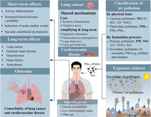
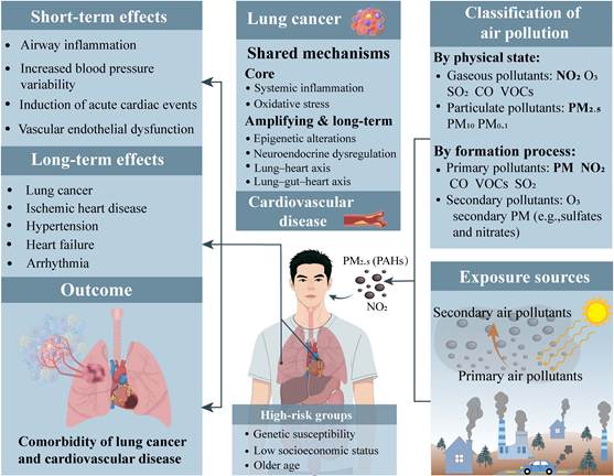
SO2 and CO are important components of the air pollution mixture and remain major air pollutants in developing countries, with their exposure also being associated with adverse health effects in the population [43]. A brief introduction to the sources and classification of air pollution is shown in Fig. 1.
Graphical abstract: conceptual framework of air pollution-related lung cancer-CVD comorbidity.
3.2 Exposure characteristics of pollutants
In recent years, the global upward trend in PM2.5 pollution has been reversed. The global population-weighted PM2.5 concentration rose from 28.3 μg/m3 in 1998 to 38.9 μg/m3 in 2011, after which it gradually declined [44]. However, studies have found that although exposure concentrations of NO2 and PM2.5 continue to decrease, their health risks have not declined correspondingly; adverse health effects can still be observed even at very low exposure levels [45]. For lung cancer and CVDs, genetic and other factors may increase population susceptibility to these conditions, and long-term exposure to air pollution may further promote the early onset of their comorbidities [46].
Although PM2.5 pollution has gradually eased, O3 pollution continues to intensify, and the issue of secondary pollution has become increasingly prominent [47]. The reduction in PM2.5 is mainly attributable to the control of precursors such as sulfur dioxide and primary particulate matter, whereas nitrogen oxides and VOCs have not yet achieved coordinated and deep emission reductions. These changes suggest a gradual transition from pollution dominated by coal-combustion-related particulate matter toward a more complex pollution pattern characterized by the coexistence of PM2.5 and O3, potentially posing additional challenges for the prevention of lung cancer and CVD [48].
3.3 Limitations of single-pollutant assessment
In real-world environments, exposures are mostly mixed and vary by region. Atmospheric NO2 can generate nitrate and secondary PM, accompanied by ozone formation; traffic sources simultaneously emit NO2 and PM; indoor biomass combustion typically releases PM and CO together in relatively stable proportions [49, 50]. These atmospheric chemical processes and real-world exposure characteristics determine that NO2, PM2.5 and O3 inevitably coexist. Furthermore, urban exposures are dominated by traffic-related components, while rural areas are significantly affected by agricultural activities and residential solid fuel combustion. Attributing comorbidities solely to a single pollutant may seriously underestimate the overall disease burden posed by air pollution [51].
4. Epidemiological evidence
4.1 Air pollution and lung cancer risk
PM2.5 has the strongest association with lung cancer. In studies from different regions published after 2020, PM2.5 is consistently linked to an increased risk of lung cancer even at very low concentrations. Specific risks are shown in the Supplementary Table S2. PAHs are one of the components of PM2.5. Studies have found that at the same PM2.5 concentration, lung cancer risk increases significantly with elevated PAHs content [52]. This suggests that regional differences in PM2.5-associated lung cancer risk may be largely attributed to variations in PM2.5 chemical composition. Multiple studies have confirmed the association between NO2 exposure and lung cancer, with its carcinogenic effect second only to PM2.5. However, this association is sometimes undetectable at low exposure concentrations [53].
The association between O3 exposure and lung cancer risk remains inconsistent, with some studies showing a positive correlation [54] or no association [55], while others, interestingly, even showing a negative correlation [56]. O3 and NO2 exhibit an inverse relationship in the natural environment. In areas with high ozone levels, the relatively low nitrogen dioxide concentrations may give rise to spurious associations, and thus the observed negative correlation should not be interpreted as a protective effect of O3. As mentioned above, O3 is a secondary pollutant, and its pollution has become increasingly prominent in recent years. Therefore, it is necessary to adjust for other pollutants (especially NOX and VOCs) before reassessing the true association of O3. Studies on SO2 and CO are relatively scarce. SO2 may be associated with lung cancer, but current evidence is still insufficient to establish a definitive link [54]. The IARC currently classifies SO2 as a Group 3 carcinogen, meaning its carcinogenicity to humans cannot be classified. In two recent studies, CO tended to show no association with lung cancer [54, 57]. In recent years, with the substantial reduction in the use of sulfur-containing fuels and household coal burning, the statistical associations of SO2 and CO with lung cancer incidence are likely to gradually weaken.
Regarding population susceptibility, individuals living in cities with low economic or educational levels, males, older adults, and those with a family history of lung cancer demonstrate higher sensitivity to air pollution exposure.
4.2 Air pollution and CVD risk
The effects of air pollution on the incidence of CVD are presented in Supplementary Table S3, which distinguishes between short-term and long-term exposure. Recent evidence shows that short-term exposure to NO2, PM2.5, SO2, O3, and CO has clear cardiovascular effects, with NO2 and PM2.5 having the greatest impact. The most significant adverse event is the triggering of acute coronary syndrome (ACS). In addition, short-term exposure increases blood pressure variability. Several meta-analyses have also found that short-term exposure to air pollution can induce atrial fibrillation (AF) (significantly associated with PM2.5, NO₂, and O₃) [58], and worsen HF (significantly associated with PM2.5, NO₂, SO₂, and CO) [59].
Long-term exposure to air pollution is associated with CHD, HF, arrhythmia, and hypertension. PM2.5 is the primary pollutant, while NO2 and O3 also pose high cardiovascular health risks.
Regarding population susceptibility, individuals with genetic risk, the elderly, obese individuals, and low-income populations in urban areas are more susceptible.
4.3 Evidence supporting the association between air pollution and lung cancer-CVD comorbidity
Current epidemiological evidence remains incomplete because no prospective cohort study has directly evaluated incident lung cancer-CVD comorbidity as a composite outcome in relation to air pollution exposure. Nevertheless, three lines of evidence consistently suggest an association between air pollution and lung cancer-CVD comorbidity.
The first line of evidence comes from studies examining the associations between air pollution and each disease separately. A large body of high-quality epidemiological evidence has consistently shown that exposure to air pollutants is associated with increased risks of both lung cancer and CVD (Supplementary Tables S2 and S3). Among the pollutants investigated, the evidence is strongest for PM2.5 and NO2. These findings indicate that air pollution represents a shared environmental risk factor for both diseases and that exposure to the same pollutants is associated with an increased risk of each. However, an important limitation of this evidence is that it cannot determine whether lung cancer and CVD develop concurrently within the same individuals as a result of the shared exposure.
The second line of evidence comes from bidirectional associations observed in single-disease cohorts (summarized in Table 2). Among cancer survivors, including those with lung cancer, air pollution has been associated with an increased risk of incident CVD [60-62]. Among patients with CVD, traffic-related air pollution has been associated with an increased risk of incident malignancies; however, in both studies, lung cancer was evaluated as a component subgroup rather than as a primary endpoint [63, 64]. These findings suggest that air pollution may exert cross-system effects in individuals with pre-existing disease and further imply a pathophysiological interplay between lung cancer and CVD. However, these findings should be interpreted with caution. Because these studies enrolled individuals with an established disease at baseline, they could not capture the entire disease trajectory from a healthy state to the development of comorbidity and therefore cannot independently demonstrate that air pollution initiates primary lung cancer-CVD comorbidity. In addition, most available studies evaluated all cancer types rather than lung cancer specifically, limiting the direct evidence for lung cancer.
Bidirectional effects of air pollution on patients with cancer and CVD
| First Author/Year | Area | Participants | Pollutants | Key Findings |
|---|---|---|---|---|
| Choi et al., 2020 [60] | Korea | 40,899 cancer survivors | PM2.5 PM10 | Patients in the highest quintile of PM2.5 had an increased risk of CVD (HR, 1.31; 95% CI, 1.07-1.59) |
| Coleman et al., 2021 [61] | US | 5,591,168 cancer patients | PM2.5 | A 10 μg/m3 increase in PM2.5 was associated with CVD mortality risk (HR, 1.32, 95% CI: 1.26-1.39). |
| Xiao et al., 2025 [62] | UK | 50,923 cancer patients | PM2.5, PM10, NO2 | Long-term exposure increases the risk of HF with HRs of 1.22 (1.07, 1.38) for PM2.5, 1.16 (1.03, 1.32) for PM10, and 1.20 (1.06, 1.36) for NO2 |
| Cohen et al., 2018 [63] | Israel | 9,816 CHD patients undergoing PCI | traffic-related air pollution (TRAP, NOX) | A 10 ppb increase in NOX was associated with an increased risk of TRAP-related cancers (including lung cancer) (HR, 1.16; 95% CI: 1.05-1.28). |
| Cohen et al., 2021 [64] | Israel | 2,040 CHD patients | NOX | A 10 ppb increase in NOX was associated with an elevated cancer risk: HR, 1.19 (1.03-1.37) |
The third line of evidence comes from disease coexistence and pathophysiological interactions. As discussed in Section 2, lung cancer and CVD frequently coexist and mutually exacerbate each other even in the absence of treatment. Subsequent sections further describe their shared molecular mechanisms, including chronic inflammation and oxidative stress. These observations provide biological plausibility for the interaction between the two diseases. However, because these studies generally investigate broad risk factors rather than air pollution specifically, they cannot establish air pollution as the direct cause of lung cancer-CVD comorbidity.
These three lines of evidence arise from different study designs but provide complementary support. Evidence from independent disease associations establishes the epidemiological basis for the relationship between air pollution exposure and each disease. Bidirectional findings from single-disease cohorts provide epidemiological evidence of cross-system health effects associated with air pollution, while studies of disease coexistence and shared biological pathways offer biological plausibility. Collectively, these findings support the cautious conclusion that air pollution is a plausible risk factor for lung cancer-CVD comorbidity. Nevertheless, current evidence remains insufficient to determine whether air pollution simultaneously initiates both diseases, triggers one disease that subsequently accelerates the development of the other, or simply exacerbates pre-existing disease, resulting in the manifestation of comorbidity. Therefore, prospective cohort studies using incident lung cancer-CVD comorbidity as a composite outcome remain an essential next step.
In addition, current evidence consistently suggests that individuals with lower socioeconomic status, older adults, and those with genetic susceptibility may be more vulnerable to the adverse health effects of air pollution. These populations should therefore be prioritized in future strategies for the prevention of lung cancer-CVD comorbidity.
4.4 High-risk populations and high-priority settings
The Wumeng Mountains area in Yunnan, particularly Xuanwei, exemplifies a region with one of the highest lung cancer incidence and mortality rates in China. Data from 2014-2016 showed that the age-standardized lung cancer mortality rate in Xuanwei was 65.75 per 100,000, which was approximately 1.5 times the average level of rural China [65]. Lung cancer here displays strong familial aggregation [66], with comparable rates between males and females, contrasting with the typical male predominance elsewhere. This pattern is closely linked to local coal distribution and usage [67, 68]. Coal combustion releases large amounts of PM and adsorbed PAHs. The annual average outdoor PM2.5 concentration in Xuanwei reached 45.3 μg/m3, far exceeding the WHO standard of 5 μg/m3 [67]. Although authoritative epidemiological data on lung cancer-CVD comorbidity in Xuanwei remain limited, existing research indicates that CVD mortality there is significantly higher than among Shanghai residents, suggesting a similarly high burden [69]. The extreme air pollution exposure in Xuanwei represents a unique regional exposure paradigm. Although it provides invaluable mechanistic proof-of-principle, its exposure characteristics may not be fully generalizable to ambient urban outdoor smog.
Northeastern China is another region with a high burden of both lung cancer and CVD. In 2021, the standardized annual rates of lung cancer incidence across provinces ranged from 59.61 to 73.48 per 100,000, exceeding the national average (44.01 per 100,000). Among various risk factors, air pollution accounted for approximately 25% of lung cancer deaths [70]. Furthermore, northeast China also bears the nation's highest burden of CVD, with a standardized high-risk rate of 12.6% (95% CI: 12.4%-12.8%), exceeding the national average of 10.3%. While unhealthy diet is a primary risk factor, long-term exposure to pollutants from coal combustion, heavy metals, and chemicals in this industrial region likely contributes to the elevated CVD incidence [71]. In a comorbidity study of 1,510 lung cancer patients in Northeast China, 18%, 7.4%, 6.8%, and 4.6% of the patients had comorbid hypertension, HF, CHD and arrhythmia, respectively [72].
These regions represent high-priority natural settings for future research on pollution-related comorbidity because they combine heavy exposure, high disease burden, and potentially distinctive susceptibility patterns.
5. A shared mechanistic framework for air pollution-related lung cancer-CVD comorbidity
5.1 Core shared pathway: systemic inflammation and immune dysregulation
Inflammation is one of the most consistently supported and mechanistically coherent shared pathways through which air pollution may contribute to both lung cancer and CVD. Following pollutant deposition in the respiratory tract, local inflammatory responses can extend beyond the lung and contribute to systemic inflammation and vascular injury. This inflammatory cascade also interacts closely with oxidative stress, epigenetic alterations, and immune dysregulation, thereby providing an important mechanistic link between pulmonary and cardiovascular injury.
5.1.1 Pulmonary deposition and local inflammatory microenvironment
Traditional models of pollutant-induced carcinogenesis have largely emphasized genotoxic mechanisms, whereby carcinogenic exposures induce DNA damage and mutations that initiate malignant transformation [73]. In recent years, this classic genotoxic model of carcinogenesis has been re-examined: the tumor-promoting effects of PM2.5 may not necessarily depend on directly inducing new DNA mutations, but rather on promoting the clonal expansion of pre-existing carcinogenically mutated cells in the lung by inducing inflammatory responses and altering the tissue microenvironment. PM2.5 deposited in the distal lung can induce the recruitment and activation of alveolar macrophages, thereby promoting the release of interleukin-1β (IL-1β) and other inflammatory mediators. IL-1β can drive alveolar type II epithelial cells harboring EGFR mutations into a progenitor-like proliferative state, thereby promoting tumorigenesis [74]. Similar tumor-promoting effects have also been observed in experimental models involving KRAS-mutant cells.
Persistent inflammatory activation further induces the production of IL-6, IL-8, tumor necrosis factor-α (TNF-α), vascular endothelial growth factor (VEGF), and matrix metalloproteinases (MMPs), establishing a chronic inflammatory microenvironment that promotes epithelial proliferation, angiogenesis, extracellular matrix remodeling, and immune dysregulation [75]. In mouse intervention studies, blockade of the IL-1β signaling pathway reduced lung tumor burden, further supporting the important role of this inflammatory pathway in lung cancer initiation and progression [76].
5.1.2 Spillover into the systemic circulation
Pollutant-induced inflammation is not restricted to the lung. Inflammatory mediators generated within the pulmonary microenvironment, including cytokines, chemokines, and ROS, can enter the systemic circulation and induce extrapulmonary effects. Animal studies have shown that intratracheal instillation of PM2.5 induces inflammatory responses beyond the lung, characterized by increased levels of pro-inflammatory cytokines and endothelial adhesion molecules in the circulation and vascular tissues, accompanied by marked macrophage infiltration into the vascular wall [77]. In the Multi-Ethnic Study of Atherosclerosis (MESA), long-term PM2.5 exposure was associated with increased circulating IL-6 levels, whereas NOX exposure was associated with elevated coagulation-related biomarkers, including D-dimer. Short-term PM2.5 exposure was also associated with increased levels of CRP, fibrinogen, and soluble E-selectin [78]. Another population-based cohort similarly reported associations between long-term exposure to PM2.5 and NO2 and elevated circulating inflammatory biomarkers [79].
This systemic inflammatory state is strongly implicated in the development and progression of CVD. In a study involving 84,399 patients with CHD, approximately 59% had CRP levels ≥2 mg/L [80], suggesting that persistent inflammatory activation is common among patients with established CVD and may contribute to residual cardiovascular risk.
5.1.3 Pathological consequences
In the lungs, sustained IL-1β signaling appears to awaken mutated epithelial cells and promote malignant proliferation. The inflammatory microenvironment promotes tumor cell survival, angiogenesis, and immune evasion, converting latent oncogenic potential into lung cancer, particularly lung adenocarcinoma.
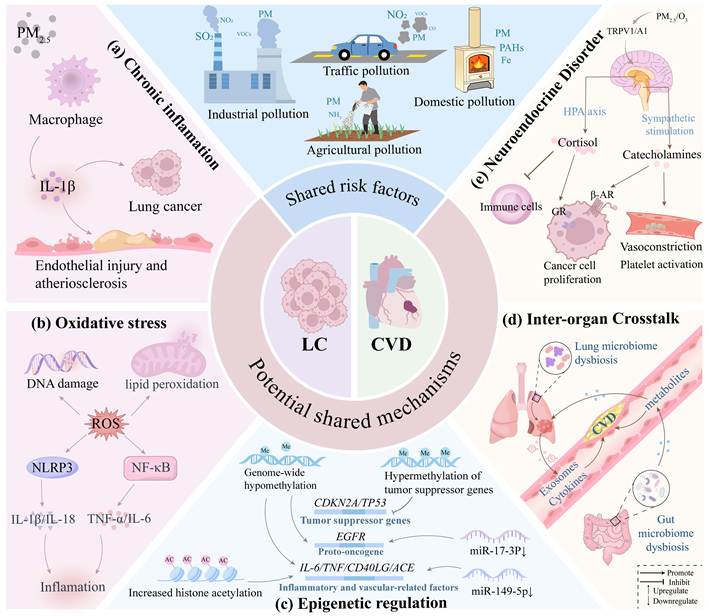
In the cardiovascular system, circulating inflammatory mediators induce endothelial activation and dysfunction, increase the expression of vascular adhesion molecules, and promote the recruitment and activation of circulating monocytes and pro-inflammatory macrophage phenotypes within the arterial wall. These inflammatory cells further amplify local inflammatory signaling, promote lipid uptake and foam cell formation, and contribute to the initiation, progression, and destabilization of atherosclerotic plaques [81]. Meanwhile, inflammatory activation can increase the expression of tissue factor (TF) and plasminogen activator inhibitor-1 (PAI-1), activate coagulation pathways, and promote platelet activation, thereby shifting the vascular environment toward a prothrombotic state (Fig. 2a) [37].
Shared mechanisms of air pollution-induced comorbidity of lung cancer (LC) and cardiovascular disease (CVD). (a) Air pollution promotes the release of IL-1β, leading to chronic inflammation, which contributes to the development of both lung cancer and CVD. (b) ROS can directly cause cellular damage and also promote inflammatory pathways. (c) Epigenetic regulation includes genome-wide hypomethylation, hypermethylation of tumor suppressor genes, increased acetylation of certain genes, and regulation by non-coding RNAs. (d) Organs interact through cytokines, exosomes, and metabolites following microbiota disruption, forming a vicious cycle. (e) Air pollution can activate the HPA axis and sympathetic nervous system, leading to metabolic and immune dysregulation. LC = Lung cancer, ROS = Reactive Oxygen Species, CD40LG = soluble CD40 Ligand, ACE = Angiotensin Converting Enzyme, TRPV1/A1 = Transient Receptor Potential Vanilloid 1 / Ankyrin 1, GR = Glucocorticoid receptor, β-AR = β-Adrenergic receptor, HPA axis = Hypothalamic-Pituitary-Adrenal axis (Italicized text represents gene symbols).
5.2 Oxidative stress: a mechanism of comorbidity amplification
Oxidative stress is closely associated with inflammation. It can amplify both upstream and downstream inflammatory signals, while simultaneously contributing to carcinogenesis and cardiovascular damage (Fig. 2b). In a population-based case-control study, levels of oxidative stress biomarkers such as 8-isoprostane and 8-OHdG were elevated in urine following air pollution exposure [82]. This provides direct evidence of oxidative stress. In another randomized controlled trial, exposure to traffic-related air pollution (PM, BC, NO2, CO) for four hours led to alterations in dozens of molecules related to systemic inflammation, oxidative stress, endothelial dysfunction, coagulation, and lipid metabolism, as measured by multi-omics approaches. Multiple pathways, including oxidative stress, were simultaneously activated systemically, and oxidative stress is more likely to contribute to pathological effects together with chronic inflammation [83].
Pollutants including PM (and adsorbed heavy metals/PAHs), NO2, O3, SO2, and high concentrations of CO can generate reactive oxygen species (ROS) through multiple convergent mechanisms: activation of NADPH oxidase in lung epithelial cells, vascular endothelial cells, and alveolar macrophages; induction of mitochondrial dysfunction leading to electron transport chain leakage; and iron-catalyzed Fenton reactions generating hydroxyl radicals [84-86]. Furthermore, ROS engage in a positive feedback loop with inflammation: ROS activate NF-κB signaling and the NOD-like receptor family pyrin domain-containing 3 (NLRP3) inflammasome, driving further release of IL-1β and IL-6 and amplifying inflammatory signals [87].
In the lungs, ROS can directly induce oxidative DNA base damage and strand breaks. Additionally, PAHs can directly form covalent DNA adducts via the AhR-Arnt-CYP1A1/1B1-BDPE axis, causing DNA damage [88]. The combination of chronic inflammation and DNA damage may be a key factor in the malignant progression of mutated epithelial cells. ROS can also disrupt cell membrane integrity through lipid peroxidation and deplete antioxidant defenses such as glutathione, creating conditions conducive to tumor development [89].
In the cardiovascular system, ROS mediate endothelial nitric oxide synthase uncoupling, reducing nitric oxide (NO) bioavailability and leading to vasomotor dysfunction, an early mechanism of hypertension and atherosclerosis [84]. ROS modify low-density lipoprotein (LDL) to form oxidized LDL (ox-LDL), promoting atherosclerotic plaque development and instability [90]. Moreover, ROS can drive myocardial fibrosis through iron-dependent mechanisms: excess iron initiates Fenton reactions, upregulates transforming growth factor-β1 (TGF-β1) secretion, drives endothelial-to-mesenchymal transition, and activates cardiac fibroblasts, ultimately potentially leading to chronic heart failure and malignant arrhythmias [91]. Cardiac MRI studies have confirmed a significant association between long-term PM2.5 exposure and diffuse myocardial fibrosis in humans [92]. In animal models, inhibition of NADPH oxidase 4 significantly reduced ROS production and effectively suppressed myocardial fibrosis, demonstrating a causal role of oxidative stress in cardiac dysfunction [93].
5.3 Epigenetic reprogramming and persistent disease memory
Air pollution induces long-lasting health effects through epigenetic modifications. PM, NO2, PAHs, heavy metals, and others have been associated with epigenetic alterations in the body [94, 95]. These alterations may sustain a state of inflammation, proliferation, and vascular damage even after exposure ceases, perpetuating disease risk.
Multiple population-based cohort studies and epigenome-wide association studies (EWAS) have demonstrated that exposure to air pollution can induce extensive epigenetic remodeling. The Multi-Ethnic Study of Atherosclerosis (MESA) reported that long-term PM2.5 exposure was associated with differential methylation at multiple CpG sites, and some of these methylation alterations were accompanied by changes in the expression of nearby genes, suggesting that DNA methylation may contribute to the development of air pollution-mediated atherosclerosis and CVD [96]. Further population-based EWAS have revealed that PM2.5-associated differentially methylated regions are primarily enriched in pathways related to inflammatory responses, oxidative stress, immune regulation, cardiovascular function-related genes, and carcinogenesis-related processes [97, 98]. Beyond DNA methylation, air pollution may regulate gene transcription through alterations in histone modifications and chromatin architecture. For example, oxidative stress induced by air pollution exposure can alter the activity of histone acetyltransferases (HATs) and histone deacetylases (HDACs), resulting in persistent dysregulation of inflammation- and metabolism-related gene expression [99, 100]. In addition, circulating microRNAs (miRNAs), which function as important post-transcriptional regulators, are also influenced by air pollution exposure. Experimental studies have shown that PM2.5 exposure can alter circulating miRNA expression profiles, thereby regulating inflammatory responses, oxidative stress, endothelial function, and tumor-related signaling pathways [101]. These findings suggest that epigenetic remodeling represents one of the key intermediate molecular mechanisms linking environmental pollutant exposure to the risk of chronic diseases.
Animal studies suggest that prenatal exposure to PM2.5 may exert persistent effects on offspring through epigenetic mechanisms and may potentially induce transgenerational epigenetic effects [102]. If this phenomenon can be confirmed in human populations, it would suggest that environmental air pollution exposure may exert long-term effects on cardiovascular and respiratory health in subsequent generations through epigenetic mechanisms and may alter susceptibility to related diseases. However, direct epidemiological evidence demonstrating that such epigenetic alterations can increase the transgenerational risk of lung cancer or CVD remains lacking.
5.3.1 DNA methylation: gene silencing and activation
The overall pattern of methylation alterations induced by air pollution exposure is characterized by global hypomethylation across the genome, accompanied by hypermethylation of specific genes [103]. PM2.5 and other air pollutants can inhibit DNA methyltransferase 1 (DNMT1) through the generation of ROS, resulting in global DNA hypomethylation and increased genomic instability, which may contribute to aberrant activation of oncogenes. Conversely, oxidative stress and inflammation-related signaling pathways can induce abnormal expression of DNMT3A/DNMT3B, leading to promoter hypermethylation and transcriptional silencing of tumor suppressor genes (e.g., TP53 and CDKN2A), thereby promoting tumor initiation and progression [104, 105]. Experimental studies have demonstrated that long-term exposure to PM components can alter DNA methylation patterns in pulmonary epithelial cells and activate pro-tumorigenic signaling pathways, including the MAPK and PI3K-AKT pathways. These alterations may have particular clinical relevance in individuals harboring oncogenic driver mutations such as EGFR and KRAS: epigenetic remodeling can establish a microenvironment conducive to tumor growth and facilitate the clonal expansion of genetically altered cells [106].
In cardiovascular tissues, air pollution-associated DNA methylation alterations can reduce the methylation levels of inflammation-related genes (CD40LG), coagulation-related genes (tissue factor F3 and SERPINE1), vasoconstriction-related genes (ACE), and endothelial repair-related genes (NOS3), thereby promoting the expression of these genes and ultimately contributing to hypercoagulability and endothelial dysfunction [107, 108]. Furthermore, long-term exposure to moderate concentrations of PM10 has been associated with hypermethylation of antioxidant pathway genes (SRXN1 and AKR7A2), which may weaken the antioxidant defense response of the organism [109]. These epigenetic alterations are closely involved in the development of CVDs. Among these, the pro-inflammatory pathways induced by DNA methylation alterations have been repeatedly validated [107, 110]. Exposure to air pollutants can modify the methylation status of regulatory regions of inflammation-related genes, including TNF, CD40LG, and ICAM1, resulting in increased inflammatory mediator release (Fig. 2c). These inflammatory pathways not only represent critical mechanisms linking air pollution exposure to CVD but also play an important role in lung cancer progression.
5.3.2 Histone modification: sustained transcriptional dysregulation
Pollutants can exert their biological effects by disrupting histone methylation, acetylation, and lactylation. PAHs can inhibit the histone methyltransferase DOT1L, thereby reducing H3K79me2 levels involved in DNA damage responses and repair, suggesting that the DOT1L-H3K79me2 axis may contribute to PAH exposure-associated genomic instability [111]. Persistent PM exposure can disrupt NAD+ metabolism in macrophages and reduce the activity of the NAD+-dependent deacetylase SIRT1, resulting in increased histone acetylation and subsequent upregulation of pro-inflammatory gene transcription [112]. In addition, animal studies have shown that gestational PM2.5 exposure increases myocardial p300/CBP expression and H3K9 acetylation in adult offspring and enhances H3K9ac enrichment at the promoter regions of cardiac hypertrophy-related genes, including GATA4 and MEF2C, ultimately contributing to cardiac hypertrophy in adulthood [113]. Recent experimental studies further demonstrated that PM2.5 promotes glycolysis and lactate accumulation in pulmonary macrophages and increases histone lactylation, whereas inhibition of glycolysis effectively attenuates pulmonary inflammation and lung tissue remodeling. These findings suggest that histone lactylation may represent one of the mechanisms underlying air pollution-induced lung tissue injury [114].
5.3.3 Non-coding RNA dysregulation: post-transcriptional regulation
A recent experimental study by Lin et al. found that PM2.5 exposure upregulated miR-21-5p in NSCLC, which subsequently targeted and suppressed Phosphodiesterase 4D interacting protein (PDE4DIP), thereby promoting NSCLC cell proliferation, migration, and invasion, suggesting a tumor-suppressive role for PDE4DIP [115]. However, another study conducted during the same period reported that PDE4DIP knockdown activated the PKA/CREB signaling pathway, induced apoptosis, and inhibited cell-cycle progression in NSCLC cells, suggesting a pro-tumorigenic role for PDE4DIP [116]. These apparently contradictory findings may be context-dependent. Under baseline tumor conditions, PDE4DIP may promote tumor growth through regulation of PKA signaling, whereas under PM2.5 exposure, PDE4DIP is downregulated by miR-21-5p, and restoration of PDE4DIP expression may attenuate the malignant phenotypes induced by PM2.5. This context-dependent mechanism warrants further investigation.
At the population level, a case-control study found that increased BaP exposure was associated with the downregulation of multiple circulating miRNAs. Among these, reduced expression of the tumor-suppressive miR-17-3p may relieve its inhibitory effect on oncogenes such as EGFR, thereby contributing to an increased risk of lung cancer [117].
In addition, experimental studies have shown that PM2.5 exposure can downregulate miR-149-5p in human bronchial epithelial cells and mouse lung tissues, thereby activating the downstream TAB2/TAK1-mediated MAPK/AP-1 and IKK/NF-κB signaling pathways and promoting the expression of pro-inflammatory cytokines, including IL-1β, IL-6, IL-8, and TNF-α [118]. Population-based studies further suggest that PM2.5-associated miRNA dysregulation may exert broader inflammatory and cardiovascular effects. Higher PM2.5 exposure has been associated with reduced circulating levels of miR-187-3p, miR-146a-5p, miR-1-3p, and miR-199a-5p, accompanied by increased expression of inflammation, coagulation, and vasoconstriction-related factors, including IL-1, IL-6, TNF, TLR2, F3, and ET-1 (Fig. 2c) [119]. These dysregulated miRNAs may act as persistent post-transcriptional regulators, thereby contributing to sustained activation of pathological signaling pathways.
5.4 Neuroendocrine and autonomic nervous system imbalance
Air pollutants activate transient receptor potential (TRP) channels in lung tissue, primarily TRPV1 and TRPA1, which trigger autonomic reflex arcs and lead to excessive sympathetic nervous system activation [120, 121]. Short-term exposure to pollutants such as PM2.5 can rapidly elevate catecholamine levels within minutes, inducing vasospasm, acute coronary syndrome, or arrhythmias [122]. A randomized controlled trial observed that short-term exposure to PM was associated with increased levels of hormones such as corticotropin-releasing hormone (CRH) and adrenocorticotropic hormone (ACTH), supporting activation of the hypothalamic-pituitary-adrenal (HPA) axis. In addition, blood pressure, insulin resistance, oxidative stress, and inflammatory markers were significantly higher following PM exposure [123]. Further research by Chen et al. found that long-term exposure to PM was closely associated with the development of metabolic syndrome, including hyperglycemia, hyperlipidemia, and hypertension [124]. Metabolic syndrome is an important risk factor for CVDs such as atherosclerosis. It is reasonable to hypothesize that if the HPA axis activation and its accompanying inflammation, oxidative stress, and insulin resistance observed in short-term exposure studies persist and gradually accumulate with long-term or repeated exposure, they may increase the risk of CVD. Furthermore, preclinical studies suggest that abnormally elevated glucocorticoids may suppress anti-tumor immunity and activate glucocorticoid receptor (GR) in cancer cells, potentially promoting proliferation and metastasis; similarly, catecholamines have been shown experimentally to enhance EGFR signaling via β-adrenergic receptors (β-AR) [125, 126]. These interconnected pathways help explain why long-term pollution exposure simultaneously amplifies both pro-tumor and cardiovascular stress pathways (Fig. 2e).
5.5 Inter-organ crosstalk: the lung-heart axis
Existing studies suggest that multiple potential forms of inter-organ signaling may mediate communication between the lungs and the cardiovascular system. As the lungs are the first organs in which inhaled air pollutants are deposited and interact with tissues, pollution-induced pulmonary inflammation, oxidative stress, and alterations in barrier function may exert distal effects through the systemic circulation. Therefore, we propose a plausible hypothesis that pulmonary inflammatory signals and extracellular vesicles induced by air pollution exposure may participate in lung-heart inter-organ crosstalk, thereby affecting vascular and myocardial function. Conversely, cardiovascular injury induced by air pollution exposure may also influence pulmonary disease progression through inflammatory mediators, tissue hypoxia, pulmonary congestion, and other circulating signals (Fig. 2d). However, current evidence regarding inter-organ interactions between lung cancer and CVD is derived predominantly from disease models, animal experiments, and single-disease cohort studies. Direct human evidence demonstrating that air pollution exposure mediates the comorbidity of lung cancer and CVD through such mechanisms remains lacking.
5.5.1 Exosome and inflammatory cytokine-mediated lung-heart crosstalk
Exosomes are a subtype of extracellular vesicles derived from the endosomal system that can carry bioactive molecules, including non-coding RNAs, proteins, and lipids, and participate in long-distance intercellular communication. They are widely considered potential diagnostic biomarkers, with non-coding RNAs being among the most extensively studied molecular cargos. Cytokines such as IL-6 and TNF-α can be present within exosomes or secreted directly by cells [127].
Previous reviews have separately summarized the effects of exosomes released by lung cancer cells and cardiovascular cells on their respective disease processes. Lung cancer cells can actively release exosomes that promote epithelial-mesenchymal transition (EMT), induce angiogenesis, and shape the tumor microenvironment [128]. Similarly, in CVDs, cardiac fibroblasts, cardiomyocytes, and macrophages release exosomes that promote inflammation, fibrosis, vascular dysfunction, and myocardial remodeling [129].
Importantly, exosomes derived from lung cancer and from the cardiovascular system may engage in a bidirectional pro-pathogenic crosstalk: in a mouse model of lung cancer, cancer-cell-secreted miR-485-3p delivered to cardiomyocytes causes mitochondrial dysfunction by suppressing PGC-1α [130]. Under hypoxic conditions, lung-cancer-derived miR-23a disrupts the endothelial barrier by inhibiting prolyl hydroxylase (PHD) and the tight junction protein (ZO-1) [131]. Lung cancer and its inflammatory microenvironment can produce inflammatory and profibrotic mediators, including IL-6 and TGF-β. Previous studies have demonstrated that IL-6 and TGF-β can act synergistically within the lung cancer microenvironment to promote fibroblast activation. These mediators are also important regulators of cardiac fibroblast activation and myocardial fibrosis and may influence distal cardiac fibroblasts through the IL-6/TGF-β pathway, thereby contributing to ventricular remodeling [132]. In lung cancer, CXCL13-mediated reprogramming of stromal macrophages within the pulmonary microenvironment may induce platelet aggregation through the secretion of prothrombotic small extracellular vesicles carrying integrin β2, a process associated with an increased risk of thrombosis [133]. Conversely, in mouse models of myocardial infarction or HF, cardiac mesenchymal stromal cells (cMSCs) have been observed to secrete exosomes carrying high levels of pro-inflammatory cytokines (IL-6, TNF-α), pro-angiogenic factors (VEGF), as well as pro-cancer proteins (osteopontin) and RNAs (miR-21). In the setting of HF complicated by pulmonary congestion, these exosomes may accumulate in the lungs and thereby accelerate tumor growth [134]. Furthermore, following myocardial infarction, cardiomyocytes can release exosomes enriched in miR-22-3p, which have been observed to suppress erastin-induced ferroptosis and thereby promote tumor cell survival (Fig. 3) [135].
Lung cancer (LC) and cardiovascular disease (CVD) mutually promote each other via exosomes and cytokines. Exosomes carrying molecules can induce endothelial injury, hypercoagulable state, myocardial fibrosis, etc. Meanwhile, CVD can promote the proliferation and metastasis of lung cancer cells through pulmonary congestion, hypoxemia, and molecules released by cardiac mesenchymal stromal cells (cMSCs) and cardiomyocytes. This process may be exacerbated by exposure to air pollution.
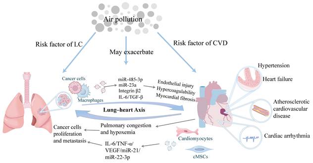
In this bidirectional pro-pathogenic process, air pollution may act as an accelerator of the vicious cycle. Experimental studies have shown that PM2.5 exposure promotes the sustained release of exosomes containing TGF-β from the lungs and increase collagen production in mouse cardiomyocytes, which is closely associated with myocardial fibrosis [136]. In addition, in cellular and animal models, PM2.5 has been shown to activate the STAT3/miR-21 signaling axis, which suppresses TIMP3 and activates MMP9, thereby disrupting the vascular barrier. This process may increase the risk of CVDs such as atherosclerosis and hypertension, although its clinical significance remains to be further validated in human populations [137].
5.5.2 Alterations of the lung microbiome: a potential mechanism of lung-heart crosstalk
The healthy lung harbors a low-biomass microbial community, whose composition is influenced primarily by factors such as microaspiration from the upper respiratory tract and may contribute to local immune regulation and inflammatory homeostasis [138]. Previous studies have shown that exposure to air pollutants, including PM2.5 and diesel exhaust, can alter the composition and functional characteristics of the pulmonary microbiome, potentially through mechanisms involving disruption of the airway barrier, impairment of mucociliary clearance, and changes in the local metabolic environment [139, 140]. However, the direction of changes in microbial diversity and specific bacterial genera varies across studies. Overall, a general pattern appears to include reduced microbial richness and diversity, enrichment of opportunistic pathogens (e.g., Staphylococcus) and pollutant-degrading bacteria (e.g., Sphingomonas), as well as enrichment of host signaling pathways related to carcinogenesis and inflammation [141, 142].
In patients with advanced lung cancer, studies have observed enrichment of oral commensal bacteria, such as Veillonella, in the lower respiratory tract microbiota, and suggested that these bacteria may influence tumor-associated inflammation and cell proliferation through signaling pathways including IL-17/MAPK/PI3K/ERK [143]. The carriage rate of Staphylococcus aureus in the respiratory tract is higher in patients with lung cancer than in healthy individuals. Its metabolite lactate may upregulate monocarboxylate transporter 1 (MCT1) in tumor cells, thereby promoting lactate uptake and activating pseudohypoxic signaling pathways, which may enhance the metastatic capacity of lung cancer cells [144]. In addition, pulmonary microbial signals such as lipopolysaccharide (LPS) and peptidoglycan may activate lung-resident γδ T cells through the TLR2/4-MYD88-IL-1β/IL-23 pathway, thereby promoting inflammatory responses [145]. These findings provide clues that the pulmonary microbiome may be involved in the development and progression of lung cancer; however, it remains unclear whether pollution-induced alterations in the pulmonary microbiome directly contribute to lung carcinogenesis.
Currently, direct causal evidence linking pulmonary microbiome dysbiosis to CVD remains limited. Recent observational studies have reported an association between reduced airway microbial diversity and subclinical atherosclerosis; however, such associations cannot establish a causal relationship [146]. Theoretically, air pollution-induced pulmonary microbiome dysbiosis may affect the cardiovascular system through two potential pathways: first, by enhancing local pulmonary inflammation and promoting its dissemination through the systemic circulation; and second, by allowing microbial-derived molecules to enter the systemic circulation. These pathways may promote endothelial activation, vascular inflammation, and thrombosis, thereby potentially contributing to the progression of CVD [147]. However, these proposed mechanisms require further validation.
5.6 Microbiota-mediated amplification: the lung-gut-heart axis
The gut microbiota has been increasingly recognized as an important regulator of systemic metabolism, immunity, and inflammatory responses, giving rise to concepts such as the gut-lung axis and the gut-heart axis. However, there is currently no direct evidence from human populations demonstrating that air pollution-induced gut microbiome dysbiosis mediates the development or progression of lung cancer-CVD comorbidity, and this remains an important direction for future research. Therefore, we propose a potential mechanism by which gut microbiota disruption contributes to comorbidities involving the lung-gut-heart axis under air pollution exposure: the lung serves as the initial site of exposure, leading to gut microbiota disruption, which in turn accelerates both lung cancer and CVD. The detailed mechanisms and potential convergence points are illustrated in Fig. 4.
Potential mechanisms by which air pollution-associated gut microbiota dysbiosis may contribute to lung cancer-CVD comorbidity. Air pollution drives gut microbiota dysregulation, characterized by reduced production of short-chain fatty acids (SCFAs), increased generation of trimethylamine N-oxide (TMAO), and elevated systemic levels of secondary bile acids (SBAs) and lipopolysaccharide (LPS). These changes promote lung tumorigenesis by modulating immune function, inducing inflammation, and directly facilitating tumor proliferation and metastasis. Concurrently, they contribute to CVD through mechanisms including chronic inflammation, endothelial injury, atherosclerosis, and myocardial fibrosis. HDAC = Histone Deacetylase, ID2/IL-12R = Inhibitor of DNA Binding 2/ Interleukin-12 Receptor, IFN-γ = Interferon-γ, GzmB = Granzyme B, FOXP3 = Forkhead Box Protein P3, TLR4 = Toll-Like Receptor 4, TGR5 = G protein-coupled bile acid receptor 1 (GPBAR1), MMP2/9 = Matrix Metalloproteinase 2/9, ox-LDL = Oxidized Low-Density Lipoprotein; CD36 = Macrophage Scavenger Receptor, SIRT3 = Sirtuin 3, SOD2 = Superoxide Dismutase 2; mtROS = mitochondrial reactive oxygen species, TGF-βR1 = Transforming Growth Factor-β Receptor 1; α-SMA = α-Smooth Muscle Actin; COL1A1 = Type I Collagen (Italicized text represents gene symbols).
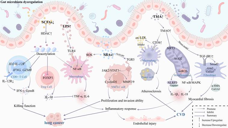
Air pollution may affect the gut microbiota through multiple pathways. PM deposited in the respiratory tract can be transported to the pharynx by the mucociliary clearance system and subsequently swallowed, thereby directly entering the gastrointestinal tract. Meanwhile, pulmonary oxidative stress and inflammatory responses may promote the release of reactive ROS, cytokines, and other inflammatory mediators into the systemic circulation, which can subsequently impair intestinal epithelial integrity and disrupt intestinal immune homeostasis [148, 149]. A systematic review has shown that exposure to PM, particularly diesel exhaust particles, can alter both the composition and metabolic functions of the gut microbiota. However, the direction and magnitude of changes in specific microbial taxa are not entirely consistent across studies, likely owing to differences in pollutant composition, exposure dose, route of exposure, and host susceptibility. Therefore, entire phyla such as Bacteroidetes, Proteobacteria, and Verrucomicrobia should not be simplistically classified as beneficial or harmful. More consistently reported alterations include disruption of gut microbial community structure, reduced microbial diversity, depletion of certain short-chain fatty acid (SCFA)-producing bacteria, and impairment of intestinal barrier function [150]. In parallel, increased circulating levels of lipopolysaccharide (LPS), TNF-α, and CVD-related biomarkers, including sICAM-1 and sP-selectin, have also been observed [148].
5.6.1 Regulatory effects of gut microbiota metabolites on lung cancer
The gut microbiota produces a wide range of metabolites, including short-chain fatty acids (SCFAs), secondary bile acids (SBAs), bacteriocins, phenylpropanoid derivatives, tryptophan metabolites, polyamines, and trimethylamine (TMA) [151]. Among these metabolites, SBAs and SCFAs may represent the most biologically plausible mediators linking gut microbiota dysbiosis to the initiation and progression of lung cancer [152].
SBAs are derivatives of primary bile acids modified by the gut microbiota. Under normal conditions, they play important roles in maintaining digestive function, gut homeostasis, regulating systemic metabolism, and immunity. Gut dysbiosis may disrupt SBA metabolism, potentially increasing their production [153]. Tang et al. found that per standard deviation increase in the concentration of taurodeoxycholic acid 3-sulfate was associated with a 33% elevated risk of lung cancer [154]. In lung cancer, SBAs can activate the TGR5 receptor, which has been associated with advanced tumor stage and poor prognosis, thereby triggering the JAK2/STAT3 signaling pathway and promoting tumor proliferation and metastasis through upregulation of downstream proteins, including Cyclin D1, MMP2/9 [155]. High concentrations of hydrophobic bile acids may induce ROS production, activate NF-κB-dependent inflammatory signaling, and cause oxidative DNA damage, with reported pro-inflammatory and pro-tumor effects. However, mechanistic studies of these effects have primarily focused on intestinal tumors and macrophages, and their relevance to lung cancer remains to be further validated [156, 157].
SCFAs, primarily including acetate, propionate, and butyrate, are key metabolites produced by gut microbiota through fermentation of dietary fiber. They provide energy for colonic epithelial cells and maintain the intestinal barrier. Deficiency of SCFAs disrupts the intestinal barrier, allowing microorganisms and their metabolites (e.g., LPS) to enter the circulatory system [158]. LPS can activate the TLR4/NF-κB pathway, leading to persistently elevated levels of inflammatory cytokines [159, 160].
SCFAs play an important role in immune regulation, primarily by inhibiting histone deacetylase (HDAC). In CD8⁺ T cells, HDAC inhibition epigenetically upregulates the expression of Interferon-γ (IFN-γ) and granzyme B (GzmB), thereby enhancing cytotoxic activity [161]. HDAC inhibition also upregulates the expression of the transcriptional regulator ID2, relieves the inhibition of IL-12 receptor (IL-12R), thereby enhancing IL-12 signaling, and promoting the production of IFN-γ and GzmB through downstream signaling pathways [162]. In regulatory T cells (Tregs), HDAC inhibition enhances FOXP3 expression, promotes Treg differentiation, and facilitates the secretion of inhibitory cytokines such as IL-10 and TGF-β, thereby preventing inflammatory damage caused by uncontrolled immune responses [163]. Furthermore, HDAC inhibition blocks NF-κB signaling, reduces the release of pro-inflammatory cytokines such as TNF-α and IL-6 [164]. Additionally, SCFAs can activate the G protein-coupled receptor GPR43 (also known as FFAR2), which triggers the mTOR/STAT3 pathway to promote Treg differentiation [165]. However, studies indicate that GPR43 expression is minimal in T cells and likely not the primary mechanism involved [166].
A decrease in SCFAs levels may weaken the aforementioned immunoregulatory and anti-inflammatory protective effects, thereby creating a more favorable environment for the development and progression of lung cancer. In vitro experiments have shown that sodium butyrate significantly reduces lung cancer cell viability by inducing apoptosis and modulating immune cell function, suggesting that restoration of SCFA levels may have therapeutic potential [167].
5.6.2 Mechanisms of cardiovascular injury mediated by gut microbiota metabolites
The relationship between the gut microbiota and its metabolites and cardiovascular health has attracted increasing research attention. Current evidence suggests that microbiota-related metabolites, including LPS, SCFAs, SBAs, and trimethylamine N-oxide (TMAO), may contribute to processes such as endothelial dysfunction and vascular remodeling through oxidative stress and inflammatory responses, thereby affecting cardiovascular health [168]. In animal studies, reduced levels of SCFAs have been shown to impair anti-inflammatory and immunoregulatory capacity. Among them, propionate has been reported to alleviate cardiac remodeling, vascular dysfunction, and hypertension in mice through regulatory T cell-mediated anti-inflammatory immunomodulation [169].
Here, the focus is on trimethylamine N-oxide (TMAO). Choline-metabolizing gut bacteria convert dietary choline into TMA, which is subsequently oxidized in the liver to form TMAO. Gut microbiota dysbiosis may be accompanied by elevated TMAO levels, which have been associated with CVDs, including atherosclerosis, HF, and hypertension [170-172]. Cellular and animal studies have shown that TMAO can upregulate the expression of scavenger receptors such as CD36 in macrophages, promoting oxLDL uptake and foam cell formation [173]. In vascular endothelial cells, TMAO can suppress the SIRT3-SOD2 antioxidant pathway, thereby increasing mitochondrial ROS (mtROS) production and activating the NLRP3 inflammasome, ultimately promoting vascular inflammation [174]. In addition, both TMAO and mtROS can activate the NF-κB and MAPK pathways, promoting vascular inflammation and endothelial injury [175]. In a mouse model, TMAO interferes with the TGF-β/Smad2 signaling by upregulating TGF-βR1, inducing fibroblast transformation and the expression of fibrotic markers α-smooth muscle actin (α-SMA) and type I collagen (COL1A1), thereby promoting myocardial fibrosis [176]. TMAO can also inhibit endothelial nitric oxide synthase activity to impair vasodilation [177], and potentiate angiotensin II-dependent pressor responses [178].
Intervention studies targeting the gut microbiota have provided further mechanistic support. Animal studies demonstrate that probiotics significantly reduce plasma TMAO and cecal TMA levels in mice [179]. Berberine inhibits TMA and TMAO synthesis; atherosclerotic patients taking Berberine experienced a 37% and 35% reduction in plasma TMA and TMAO, respectively [180]. Puerarin also ameliorates atherosclerosis by reducing TMAO production [181]. These findings offer new therapeutic avenues for microbiome-related CVD.
Overall, air pollution-associated gut microbiota dysbiosis may amplify the risk of lung cancer-CVD comorbidity through four interconnected processes: impaired SCFA-mediated intestinal barrier protection and immune regulation, LPS translocation, and increased TMAO production. These processes may converge on several shared pathological mechanisms, including ROS generation, activation of NF-κB and NLRP3 signaling, chronic systemic inflammation, endothelial dysfunction, immune remodeling, and tissue fibrosis. It is worth noting that the evidence supporting the above pathways is derived predominantly from in vitro cellular experiments and animal models, while existing human studies are mainly observational, with some interventional studies providing only indirect support. The causal relationships underlying these pathways remain to be further validated.
5.7 Additional modifiers and vicious cycles
Other pathological states induced by air pollution in the lungs also play significant roles in lung cancer-CVD comorbidity. Alveolar deposition of PM2.5 triggers local inflammation and protease-antiprotease imbalance, while long-term exposure is associated with the development of chronic obstructive pulmonary disease (COPD) and progressive decline in lung function [182]. The chronic hypoxia accompanying COPD progression may further activate the HIF-1α signaling pathway, contributing to pulmonary hypertension and subsequently leading to right ventricular remodeling and impaired cardiac function [183]. Hypoxia accelerates tumor growth through multiple mechanisms. Wang et al. found that under hypoxic conditions, NSCLC cells release exosomes enriched in circPLEKHM1, which induces M2 macrophage polarization and promotes metastasis [184]. Many CVDs, such as hypertension and CHD, can progress to HF in later stages, which leads to pulmonary congestion, edema, and hypoxia. Similarly, advanced lung cancer causes respiratory dysfunction and hypoxemia, thereby promoting the development of both conditions [134]. This cross-organ influence creates a vicious cycle, typically resulting in poorer prognosis for comorbid patients. Pre-existing metabolic conditions such as hyperglycemia may exacerbate the aforementioned inflammatory responses. Studies indicate that high glucose pre-treatment enhances PM2.5-induced IL-1β and ROS generation in these metabolic pathways [185].
The comorbidity state is susceptible to other factors, such as the most common cardiotoxicity associated with cancer therapies. Cytotoxic chemotherapy agents significantly increase the risk of developing CVD [186]. This represents one of the non-negligible factors but must be strictly distinguished from lung cancer-CVD comorbidity under air pollution exposure.
6. Limitations of current evidence: what remains unresolved
The medical costs increase rapidly with each additional chronic disease, highlighting the need for greater attention to the phenomenon of comorbidity [187]. Air pollution is an inevitable consequence of human activity, and its health impacts, as discussed, have been repeatedly confirmed. However, in environmental epidemiology, air pollution exposure cannot be randomly assigned, and lung cancer-CVD comorbidity is characterized by a long latency period. Prospective cohort studies that directly evaluate incident comorbidity as the primary outcome are inherently challenging to conduct. The inference presented here is therefore based on the integration of multiple independent and complementary lines of indirect evidence and should be interpreted with appropriate caution. Current evidence is insufficient to determine whether air pollution simultaneously initiates both diseases, first triggers one disease and subsequently accelerates the development of the other, or merely exacerbates pre-existing disease, ultimately manifesting as comorbidity. Nevertheless, the available evidence provides an important basis for identifying high-risk populations, informing preventive strategies, and guiding future study design. Direct prospective studies using incident lung cancer-CVD comorbidity as the outcome remain necessary to establish causality and to provide a stronger evidence base for prioritizing preventive and intervention strategies.
However, a practical challenge arises in conducting such studies: most patients begin anticancer treatment shortly after being diagnosed with lung cancer, and these treatments themselves may contribute to cardiovascular injury. An important challenge for future cohort studies is how to distinguish cardiovascular risks associated with air pollution exposure from cardiovascular toxicity induced by lung cancer treatment. A feasible approach would be to establish or utilize existing prospective or retrospective population-based cohorts, integrating cancer registry data, medical records, and long-term air pollution exposure data. Following lung cancer diagnosis, patients' cardiovascular status should be assessed, whenever possible, before the initiation of anticancer treatment. If long-term air pollution exposure is associated with pre-treatment CVD status and this association remains after adjustment for smoking, age, and other traditional cardiovascular risk factors, this would provide more direct population-level evidence that air pollution may independently contribute to the development of lung cancer-CVD comorbidity beyond the effects of anticancer treatment. For CVD events occurring after treatment initiation, the timing and type of anticancer treatment should be systematically recorded. After adjustment for long-term air pollution exposure and major cardiovascular risk factors, the risk of CVD should be further compared across different treatment exposure states to determine whether CVD risk increases following the initiation of specific treatments and whether air pollution exposure and treatment-related factors have additive or interactive effects. This approach may help address another important research question: whether environmental exposures can exacerbate treatment-related cardiotoxicity.
Currently, there are technical limitations in air pollution monitoring. In large-scale epidemiological studies, it is often unavoidable to use fixed-site monitoring values or model-estimated values as proxies for individual actual exposure levels. This error can mislead conclusions when assessing complex outcomes like comorbidity, potentially leading to a severe underestimation of the health effects of air pollution [51]. Additionally, current animal models fail to recapitulate the multifactorial exposure environments experienced by humans or the background of age-related immune senescence, limiting the clinical translation of mechanistic findings.
Effective management of comorbidity relies on early identification. However, the currently proposed biomarkers or predictive models remain mostly candidate directions rather than mature clinical solutions. IL-6, a key inflammatory mediator, has shown potential diagnostic and prognostic monitoring value for lung cancer and CVDs in studies, and theoretically could also serve as a predictor for lung cancer-CVD comorbidity, but its specificity is extremely low [188, 189]. 1-OHP has long been widely used to assess exposure to PAHs. 8-OHdG is a classic product of oxidative damage caused by ROS and is suitable as a risk assessment marker [190]. There have been many studies on epigenetic alterations and exosomes, however, most of these findings remain at the mechanistic level, and evidence for their clinical application is very limited. Some studies have confirmed their potential as biomarkers and therapeutic targets. For example, miR-21 has a dual role in promoting both cancer and vascular injury, and its expression is upregulated upon exposure to air pollution. It has shown good diagnostic performance in trials for lung cancer and CVD, making it a potential biomarker [191, 192].
It is noteworthy that the limited specificity of individual circulating biomarkers may be particularly pronounced in older adults and patients with multiple comorbidities. These populations commonly exhibit inflammaging, metabolic dysregulation, age-related oxidative stress, and exposure to various environmental or lifestyle factors, all of which can generate substantial background signals that interfere with biomarker detection. In this context, the major challenge for the early prediction and clinical translation of comorbidity risk is not simply to identify more powerful biomarkers, but rather to distinguish pollution-related biological perturbations from nonspecific systemic inflammation or oxidative stress. Such discrimination is unlikely to be achieved by any single circulating molecule. A more promising strategy may involve integrating multiple biomarkers or multi-omics profiles that capture distinct pathological processes involved in air pollution-related disease progression. At a minimum, such an approach should encompass two complementary dimensions. The first is the exposure profile, including the intensity, composition, and temporal pattern of air pollution exposure. The second is the biological response profile, including inflammation, oxidative stress, vascular injury, epigenomic alterations, and extracellular vesicle characteristics as potential domains [193]. For example, the lung immune prognostic index (LIPI), composed of routine clinical indicators, can be assessed before treatment. This index reflects the baseline host inflammatory status and has been shown in a meta-analysis to have prognostic predictive value in NSCLC patients receiving immunotherapy [194]. Building on this, combining pretreatment LIPI with air pollution exposure indicators and biomarkers of oxidative stress and vascular injury, together with longitudinal assessment of changes in these parameters, may help facilitate risk stratification and aid in distinguishing treatment-related cardiovascular events.
Emerging disease risk prediction models also provide methodological insights into the potential for clinical translation. For example, Jiang et al. developed the UKB-MDRMF model, which integrates multiple types of information, including environmental factors, to enable joint disease prediction and risk stratification across multiple conditions, demonstrating the methodological feasibility of comorbidity prediction models [195]. However, further improvement in the accuracy of air pollution-related comorbidity prediction will require more targeted incorporation of both exposure characteristics and shared pathogenic mechanisms, such as inflammation, oxidative stress, and DNA methylation patterns, as predictive factors. This may facilitate a shift from identifying disease-associated biomarkers alone toward pollution-attributable risk stratification, thereby enhancing the potential clinical utility of such approaches, particularly in older adults and individuals with multiple comorbidities.
The management of CVDs associated with lung cancer treatment typically involves adjusting the anticancer regimen based on the patient's cardiovascular status during therapy. In contrast, comorbidities induced by air pollution require balancing the treatment of both chronic conditions from the outset, with an emphasis on cardiovascular risk monitoring throughout the entire course of lung cancer care.
Currently, drug development targeting air pollution-induced cardiopulmonary injury remains largely focused on anti-inflammatory and antioxidant strategies, and is still in animal experiments or preclinical exploratory stages. There is a substantial gap in clinical application evidence, and extensive research is needed to evaluate benefits and risks and to expand indications. The classic antidiabetic drug metformin has been found in mouse models to inhibit systemic and pulmonary inflammation, reduce oxidative stress levels, and effectively alleviate PM2.5-induced lung injury and cardiac dysfunction [196]. In randomized controlled trials involving patients with atherosclerosis, treatment with the anti-inflammatory drug canakinumab to inhibit the IL-1β inflammatory pathway appeared to reduce the incidence of lung cancer and lung cancer mortality [197]. However, a subsequent phase III clinical trial indicated that canakinumab did not improve progression-free survival or overall survival in patients with lung cancer [198]. Therefore, current evidence does not support the use of canakinumab for the treatment of lung cancer, although its potential role in preventing comorbidities may warrant further investigation.
Furthermore, antioxidant-based interventions targeting oxidative stress present a well-recognized paradox in respiratory oncology. Although antioxidants are theoretically expected to be beneficial, randomized controlled trials have shown that supplementation with exogenous antioxidants, such as β-carotene and vitamin E, can paradoxically increase lung cancer incidence in high-risk populations, including smokers, and has also been associated with an increased risk of cardiovascular mortality [199, 200]. Preclinical studies have further demonstrated that, in KRAS-mutant mouse models of lung cancer, antioxidants such as N-acetylcysteine and vitamin E can promote lung cancer cell proliferation by reducing oxidative stress and suppressing p53 expression [201]. In addition, antioxidant supplementation may promote lung cancer cell metastasis by stabilizing the transcription factor BACH1 and activating glycolysis [202]. Therefore, nonspecific systemic antioxidant supplementation is not recommended as a routine intervention for lung cancer-CVD comorbidity. In particular, indiscriminate antioxidant use should be avoided in high-risk populations.
Based on the shared molecular pathways induced by air pollution that we have summarized, including chronic inflammation, oxidative stress, epigenetic alterations, organ crosstalk, and microbial dysbiosis, from a theoretical perspective, some specific pathways such as IL-6, TNF-α, IL-17/23, MMP-9/12, NF-κB signaling, ROS generation pathways, and gut microbiota dysbiosis may represent potential candidates for therapeutic investigation in air pollution-related lung cancer-CVD comorbidity. Given the antioxidant paradox and the potential risk that antioxidant supplementation may promote the survival and metastasis of tumor cells, interventions targeting ROS should no longer rely on the indiscriminate use of broad-spectrum exogenous antioxidants, but should instead further explore the selective modulation of specific redox signaling pathways. However, to date, no studies have confirmed that targeting these pathways can simultaneously improve clinical outcomes for both diseases, particularly in the context of air pollution exposure.
7. Clinical and public health implications
Regarding air pollution, there are currently well-established recommended measures that may simultaneously reduce the comorbidity burden of lung cancer and CVD, thereby achieving dual health benefits. At the individual level, promoting the use of indoor air purifiers, wearing filtering masks during high-pollution weather, and adopting clean energy stoves can effectively reduce individual exposure risks [203]. Effective source control and policy interventions include establishing low-emission zones, expanding urban green spaces, optimizing urban planning to enhance atmospheric circulation, timely issuing air quality forecasts and warnings, and implementing strict emission controls on industry and vehicles [204].
Several targeted interventions show great promise in the prevention and management of comorbidity but have not yet been incorporated into routine practice: in high-risk residential areas, such as the Wumeng Mountain region with a high prevalence of familial lung cancer, northeastern China where both lung cancer and CVDs are highly prevalent, and heavily polluted regions, there is an urgent need for continuous environmental monitoring and early warnings, along with the use of more precise exposure assessment methods, such as equipping study subjects with personal exposure monitors. High-risk populations should be encouraged to participate in joint screening programs for lung cancer and CVD, especially those with genetic susceptibility. For instance, a single low-dose chest CT scan can simultaneously detect lung nodules and preliminarily assess coronary artery calcification (CAC), offering the potential for early joint screening of lung cancer and CVDs [205]. However, in real-world clinical practice, lung nodule screening itself carries risks of false-positive findings and overdiagnosis. Incidentally detected CAC may prompt additional imaging, specialist referrals, or even invasive investigations, thereby increasing the burden on patients and the consumption of healthcare resources. To date, there is insufficient evidence that incorporating CAC assessment into low-dose CT for lung cancer screening further reduces all-cause mortality. In addition, standardized reporting criteria, indications for further investigations, and subsequent management pathways for incidentally detected CAC remain insufficiently established. These challenges need to be addressed before this approach can be routinely implemented in clinical practice. Furthermore, tracking the incidence of comorbidities through community health records and improving data on these conditions in these regions is essential.
To translate key research findings into clinical practice in the future, prospective cohort studies in high-exposure populations are needed to systematically track the incidence of comorbidity. Further investigations should also explore the potential value of anti-inflammatory drugs, selective redox pathway modulators, or microbiota-modulating interventions in reducing the risk of air pollution-related comorbidity. It is necessary to leverage multi-omics technologies to further identify biomarkers with high sensitivity and specificity, and to incorporate shared pathogenic mechanisms into comorbidity prediction models, thereby supporting early warning of disease.
In summary, addressing comorbidities under air pollution exposure requires integrating public health policies with clinical diagnosis and treatment, shifting from a linear mindset focused on single diseases and single factors to a systems thinking approach centered on comorbidities and multiple factors, thereby alleviating the burden of comorbidities.
8. Conclusion
Air pollution not only increases the risks of lung cancer and CVD individually, but may also create a shared biological and epidemiological environment that promotes the co-occurrence and mutual aggravation of these two diseases. PM2.5 and NO2 are the most extensively studied pollutants and currently have the strongest evidence suggesting a potential role in promoting lung cancer-CVD comorbidity. Other common pollutants also have some mechanistic and epidemiological support; however, validation from cohort studies remains limited, and findings across studies are not entirely consistent. It should be emphasized that the current evidence mainly supports the biological plausibility and epidemiological possibility that air pollution contributes to lung cancer-CVD comorbidity, while direct evidence is still needed to establish causality. With regard to susceptible populations, individuals of lower socioeconomic status, older age, and those with a genetic predisposition to lung cancer or CVD may represent high-risk groups for comorbidity. Mechanistically, the most well-established pathways involve systemic inflammation, oxidative stress, and tumor-promoting angiogenesis. Organ crosstalk, exosome-mediated signaling, and microbiota dysbiosis, particularly gut microbiota dysbiosis, represent promising mechanisms but require further validation and integration into clinical practice. Future studies are strongly encouraged to investigate the risk of CVD associated with air pollution exposure in lung cancer cohorts, particularly in highly polluted regions or areas with a high incidence of lung cancer, and to conduct prospective cohort studies using comorbidity as a predefined endpoint. In addition, the shared pathogenic pathways underlying these two diseases require further validation, targeted interventions should be explored, and precision strategies for air pollution control and disease prevention should be developed to effectively reduce the overall disease burden.
Abbreviations
AF: Atrial Fibrillation
ACE: Angiotensin Converting Enzyme
BaP: Benzo[a]pyrene
α-SMA: α-Smooth Muscle Actin
CAC: Coronary Artery Calcification
CO: Carbon Monoxide
CVD: Cardiovascular Disease
COL1A1: Type I Collagen
cMSCs: Cardiac Mesenchymal Stromal Cells
CD36: Macrophage Scavenger Receptor CD36
DNMT: DNA Methyltransferase
CRP: C-Reactive Protein
DALYs: Disability-Adjusted Life Years
GPBAR1 (TGR5): G protein-Coupled Bile Acid Receptor 1
GR: Glucocorticoid Receptor
GzmB: Granzyme B
EGFR: Epidermal Growth Factor Receptor
EMT: Epithelial-Mesenchymal Transition
FOXP3: Forkhead Box Protein P3
HPA axis: Hypothalamic-Pituitary-Adrenal Axis
HDAC: Histone Deacetylase
HF: Heart Failure
HR: Hazard Ratio
IL: Interleukin
IQR: Interquartile Range
ID2: Inhibitor of DNA Binding 2
IFN-γ: Interferon-γ
LPS: Lipopolysaccharide
LC: Lung Cancer
LIPI: Lung Immune Prognostic Index
mtROS: Mitochondrial Reactive Oxygen Species
MMP: Matrix Metalloproteinases
NF-κB: Nuclear Factor-κB
NLRP3: NOD-like Receptor Family Pyrin Domain Containing 3
NO2: Nitrogen Dioxide
NSCLC: Non-Small Cell Lung Cancer
ox-LDL: Oxidized Low-Density Lipoprotein
O3: Ozone
PAHs: Polycyclic Aromatic Hydrocarbons
PDE4DIP: Phosphodiesterase 4D Interacting Protein
PM: Particulate Matter
ROS: Reactive Oxygen Species
RR: Relative Risk
SBAs: Secondary Bile Acids
SCFAs: Short-Chain Fatty Acids
SO2: Sulfur Dioxide
CD40LG: Soluble CD40 Ligand
SIRT3: Sirtuin 3
SOD2: Superoxide Dismutase 2
TMAO: Trimethylamine N-Oxide
TLR4: Toll-Like Receptor 4
TRPV1/A1: Transient Receptor Potential Vanilloid 1 / Ankyrin 1
TRAP: Traffic-Related Air Pollution
TNF: Tumor Necrosis Factor
VOCs: Volatile Organic Compounds
Supplementary Material
Literature search strategy for this review and the epidemiological evidence for the incidence rates of lung cancer and CVDs associated with air pollution.

Acknowledgements
Funding
This work was supported by the National Natural Science Foundation of China (Nos. 82060426, 82272980, and 82360562); the Yunnan Provincial Key Research and Development Program (No. 202403AC100015); the First-Class Discipline Team Construction Project of Kunming Medical University for the Environmental, Genetic, and Lung Cancer Pathogenesis Research Team (Nos. 2024XKTDPY07); the First-Class Discipline Team Construction Project of Kunming Medical University (No. 2024XKTDYS07); the Yunnan Provincial Chest Tumor Prevention and Treatment Research Innovation Team (No. 202405AS350015); the Yunnan Provincial Medical Academic Leader Program (No. D-2025006); the Medical and Health Talent Project of the Yunnan “Xingdian Talent Support Program” (No. XDYC-YLWS-2025-0083); and the Development of Precision Prevention and Full-Cycle Intelligent Management System for Regionally High-Incidence Lung Cancer in Yunnan (No. 202303AC100203).
Author contributions
Y C contributed to conceptualization and funding acquisition; K Y supervised the whole study and contributed to funding acquisition. H Y was responsible for writing the original draft and formal analysis; P L participated in writing the original draft, data curation and literature screening; J Z and D S were involved in visualization and data curation; all authors contributed to writing - review & editing and approved the final manuscript.
Competing Interests
The authors have declared that no competing interest exists.
References
1. WHO. Ambient (outdoor) air pollution. https://www.who.int/news-room/fact-sheets/detail/ambient-(outdoor)-air-quality-and-health
2. HEI. State of Global Air Report 2024. https://www.stateofglobalair.org/resources/report/state-global-air-report-2024
3. Brauer M, Roth GA, Aravkin AY, Zheng P, Abate KH, Abate YH. et al. Global burden and strength of evidence for 88 risk factors in 204 countries and 811 subnational locations, 1990-2021: a systematic analysis for the Global Burden of Disease Study 2021. Lancet. 2024;403:2162-203
4. Clark SN, Anenberg SC, Brauer M. Global Burden of Disease from Environmental Factors. Annu Rev Public Health. 2025;46:233-51
5. Bray F, Laversanne M, Sung H, Ferlay J, Siegel RL, Soerjomataram I. et al. Global cancer statistics 2022: GLOBOCAN estimates of incidence and mortality worldwide for 36 cancers in 185 countries. CA Cancer J Clin. 2024;74:229-63
6. Collaborators GCD. Global, Regional, and National Burden of Cardiovascular Diseases and Risk Factors in 204 Countries and Territories, 1990-2023. J Am Coll Cardiol. 2025;86:2167-243
7. Zhang Y, Wang W, Dai K, Huang Y, Wang R, He D. et al. Global lung cancer burden attributable to air fine particulate matter and tobacco smoke exposure: spatiotemporal patterns, sociodemographic characteristics, and transnational inequalities from 1990 to 2021. BMC Public Health. 2025;25:1260
8. Stephens EKH, Marshall HM, Chin V, Fong KM. Air pollution and lung cancer-A new era. Respirology. 2023;28:313-5
9. Berg CD, Schiller JH, Boffetta P, Cai J, Connolly C, Kerpel-Fronius A. et al. Air Pollution and Lung Cancer: A Review by International Association for the Study of Lung Cancer Early Detection and Screening Committee. J Thorac Oncol. 2023;18:1277-89
10. Mao Q, Zhu X, Zhang X, Kong Y. Effect of air pollution on the global burden of cardiovascular diseases and forecasting future trends of the related metrics: a systematic analysis from the Global Burden of Disease Study 2021. Front Med (Lausanne). 2024;11:1472996
11. Zhang S, Liu L, Shi S, He H, Shen Q, Wang H. et al. Bidirectional Association Between Cardiovascular Disease and Lung Cancer in a Prospective Cohort Study. J Thorac Oncol. 2024;19:80-93
12. Zhu W, Al-Kindi SG, Rajagopalan S, Rao X. Air Pollution in Cardio-Oncology and Unraveling the Environmental Nexus: JACC: CardioOncology State-of-the-Art Review. JACC CardioOncol. 2024;6:347-62
13. Zhu D, Ding R, Ma Y, Chen Z, Shi X, He P. Comorbidity in lung cancer patients and its association with hospital readmission and fatality in China. BMC Cancer. 2021;21:557
14. Wang Z, Fan Z, Yang L, Liu L, Sheng C, Song F. et al. Higher risk of cardiovascular mortality than cancer mortality among long-term cancer survivors. Front Cardiovasc Med. 2023;10:1014400
15. Battisti NML, Welch CA, Sweeting M, de Belder M, Deanfield J, Weston C. et al. Prevalence of Cardiovascular Disease in Patients With Potentially Curable Malignancies: A National Registry Dataset Analysis. JACC CardioOncol. 2022;4:238-53
16. Mitchell JD, Laurie M, Xia Q, Dreyfus B, Jain N, Jain A. et al. Risk profiles and incidence of cardiovascular events across different cancer types. ESMO Open. 2023;8:101830
17. He D, Qin K, Li J, Li Y, Chen Z, Xu J. et al. Increased incidence risks of cardiovascular disease among cancer patients: Evidence from a population-based cohort study in China. Int J Cardiol. 2024;396:131362
18. Gon Y, Zha L, Sasaki T, Morishima T, Ohno Y, Mochizuki H. et al. Heart Disease Mortality in Cancer Survivors: A Population-Based Study in Japan. J Am Heart Assoc. 2023;12:e029967
19. Bell CF, Lei X, Haas A, Baylis RA, Gao H, Luo L. et al. Risk of Cancer After Diagnosis of Cardiovascular Disease. JACC CardioOncol. 2023;5:431-40
20. Luo H, Zhang Q, Niu Y, Kan H, Chen R. Fine particulate matter and cardiorespiratory health in China: A systematic review and meta-analysis of epidemiological studies. J Environ Sci (China). 2023;123:306-16
21. Yang JJ, Yu D, Shu XO, Freedman ND, Wen W, Rahman S. et al. Quantifying the association of low-intensity and late initiation of tobacco smoking with total and cause-specific mortality in Asia. Tob Control. 2021;30:328-35
22. Jain V, Rifai MA, Brinzevich D, Taj M, Saleh M, Krittanawong C. et al. Association of premature atherosclerotic cardiovascular disease with higher risk of cancer: a behavioral risk factor surveillance system study. Eur J Prev Cardiol. 2022;29:493-501
23. Zhao HQ, Lv JL, Gao YZ, Hu B, Du ZD, Wang Y. et al. Association between inflammatory score, healthy lifestyle, and cardiovascular disease: a national cohort study. Front Nutr. 2025;12:1534458
24. Wang C, Wang Z, Yang J, Zhang S, Zhang P, Yang Y. Lung cancer and risk of cardiovascular mortality. Front Cardiovasc Med. 2024;11:1491912
25. Alhuneafat L, Guha A, Blaes A, Konety SH. Cancer and Cardiovascular Disease: Shared Risk Factors, Mechanisms, and Clinical Implications: JACC: CardioOncology State-of-the-Art Review. JACC CardioOncol. 2025;7:453-69
26. Nong S, Han X, Xiang Y, Qian Y, Wei Y, Zhang T. et al. Metabolic reprogramming in cancer: Mechanisms and therapeutics. MedComm (2020). 2023;4:e218
27. Hua J, Lin H, Wang X, Qian ZM, Vaughn MG, Tabet M. et al. Associations of glycosylated hemoglobin, pre-diabetes, and type 2 diabetes with incident lung cancer: A large prospective cohort study. Diabetes Metab Syndr. 2024;18:102968
28. Liu J, Wang R, Tan S, Zhao X, Hou A. Association between insulin resistance, metabolic syndrome and its components and lung cancer: a systematic review and meta-analysis. Diabetol Metab Syndr. 2024;16:63
29. Thyagarajan A, Gajjar V, Sahu RP. Are metformin-based combination approaches beneficial for non-small cell lung cancer: evidence from experimental and clinical studies. Mil Med Res. 2025;12:61
30. Tan Z, Xue H, Sun Y, Zhang C, Song Y, Qi Y. The Role of Tumor Inflammatory Microenvironment in Lung Cancer. Front Pharmacol. 2021;12:688625
31. Sengupta D, Banerjee S, Mukhopadhyay P, Mitra R, Chaudhuri T, Sarkar A. et al. A comprehensive meta-analysis and a case-control study give insights into genetic susceptibility of lung cancer and subgroups. Sci Rep. 2021;11:14572
32. Mao Y, Yang L, Chen Q, Li G, Sun Y, Wu J. et al. The influence of CYP1A1 and CYP1A2 polymorphisms on stroke risk in the Chinese population. Lipids Health Dis. 2020;19:221
33. Lin CY, Chang WT, Su PL, Kuo CW, Yang J, Lin CC. et al. Cardiac Events and Survival in Patients With EGFR-Mutant Non-Small Cell Lung Cancer Treated With Osimertinib. JAMA Netw Open. 2024;7:e2448364
34. Liu E, Guan X, Wei R, Jiang Z, Liu Z, Wang G. et al. Association Between Radiotherapy and Death From Cardiovascular Disease Among Patients With Cancer: A Large Population-Based Cohort Study. J Am Heart Assoc. 2022;11:e023802
35. Curigliano G, Cardinale D, Dent S, Criscitiello C, Aseyev O, Lenihan D. et al. Cardiotoxicity of anticancer treatments: Epidemiology, detection, and management. CA Cancer J Clin. 2016;66:309-25
36. Yoon DW, Shin DW, Cho JH, Yang JH, Jeong SM, Han K. et al. Increased risk of coronary heart disease and stroke in lung cancer survivors: A Korean nationwide study of 20,458 patients. Lung Cancer. 2019;136:115-21
37. Bae HR, Chandy M, Aguilera J, Smith EM, Nadeau KC, Wu JC. et al. Adverse effects of air pollution-derived fine particulate matter on cardiovascular homeostasis and disease. Trends Cardiovasc Med. 2022;32:487-98
38. Keyte IJ, Harrison RM, Lammel G. Chemical reactivity and long-range transport potential of polycyclic aromatic hydrocarbons-a review. Chem Soc Rev. 2013;42:9333-91
39. Wong JYY, Bassig BA, Hu W, Seow WJ, Shiels MS, Ji BT. et al. Household coal combustion, indoor air pollutants, and circulating immunologic/inflammatory markers in rural China. J Toxicol Environ Health A. 2019;82:411-21
40. Gray NR, Lewis AC, Moller SJ. Evaluating disparities in air pollution as a function of ethnicity, deprivation and sectoral emissions in England. Environ Int. 2024;194:109146
41. WHO. WHO Global Air Quality Guidelines: Particulate Matter (PM2.5 and PM10), Ozone, Nitrogen Dioxide, Sulfur Dioxide and Carbon Monoxide. http://www.who.int/publications/i/item/9789240034228
42. Zhang JJ, Wei Y, Fang Z. Ozone Pollution: A Major Health Hazard Worldwide. Front Immunol. 2019;10:2518
43. Newell K, Kartsonaki C, Lam KBH, Kurmi O. Cardiorespiratory health effects of gaseous ambient air pollution exposure in low and middle income countries: a systematic review and meta-analysis. Environ Health. 2018;17:41
44. Li C, van Donkelaar A, Hammer MS, McDuffie EE, Burnett RT, Spadaro JV. et al. Reversal of trends in global fine particulate matter air pollution. Nat Commun. 2023;14:5349
45. Schwarz M, Peters A, Stafoggia M, de'Donato F, Sera F, Bell ML. et al. Temporal variations in the short-term effects of ambient air pollution on cardiovascular and respiratory mortality: a pooled analysis of 380 urban areas over a 22-year period. Lancet Planet Health. 2024;8:e657-e65
46. Zeng J, Lin H, Shi D, Li P, Zhang J, Yang H. et al. Unique clinical features and transcriptomic profiling of carcinogenesis in patients with familial lung cancer in Yunnan Province, Wumeng mountains, China. Respir Res. 2026;27:130
47. Lei R, Nie D, Zhang S, Yu W, Ge X, Song N. Spatial and temporal characteristics of air pollutants and their health effects in China during 2019-2020. J Environ Manage. 2022;317:115460
48. Sicard P, Agathokleous E, Anenberg SC, De Marco A, Paoletti E, Calatayud V. Trends in urban air pollution over the last two decades: A global perspective. Sci Total Environ. 2023;858:160064
49. Harrison RM, Vu TV, Jafar H, Shi Z. More mileage in reducing urban air pollution from road traffic. Environ Int. 2021;149:106329
50. Jin Q, Ma X, Wang G, Yang X, Guo F. Dynamics of major air pollutants from crop residue burning in mainland China, 2000-2014. J Environ Sci (China). 2018;70:190-205
51. Evangelopoulos D, Wood D, Barratt B, Zhang H, de Nazelle A, Beevers S. et al. Exposure measurement error in air-pollution epidemiology and its determinants: results from the MELONS study. Int J Epidemiol. 2026;55:dyaf214
52. Yu Z, Wang H, Zhang X, Gong S, Liu Z, Zhao N. et al. Long-term environmental surveillance of PM2.5-bound polycyclic aromatic hydrocarbons in Jinan, China (2014-2020): Health risk assessment. J Hazard Mater. 2022;425:127766
53. Lim EH, Franklin P, Trevenen ML, Nieuwenhuijsen M, Yeap BB, Almeida OP. et al. Exposure to low-level ambient air pollution and the relationship with lung and bladder cancer in older men, in Perth, Western Australia. Br J Cancer. 2023;129:1500-9
54. Diver WR, Teras LR, Deubler EL, Flanders WD, Wang Y, Liang D. et al. Association of ambient air pollutants with risk of lung cancer subtypes and survival after diagnosis. Environ Pollut. 2026;390:127456
55. Bai L, Shin S, Burnett RT, Kwong JC, Hystad P, van Donkelaar A. et al. Exposure to ambient air pollution and the incidence of lung cancer and breast cancer in the Ontario Population Health and Environment Cohort. Int J Cancer. 2020;146:2450-9
56. Chen J, Atkinson RW, Andersen ZJ, Oftedal B, Stafoggia M, Lim YH. et al. Long-term exposure to ambient air pollution and risk of lung cancer - A comparative analysis of incidence and mortality in four administrative cohorts in the ELAPSE study. Environ Res. 2024;263:120236
57. Cheng I, Yang J, Tseng C, Wu J, Shariff-Marco S, Park SL. et al. Traffic-related Air Pollution and Lung Cancer Incidence: The California Multiethnic Cohort Study. Am J Respir Crit Care Med. 2022;206:1008-18
58. Yue C, Yang F, Li F, Chen Y. Association between air pollutants and atrial fibrillation in general population: A systematic review and meta-analysis. Ecotoxicol Environ Saf. 2021;208:111508
59. Jia Y, Lin Z, He Z, Li C, Zhang Y, Wang J. et al. Effect of Air Pollution on Heart Failure: Systematic Review and Meta-Analysis. Environ Health Perspect. 2023;131:76001
60. Choi S, Kim KH, Kim K, Chang J, Kim SM, Kim SR. et al. Association between Post-Diagnosis Particulate Matter Exposure among 5-Year Cancer Survivors and Cardiovascular Disease Risk in Three Metropolitan Areas from South Korea. Int J Environ Res Public Health. 2020;17:2841
61. Coleman NC, Ezzati M, Marshall JD, Robinson AL, Burnett RT, Pope CA 3rd. Fine Particulate Matter Air Pollution and Mortality Risk Among US Cancer Patients and Survivors. JNCI Cancer Spectr. 2021;5:pkab001
62. Xiao X, Yang Y, Zhang B, Chi K, He H, Guo L. et al. Joint Exposure to Multiple Air Pollutants, Genetic Susceptibility, and Risk of Heart Failure in Cancer Patients: A Prospective Study in UK Biobank. Clin Cardiol. 2025;48:e70235
63. Cohen G, Levy I, Yuval, Kark JD, Levin N, Witberg G. et al. Chronic exposure to traffic-related air pollution and cancer incidence among 10,000 patients undergoing percutaneous coronary interventions: A historical prospective study. Eur J Prev Cardiol. 2018;25:659-70
64. Cohen G, Steinberg DM, Keinan-Boker L, Yuval, Levy I, Chen S. et al. Preexisting coronary heart disease and susceptibility to long-term effects of traffic-related air pollution: a matched cohort analysis. Eur J Prev Cardiol. 2021;28:1475-86
65. Liu XY, Liu LQ, Zou XN, Ma XY, Ning BF, Ning YF. et al. Epidemiological Features of Lung Cancer Mortality between 1990 and 2016 in Xuanwei City,Yunnan Province. Acta Acad Med Sin. 2019;41:338-43
66. Lv L, Liu Z, Liu Y, Zhang W, Jiang L, Li T. et al. Distinct EGFR Mutation Pattern in Patients With Non-Small Cell Lung Cancer in Xuanwei Region of China: A Systematic Review and Meta-Analysis. Front Oncol. 2020;10:519073
67. Hu W, Downward G, Wong JYY, Reiss B, Rothman N, Portengen L. et al. Characterization of outdoor air pollution from solid fuel combustion in Xuanwei and Fuyuan, a rural region of China. Sci Rep. 2020;10:11335
68. Portengen L, Downward G, Bassig BA, Blechter B, Hu W, Wong JYY. et al. Methylated polycyclic aromatic hydrocarbons from household coal use across the life course and risk of lung cancer in a large cohort of 42,420 subjects in Xuanwei, China. Environ Int. 2023;173:107870
69. Nagaradona T, Bassig BA, Hosgood D, Vermeulen RCH, Ning B, Seow WJ. et al. Overall and cause-specific mortality rates among men and women with high exposure to indoor air pollution from the use of smoky and smokeless coal: a cohort study in Xuanwei, China. BMJ Open. 2022;12:e058714
70. Liu X, Yang Q, Pan L, Ye Y, Kuang L, Xu D. et al. Burden of respiratory tract cancers in China and its provinces, 1990-2021: a systematic analysis of the Global Burden of Disease Study 2021. Lancet Reg Health West Pac. 2025;55:101485
71. Li X, Wu C, Lu J, Chen B, Li Y, Yang Y. et al. Cardiovascular risk factors in China: a nationwide population-based cohort study. Lancet Public Health. 2020;5:e672-e81
72. Feng J, Mu XM, Ma LL, Wang W. Comorbidity Patterns of Older Lung Cancer Patients in Northeast China: An Association Rules Analysis Based on Electronic Medical Records. Int J Environ Res Public Health. 2020;17:9119
73. Yu XJ, Yang MJ, Zhou B, Wang GZ, Huang YC, Wu LC. et al. Characterization of Somatic Mutations in Air Pollution-Related Lung Cancer. EBioMedicine. 2015;2:583-90
74. Hill W, Lim EL, Weeden CE, Lee C, Augustine M, Chen K. et al. Lung adenocarcinoma promotion by air pollutants. Nature. 2023;616:159-67
75. Omran K, Jiang YJ, Ho TL, Kousar I, Tang CH, Tan M. Long-Term Particulate Matter (PM) Exposure Promotes Non-Small-Cell Lung Cancer (NSCLC) Angiogenesis Through Up-Regulation of VEGFA. Cancers (Basel). 2025;17:2868
76. Yuan B, Clowers MJ, Velasco WV, Peng S, Peng Q, Shi Y. et al. Targeting IL-1β as an immunopreventive and therapeutic modality for K-ras-mutant lung cancer. JCI Insight. 2022;7:e157788
77. Hu T, Zhu P, Liu Y, Zhu H, Geng J, Wang B. et al. PM2.5 induces endothelial dysfunction via activating NLRP3 inflammasome. Environ Toxicol. 2021;36:1886-93
78. Hajat A, Allison M, Diez-Roux AV, Jenny NS, Jorgensen NW, Szpiro AA. et al. Long-term exposure to air pollution and markers of inflammation, coagulation, and endothelial activation: a repeat-measures analysis in the Multi-Ethnic Study of Atherosclerosis (MESA). Epidemiology. 2015;26:310-20
79. Iyer HS, Hart JE, Fiffer MR, Elliott EG, Yanosky JD, Kaufman JD. et al. Impacts of long-term ambient particulate matter and gaseous pollutants on circulating biomarkers of inflammation in male and female health professionals. Environ Res. 2022;214:113810
80. Mazhar F, Faucon A-L, Fu EL, Szummer KE, Mathisen J, Gerward S. et al. Systemic inflammation and health outcomes in patients receiving treatment for atherosclerotic cardiovascular disease. Eur Heart J. 2024;45:4719-30
81. Ding R, Huang L, Yan K, Sun Z, Duan J. New insight into air pollution-related cardiovascular disease: an adverse outcome pathway framework of PM2.5-associated vascular calcification. Cardiovasc Res. 2024;120:699-707
82. Squillacioti G, Bellisario V, Ghelli F, Marcon A, Marchetti P, Corsico AG. et al. Air pollution and oxidative stress in adults suffering from airway diseases. Insights from the Gene Environment Interactions in Respiratory Diseases (GEIRD) multi-case control study. Sci Total Environ. 2024;909:168601
83. Zhang Q, Du X, Li H, Jiang Y, Zhu X, Zhang Y. et al. Cardiovascular effects of traffic-related air pollution: A multi-omics analysis from a randomized, crossover trial. J Hazard Mater. 2022;435:129031
84. Gangwar RS, Bevan GH, Palanivel R, Das L, Rajagopalan S. Oxidative stress pathways of air pollution mediated toxicity: Recent insights. Redox Biol. 2020;34:101545
85. Sepehri B, Darbani R, Mesgari-Abbasi M, Kheirouri S, Shanehbandi D, Khordadmehr M. et al. The effects of short-time air pollution, SO2, and ozone on biochemical, histo-pathological, oxidative stress, and carcinogenesis related genes expressions in the liver of the rats. Hum Exp Toxicol. 2024;43:9603271241263569
86. Angelova PR, Myers I, Abramov AY. Carbon monoxide neurotoxicity is triggered by oxidative stress induced by ROS production from three distinct cellular sources. Redox Biol. 2023;60:102598
87. Wang F, Liang Q, Ma Y, Sun M, Li T, Lin L. et al. Silica nanoparticles induce pyroptosis and cardiac hypertrophy via ROS/NLRP3/Caspase-1 pathway. Free Radic Biol Med. 2022;182:171-81
88. Holme JA, Vondráček J, Machala M, Lagadic-Gossmann D, Vogel CFA, Le Ferrec E. et al. Lung cancer associated with combustion particles and fine particulate matter (PM2.5) - The roles of polycyclic aromatic hydrocarbons (PAHs) and the aryl hydrocarbon receptor (AhR). Biochem Pharmacol. 2023;216:115801
89. Su LJ, Zhang JH, Gomez H, Murugan R, Hong X, Xu D. et al. Reactive Oxygen Species-Induced Lipid Peroxidation in Apoptosis, Autophagy, and Ferroptosis. Oxid Med Cell Longev. 2019;2019:5080843
90. Wang S, Wang F, Yang L, Li Q, Huang Y, Cheng Z. et al. Effects of coal-fired PM2.5 on the expression levels of atherosclerosis-related proteins and the phosphorylation level of MAPK in ApoE-/- mice. BMC Pharmacol Toxicol. 2020;21:34
91. Sun Q, Wang M, Liu L, Ding R, Yan K, Liu S. et al. PM2.5 Induce Endothelial-Mesenchymal Transition and Cardiac Fibrosis via the NCOA4-Mediated Ferritinophagy. Adv Sci (Weinh). 2025;12:e07536
92. Du Plessis J, DesRoche C, Delaney S, Nethery RC, Hong R, Thavendiranathan P. et al. Association between Long-term Exposure to Ambient Air Pollution and Myocardial Fibrosis Assessed with Cardiac MRI. Radiology. 2025;316:e250331
93. Wu M, Xing Q, Duan H, Qin G, Sang N. Suppression of NADPH oxidase 4 inhibits PM(2.5)-induced cardiac fibrosis through ROS-P38 MAPK pathway. Sci Total Environ. 2022;837:155558
94. Chen CS, Yuan TH, Lu TP, Lee HY, Chen YH, Lai LC. et al. Exposure-associated DNA methylation among people exposed to multiple industrial pollutants. Clin Epigenetics. 2024;16:111
95. Plusquin M, Guida F, Polidoro S, Vermeulen R, Raaschou-Nielsen O, Campanella G. et al. DNA methylation and exposure to ambient air pollution in two prospective cohorts. Environ Int. 2017;108:127-36
96. Chi GC, Liu Y, MacDonald JW, Barr RG, Donohue KM, Hensley MD. et al. Long-term outdoor air pollution and DNA methylation in circulating monocytes: results from the Multi-Ethnic Study of Atherosclerosis (MESA). Environ Health. 2016;15:119
97. Gondalia R, Baldassari A, Holliday KM, Justice AE, Méndez-Giráldez R, Stewart JD. et al. Methylome-wide association study provides evidence of particulate matter air pollution-associated DNA methylation. Environ Int. 2019;132:104723
98. Cai X, Song Q, Meng X, Li K, Shi S, Jin L. et al. Epigenome-wide association study on ambient PM(2.5) exposure in Han Chinese, the NSPT study. Environ Res. 2024;247:118276
99. Palanivel R, Vinayachandran V, Biswal S, Deiuliis JA, Padmanabhan R, Park B. et al. Exposure to Air Pollution Disrupts Circadian Rhythm through Alterations in Chromatin Dynamics. iScience. 2020;23:101728
100. Liu C, Xu J, Chen Y, Guo X, Zheng Y, Wang Q. et al. Characterization of genome-wide H3K27ac profiles reveals a distinct PM2.5-associated histone modification signature. Environ Health. 2015;14:65
101. Shang Y, Liu Q, Wang L, Qiu X, Chen Y, An J. microRNA-146a-5p negatively modulates PM(2.5) caused inflammation in THP-1 cells via autophagy process. Environ Pollut. 2021;268:115961
102. Lee CW, Chen KL, Yuan CS, Lai CS, Tsai XY, Wu PH. et al. Epigenetic transgenerational effects of PM2.5 collected from southern Taiwan on sperm functions and DNA methylation in mouse offspring. Ecotoxicol Environ Saf. 2024;269:115802
103. Wu H, Eckhardt CM, Baccarelli AA. Molecular mechanisms of environmental exposures and human disease. Nat Rev Genet. 2023;24:332-44
104. Soberanes S, Gonzalez A, Urich D, Chiarella SE, Radigan KA, Osornio-Vargas A. et al. Particulate matter Air Pollution induces hypermethylation of the p16 promoter Via a mitochondrial ROS-JNK-DNMT1 pathway. Sci Rep. 2012;2:275
105. Zhou W, Tian D, He J, Wang Y, Zhang L, Cui L. et al. Repeated PM2.5 exposure inhibits BEAS-2B cell P53 expression through ROS-Akt-DNMT3B pathway-mediated promoter hypermethylation. Oncotarget. 2016;7:20691-703
106. Huang SK, Tripathi P, Koneva LA, Cavalcante RG, Craig N, Scruggs AM. et al. Effect of concentration and duration of particulate matter exposure on the transcriptome and DNA methylome of bronchial epithelial cells. Environ Epigenet. 2021;7:dvaa022
107. Chen R, Meng X, Zhao A, Wang C, Yang C, Li H. et al. DNA hypomethylation and its mediation in the effects of fine particulate air pollution on cardiovascular biomarkers: A randomized crossover trial. Environ Int. 2016;94:614-9
108. Tarantini L, Bonzini M, Tripodi A, Angelici L, Nordio F, Cantone L. et al. Blood hypomethylation of inflammatory genes mediates the effects of metal-rich airborne pollutants on blood coagulation. Occup Environ Med. 2013;70:418-25
109. Messingschlager M, Bartel-Steinbach M, Mackowiak SD, Denkena J, Bieg M, Klös M. et al. Genome-wide DNA methylation sequencing identifies epigenetic perturbations in the upper airways under long-term exposure to moderate levels of ambient air pollution. Environ Res. 2023;233:116413
110. Lei X, Chen R, Wang C, Shi J, Zhao Z, Li W. et al. Personal Fine Particulate Matter Constituents, Increased Systemic Inflammation, and the Role of DNA Hypomethylation. Environ Sci Technol. 2019;53:9837-44
111. Zhang Z, Xing X, Jiang S, Qiu C, Mo Z, Chen S. et al. Global H3K79 di-methylation mediates DNA damage response to PAH exposure in Chinese coke oven workers. Environ Pollut. 2021;268:115956
112. Noh M, Sim JY, Kim J, Ahn JH, Min HY, Lee JU. et al. Particulate matter-induced metabolic recoding of epigenetics in macrophages drives pathogenesis of chronic obstructive pulmonary disease. J Hazard Mater. 2024;464:132932
113. Wu X, Pan B, Liu L, Zhao W, Zhu J, Huang X. et al. In utero exposure to PM2.5 during gestation caused adult cardiac hypertrophy through histone acetylation modification. J Cell Biochem. 2019;120:4375-84
114. Li J, Zeng G, Zhang Z, Wang Y, Shao M, Li C. et al. Urban airborne PM(2.5) induces pulmonary fibrosis through triggering glycolysis and subsequent modification of histone lactylation in macrophages. Ecotoxicol Environ Saf. 2024;273:116162
115. Lin YJ, Wu YH, Ma J, Fang L, Liu B, Meng S. PM2.5 promotes non-small cell lung cancer tumorigenesis by miR-21-5p targeting PDE4DIP accumulated. Transl Cancer Res. 2025;14:7580-97
116. Fu Y, Huang S, Pan R, Chen X, Liu T, Zhang R. et al. The PDE4DIP-AKAP9 axis promotes lung cancer growth through modulation of PKA signalling. Commun Biol. 2025;8:178
117. Xiao Y, Liu C, Fu Y, Zhong G, Guan X, Li W. et al. Mediation of association between benzo[a]pyrene exposure and lung cancer risk by plasma microRNAs: A Chinese case-control study. Ecotoxicol Environ Saf. 2024;271:115980
118. Li Q, Li S, Xu C, Zhao J, Hou L, Jiang F. et al. microRNA-149-5p mediates the PM(2.5)-induced inflammatory response by targeting TAB2 via MAPK and NF-κB signaling pathways in vivo and in vitro. Cell Biol Toxicol. 2023;39:703-17
119. Chen R, Li H, Cai J, Wang C, Lin Z, Liu C. et al. Fine Particulate Air Pollution and the Expression of microRNAs and Circulating Cytokines Relevant to Inflammation, Coagulation, and Vasoconstriction. Environ Health Perspect. 2018;126:017007
120. De Logu F, Patacchini R, Fontana G, Geppetti P. TRP functions in the broncho-pulmonary system. Semin Immunopathol. 2016;38:321-9
121. Liviero F, Scarpa MC, De Stefani D, Folino F, Campisi M, Mason P. et al. Modulation of TRPV-1 by prostaglandin-E(2) and bradykinin changes cough sensitivity and autonomic regulation of cardiac rhythm in healthy subjects. Sci Rep. 2020;10:15163
122. Brook RD, Rajagopalan S. Getting Sympathetic About Air Pollution Exposure. J Am Heart Assoc. 2021;10:e021675
123. Li H, Cai J, Chen R, Zhao Z, Ying Z, Wang L. et al. Particulate Matter Exposure and Stress Hormone Levels: A Randomized, Double-Blind, Crossover Trial of Air Purification. Circulation. 2017;136:618-27
124. Chen YC, Chin WS, Pan SC, Wu CD, Guo YL. Long-Term Exposure to Air Pollution and the Occurrence of Metabolic Syndrome and Its Components in Taiwan. Environ Health Perspect. 2023;131:17001
125. Al-Wadei HA, Ullah MF, Al-Wadei MH. Intercepting neoplastic progression in lung malignancies via the beta adrenergic (β-AR) pathway: implications for anti-cancer drug targets. Pharmacol Res. 2012;66:33-40
126. Papavassiliou KA, Anagnostopoulos N, Papavassiliou AG. Glucocorticoid Receptor Signaling in NSCLC: Mechanistic Aspects and Therapeutic Perspectives. Biomolecules. 2023;13:1286
127. Kalluri R, LeBleu VS. The biology, function, and biomedical applications of exosomes. Science. 2020;367:eaau6977
128. Jiang C, Zhang N, Hu X, Wang H. Tumor-associated exosomes promote lung cancer metastasis through multiple mechanisms. Mol Cancer. 2021;20:117
129. Fu S, Zhang Y, Li Y, Luo L, Zhao Y, Yao Y. Extracellular vesicles in cardiovascular diseases. Cell Death Discov. 2020;6:68
130. Mei Z, Liu Q, Liu G, Zhang M, Fang J, He X. et al. Tumor Extracellular Vesicles Aggravate Mitochondrial Damage in Myocardial Ischemia/Reperfusion Injury. Adv Sci (Weinh). 2025;12:e17493
131. Hsu YL, Hung JY, Chang WA, Lin YS, Pan YC, Tsai PH. et al. Hypoxic lung cancer-secreted exosomal miR-23a increased angiogenesis and vascular permeability by targeting prolyl hydroxylase and tight junction protein ZO-1. Oncogene. 2017;36:4929-42
132. Abulaiti A, Shintani Y, Funaki S, Nakagiri T, Inoue M, Sawabata N. et al. Interaction between non-small-cell lung cancer cells and fibroblasts via enhancement of TGF-β signaling by IL-6. Lung Cancer. 2013;82:204-13
133. Lucotti S, Ogitani Y, Kenific CM, Geri J, Kim YH, Gu J. et al. Extracellular vesicles from the lung pro-thrombotic niche drive cancer-associated thrombosis and metastasis via integrin beta 2. Cell. 2025;188:1642-61.e24
134. Caller T, Rotem I, Shaihov-Teper O, Lendengolts D, Schary Y, Shai R. et al. Small Extracellular Vesicles From Infarcted and Failing Heart Accelerate Tumor Growth. Circulation. 2024;149:1729-48
135. Yuan Y, Mei Z, Qu Z, Li G, Yu S, Liu Y. et al. Exosomes secreted from cardiomyocytes suppress the sensitivity of tumor ferroptosis in ischemic heart failure. Signal Transduct Target Ther. 2023;8:121
136. Hu X, Chen M, Cao X, Yuan X, Zhang F, Ding W. TGF-β-Containing Small Extracellular Vesicles From PM(2.5)-Activated Macrophages Induces Cardiotoxicity. Front Cardiovasc Med. 2022;9:917719
137. Dai J, Chen W, Lin Y, Wang S, Guo X, Zhang QQ. Exposure to Concentrated Ambient Fine Particulate Matter Induces Vascular Endothelial Dysfunction via miR-21. Int J Biol Sci. 2017;13:868-77
138. Whiteside SA, McGinniss JE, Collman RG. The lung microbiome: progress and promise. J Clin Invest. 2021;131:e150473
139. Xu R, Zhang Y, Wu T, Liu H, Peng J, Wang Z. et al. Traffic-related air pollution (TRAP) exposure, lung function, airway inflammation and expiratory microbiota: A randomized crossover study. Ecotoxicol Environ Saf. 2025;289:117545
140. Ma J, Chiu YF, Kao CC, Chuang CN, Chen CY, Lai CH. et al. Fine particulate matter manipulates immune response to exacerbate microbial pathogenesis in the respiratory tract. Eur Respir Rev. 2024;33:230259
141. Ryu MH, Soumana IH, Wooding DJ, Leitao Filho FS, Yang J, Nislow C. et al. Relationship between Airway Microbiome and the Immune Response to Diesel Exhaust: A Randomized Crossover Controlled Exposure Study. Environ Health Perspect. 2024;132:77701
142. Chen Y, Huang Y, Ding X, Yang Z, He L, Ning M. et al. A Multi-Omics Study of Familial Lung Cancer: Microbiome and Host Gene Expression Patterns. Front Immunol. 2022;13:827953
143. Tsay JJ, Wu BG, Sulaiman I, Gershner K, Schluger R, Li Y. et al. Lower Airway Dysbiosis Affects Lung Cancer Progression. Cancer Discov. 2021;11:293-307
144. Yu H, Du Y, He Y, Sun Y, Li J, Jia B. et al. Lactate production by tumor-resident Staphylococcus promotes metastatic colonization in lung adenocarcinoma. Cell Host Microbe. 2025;33:1089-105
145. Jin C, Lagoudas GK, Zhao C, Bullman S, Bhutkar A, Hu B. et al. Commensal Microbiota Promote Lung Cancer Development via γδ T Cells. Cell. 2019;176:998-1013
146. Mena-Yi D, Puerto A, Mestra S, Zakzuk J, Munera M, Manzur-Jattin F. et al. Airway Microbiome Is Associated with Atherosclerosis in Adults from Southern Caribbean. Microorganisms. 2026;14:1699
147. Li R, Li J, Zhou X. Lung microbiome: new insights into the pathogenesis of respiratory diseases. Signal Transduct Target Ther. 2024;9:19
148. Phillippi DT, Daniel S, Pusadkar V, Youngblood VL, Nguyen KN, Azad RK. et al. Inhaled diesel exhaust particles result in microbiome-related systemic inflammation and altered cardiovascular disease biomarkers in C57Bl/6 male mice. Part Fibre Toxicol. 2022;19:10
149. Liu B, Wang G, Wang L, Yan J, Zhu K, Liu Q. et al. Unraveling Cross-Organ Impacts of Airborne Pollutants: A Multiomics Study on Respiratory Exposure and Gastrointestinal Health. Environ Sci Technol. 2024;58:15511-21
150. Van Pee T, Nawrot TS, van Leeuwen R, Hogervorst J. Ambient particulate air pollution and the intestinal microbiome; a systematic review of epidemiological, in vivo and, in vitro studies. Sci Total Environ. 2023;878:162769
151. Li X, Shang S, Wu M, Song Q, Chen D. Gut microbial metabolites in lung cancer development and immunotherapy: Novel insights into gut-lung axis. Cancer Lett. 2024;598:217096
152. Jaye K, Li CG, Chang D, Bhuyan DJ. The role of key gut microbial metabolites in the development and treatment of cancer. Gut Microbes. 2022;14:2038865
153. Kondapalli N, Katari V, Dalal KK, Paruchuri S, Thodeti CK. Microbiota in Gut-Heart Axis: Metabolites and Mechanisms in Cardiovascular Disease. Compr Physiol. 2025;15:e70024
154. Tang Z, Liang D, Deubler EL, Sarnat JA, Chow SS, Diver WR. et al. Lung cancer metabolomics: a pooled analysis in the Cancer Prevention Studies. BMC Med. 2024;22:262
155. Liu X, Chen B, You W, Xue S, Qin H, Jiang H. The membrane bile acid receptor TGR5 drives cell growth and migration via activation of the JAK2/STAT3 signaling pathway in non-small cell lung cancer. Cancer Lett. 2018;412:194-207
156. Cai J, Sun L, Gonzalez FJ. Gut microbiota-derived bile acids in intestinal immunity, inflammation, and tumorigenesis. Cell Host Microbe. 2022;30:289-300
157. Wu Z, Sun Y, Huang W, Jin Z, You F, Li X. et al. Direct and indirect effects of estrogens, androgens and intestinal microbiota on colorectal cancer. Front Cell Infect Microbiol. 2024;14:1458033
158. Hays KE, Pfaffinger JM, Ryznar R. The interplay between gut microbiota, short-chain fatty acids, and implications for host health and disease. Gut Microbes. 2024;16:2393270
159. Liu X, Cheng Y, Zang D, Zhang M, Li X, Liu D. et al. The Role of Gut Microbiota in Lung Cancer: From Carcinogenesis to Immunotherapy. Front Oncol. 2021;11:720842
160. Ivleva EA, Grivennikov SI. Microbiota-driven mechanisms at different stages of cancer development. Neoplasia. 2022;32:100829
161. Luu M, Riester Z, Baldrich A, Reichardt N, Yuille S, Busetti A. et al. Microbial short-chain fatty acids modulate CD8(+) T cell responses and improve adoptive immunotherapy for cancer. Nat Commun. 2021;12:4077
162. He Y, Fu L, Li Y, Wang W, Gong M, Zhang J. et al. Gut microbial metabolites facilitate anticancer therapy efficacy by modulating cytotoxic CD8(+) T cell immunity. Cell Metab. 2021;33:988-1000
163. Rangan P, Mondino A. Microbial short-chain fatty acids: a strategy to tune adoptive T cell therapy. J Immunother Cancer. 2022;10:e004147
164. Mirzaei R, Afaghi A, Babakhani S, Sohrabi MR, Hosseini-Fard SR, Babolhavaeji K. et al. Role of microbiota-derived short-chain fatty acids in cancer development and prevention. Biomed Pharmacother. 2021;139:111619
165. Feitelson MA, Arzumanyan A, Medhat A, Spector I. Short-chain fatty acids in cancer pathogenesis. Cancer Metastasis Rev. 2023;42:677-98
166. Park J, Kim M, Kang SG, Jannasch AH, Cooper B, Patterson J. et al. Short-chain fatty acids induce both effector and regulatory T cells by suppression of histone deacetylases and regulation of the mTOR-S6K pathway. Mucosal Immunol. 2015;8:80-93
167. Thome CD, Tausche P, Hohenberger K, Yang Z, Krammer S, Trufa DI. et al. Short-chain fatty acids induced lung tumor cell death and increased peripheral blood CD4+ T cells in NSCLC and control patients ex vivo. Front Immunol. 2024;15:1328263
168. Rahman MM, Islam F, Or-Rashid MH, Mamun AA, Rahaman MS, Islam MM. et al. The Gut Microbiota (Microbiome) in Cardiovascular Disease and Its Therapeutic Regulation. Front Cell Infect Microbiol. 2022;12:903570
169. Bartolomaeus H, Balogh A, Yakoub M, Homann S, Markó L, Höges S. et al. Short-Chain Fatty Acid Propionate Protects From Hypertensive Cardiovascular Damage. Circulation. 2019;139:1407-21
170. Talmor-Barkan Y, Yu J, Yacovzada NS, Pravda NS, Ayers C, de Lemos JA. et al. Trimethylamine-N-Oxide and Related Metabolites: Assessing Cardiovascular Risk in the Dallas Heart Study. Mayo Clin Proc. 2024;99:1606-14
171. Tang WHW, Lemaitre RN, Jensen PN, Wang M, Wang Z, Li XS. et al. Trimethylamine N-Oxide and Related Gut Microbe-Derived Metabolites and Incident Heart Failure Development in Community-Based Populations. Circ Heart Fail. 2024;17:e011569
172. Ge X, Zheng L, Zhuang R, Yu P, Xu Z, Liu G. et al. The Gut Microbial Metabolite Trimethylamine N-Oxide and Hypertension Risk: A Systematic Review and Dose-Response Meta-analysis. Adv Nutr. 2020;11:66-76
173. Geng J, Yang C, Wang B, Zhang X, Hu T, Gu Y. et al. Trimethylamine N-oxide promotes atherosclerosis via CD36-dependent MAPK/JNK pathway. Biomed Pharmacother. 2018;97:941-7
174. Chen ML, Zhu XH, Ran L, Lang HD, Yi L, Mi MT. Trimethylamine-N-Oxide Induces Vascular Inflammation by Activating the NLRP3 Inflammasome Through the SIRT3-SOD2-mtROS Signaling Pathway. J Am Heart Assoc. 2017;6:e006347
175. Seldin MM, Meng Y, Qi H, Zhu W, Wang Z, Hazen SL. et al. Trimethylamine N-Oxide Promotes Vascular Inflammation Through Signaling of Mitogen-Activated Protein Kinase and Nuclear Factor-κB. J Am Heart Assoc. 2016;5:e002767
176. Yang W, Zhang S, Zhu J, Jiang H, Jia D, Ou T. et al. Gut microbe-derived metabolite trimethylamine N-oxide accelerates fibroblast-myofibroblast differentiation and induces cardiac fibrosis. J Mol Cell Cardiol. 2019;134:119-30
177. Querio G, Antoniotti S, Geddo F, Levi R, Gallo MP. Trimethylamine N-Oxide (TMAO) Impairs Purinergic Induced Intracellular Calcium Increase and Nitric Oxide Release in Endothelial Cells. Int J Mol Sci. 2022;23:3982
178. Liu M, Han Q, Yang J. Trimethylamine-N-oxide (TMAO) increased aquaporin-2 expression in spontaneously hypertensive rats. Clin Exp Hypertens. 2019;41:312-22
179. Wang Q, Guo M, Liu Y, Xu M, Shi L, Li X. et al. Bifidobacterium breve and Bifidobacterium longum Attenuate Choline-Induced Plasma Trimethylamine N-Oxide Production by Modulating Gut Microbiota in Mice. Nutrients. 2022;14:1222
180. Ma SR, Tong Q, Lin Y, Pan LB, Fu J, Peng R. et al. Berberine treats atherosclerosis via a vitamine-like effect down-regulating Choline-TMA-TMAO production pathway in gut microbiota. Signal Transduct Target Ther. 2022;7:207
181. Li ZH, Weng J, Yan J, Zeng YH, Hao QY, Sheng HF. et al. Puerarin alleviates atherosclerosis via the inhibition of Prevotella copri and its trimethylamine production. Gut. 2024;73:1934-43
182. Zhao J, Li M, Wang Z, Chen J, Zhao J, Xu Y. et al. Role of PM(2.5) in the development and progression of COPD and its mechanisms. Respir Res. 2019;20:120
183. Ball MK, Waypa GB, Mungai PT, Nielsen JM, Czech L, Dudley VJ. et al. Regulation of hypoxia-induced pulmonary hypertension by vascular smooth muscle hypoxia-inducible factor-1α. Am J Respir Crit Care Med. 2014;189:314-24
184. Wang D, Wang S, Jin M, Zuo Y, Wang J, Niu Y. et al. Hypoxic Exosomal circPLEKHM1-Mediated Crosstalk between Tumor Cells and Macrophages Drives Lung Cancer Metastasis. Adv Sci (Weinh). 2024;11:e2309857
185. Mo Y, Mo L, Zhang Y, Zhang Y, Yuan J, Zhang Q. High glucose enhances the activation of NLRP3 inflammasome by ambient fine particulate matter in alveolar macrophages. Part Fibre Toxicol. 2023;20:41
186. Muhandiramge J, Zalcberg JR, Warner ET, Polekhina G, Gibbs P, van Londen GJ. et al. Cardiovascular disease and stroke following cancer and cancer treatment in older adults. Cancer. 2024;130:4138-48
187. Zhao Y, Atun R, Anindya K, McPake B, Marthias T, Pan T. et al. Medical costs and out-of-pocket expenditures associated with multimorbidity in China: quantile regression analysis. BMJ Glob Health. 2021;6:e004042
188. Ridker PM, MacFadyen JG, Glynn RJ, Bradwin G, Hasan AA, Rifai N. Comparison of interleukin-6, C-reactive protein, and low-density lipoprotein cholesterol as biomarkers of residual risk in contemporary practice: secondary analyses from the Cardiovascular Inflammation Reduction Trial. Eur Heart J. 2020;41:2952-61
189. Ryan BM, Pine SR, Chaturvedi AK, Caporaso N, Harris CC. A combined prognostic serum interleukin-8 and interleukin-6 classifier for stage 1 lung cancer in the prostate, lung, colorectal, and ovarian cancer screening trial. J Thorac Oncol. 2014;9:1494-503
190. Hua L, Gao Y, Guo S, Zhu H, Yao Y, Wang B. et al. Urinary Metabolites of Polycyclic Aromatic Hydrocarbons of Rural Population in Northwestern China: Oxidative Stress and Health Risk Assessment. Environ Sci Technol. 2024;58:7758-69
191. Wang K, Li K, Li Z, Yan X. Circulating miRNA-21 as early potential diagnostic biomarker for acute myocardial infarction: a meta-analysis. Front Cardiovasc Med. 2024;11:1330884
192. Li R, Chen G, Shao Y, Jin X, Zhang Z, Wu W. et al. Evaluating the value of circulating miR-21, miR-210 and miR-942 in the diagnosis of early-stage lung adenocarcinoma. BMC Cancer. 2025;25:1887
193. Jiang Z, Zhang H, Gao Y, Sun Y. Multi-omics strategies for biomarker discovery and application in personalized oncology. Mol Biomed. 2025;6:115
194. Wang Y, Lei Y, Zheng D, Yang Y, Luo L, Li J. et al. Prognostic value of lung immune prognostic index in non-small cell lung cancer patients receiving immune checkpoint inhibitors: a meta-analysis. Pathol Oncol Res. 2024;30:1611773
195. Jiang Y, Zhao B, Wang X, Tang B, Peng H, Luo Z. et al. UKB-MDRMF: a multi-disease risk and multimorbidity framework based on UK biobank data. Nat Commun. 2025;16:3767
196. Gao J, Yuan J, Wang Q, Lei T, Shen X, Cui B. et al. Metformin protects against PM(2.5)-induced lung injury and cardiac dysfunction independent of AMP-activated protein kinase α2. Redox Biol. 2020;28:101345
197. Ridker PM, MacFadyen JG, Thuren T, Everett BM, Libby P, Glynn RJ. Effect of interleukin-1β inhibition with canakinumab on incident lung cancer in patients with atherosclerosis: exploratory results from a randomised, double-blind, placebo-controlled trial. Lancet. 2017;390:1833-42
198. Tan DSW, Felip E, de Castro G, Solomon BJ, Greystoke A, Cho BC. et al. Canakinumab Versus Placebo in Combination With First-Line Pembrolizumab Plus Chemotherapy for Advanced Non-Small-Cell Lung Cancer: Results From the CANOPY-1 Trial. J Clin Oncol. 2024;42:192-204
199. Albanes D, Heinonen OP, Taylor PR, Virtamo J, Edwards BK, Rautalahti M. et al. Alpha-Tocopherol and beta-carotene supplements and lung cancer incidence in the alpha-tocopherol, beta-carotene cancer prevention study: effects of base-line characteristics and study compliance. J Natl Cancer Inst. 1996;88:1560-70
200. Omenn GS, Goodman GE, Thornquist MD, Balmes J, Cullen MR, Glass A. et al. Effects of a combination of beta carotene and vitamin A on lung cancer and cardiovascular disease. N Engl J Med. 1996;334:1150-5
201. Sayin VI, Ibrahim MX, Larsson E, Nilsson JA, Lindahl P, Bergo MO. Antioxidants accelerate lung cancer progression in mice. Sci Transl Med. 2014;6:221ra15
202. Wiel C, Le Gal K, Ibrahim MX, Jahangir CA, Kashif M, Yao H. et al. BACH1 Stabilization by Antioxidants Stimulates Lung Cancer Metastasis. Cell. 2019;178:330-45.e22
203. Allen RW, Barn P. Individual- and Household-Level Interventions to Reduce Air Pollution Exposures and Health Risks: a Review of the Recent Literature. Curr Environ Health Rep. 2020;7:424-40
204. Baumgartner J, Brauer M, Ezzati M. The role of cities in reducing the cardiovascular impacts of environmental pollution in low- and middle-income countries. BMC Med. 2020;18:39
205. Chiles C, Duan F, Gladish GW, Ravenel JG, Baginski SG, Snyder BS. et al. Association of Coronary Artery Calcification and Mortality in the National Lung Screening Trial: A Comparison of Three Scoring Methods. Radiology. 2015;276:82-90
Author contact
Corresponding authors: Kaiyun Yang, yangky2214com; Ying Chen, Chenying12edu.cn.