Impact Factor ISSN: 1449-1907




Int J Med Sci 2026; 23(12):3768-3780. doi:10.7150/ijms.137933 This issue Cite
Research Paper
Vismodegib Induces Quiescence-Like Responses in Gastric Cancer Cells While Inhibiting the Growth of Patient-Derived Gastric Cancer Organoids
Yi-Hsuan Yin1#, Hsin-Hsien Yu2,3#, Xu-Chen Liu1, Chia-Chi Tsai4, Kai-Chun Hong4, Jen-Jui Chang1, Chian-Ju Hsu1, Tzu-Cheng Chien1, Lei-Ni Liang1, You-Syuan Lou1, Heng-Sheng Chiu1, Chih-Cheng Cheng2, Bor-Chyuan Su5 
1. School of Medicine, College of Medicine, Taipei Medical University, Taipei, Taiwan.
2. Division of General Surgery, Department of Surgery, Wan Fang Hospital, Taipei Medical University, Taipei, Taiwan.
3. Division of General Surgery, Department of Surgery, School of Medicine, College of Medicine, Taipei Medical University, Taipei, Taiwan.
4. School of Pharmacy, College of Pharmacy, Taipei Medical University, Taipei, Taiwan.
5. Department of Anatomy and Cell Biology, School of Medicine, College of Medicine, Taipei Medical University, Taipei, Taiwan.
# These authors contributed equally.
Received 2026-5-18; Accepted 2026-10-23; Published 2026-11-13
Abstract
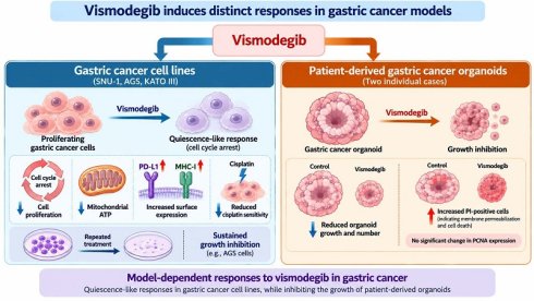
Gastric cancer is a prevalent and deadly disease, particularly in East Asia. Although new treatment options have emerged in recent years, the five-year survival rate for patients with advanced gastric cancer remains below 25%. Numerous studies have identified abnormally activated hedgehog signaling as a major contributor to gastric cancer development, suggesting that inhibitors of this pathway may have therapeutic benefit. Vismodegib (GDC-0449), a hedgehog pathway inhibitor, has demonstrated excellent therapeutic effects in advanced basal cell carcinoma. However, its efficacy against gastric cancer cells remains unclear. In this study, we show that vismodegib does not induce apoptosis or cell death in gastric cancer cells. Rather, it inhibits cell proliferation by causing cell cycle arrest in the G0/G1 phase. Additionally, vismodegib reduces the cytotoxic effect of cisplatin on gastric cancer cells and increases PD-L1 expression. Taken together, these results suggest that vismodegib induces a quiescent-like response in gastric cancer cells. Interestingly, vismodegib induced quiescence-like responses in gastric cancer cell lines exhibiting different baseline GLI-1 protein expression levels, including cells in which GLI-1 was not detectable by western blotting. In addition, repeated treatment of gastric cancer cells with vismodegib effectively inhibited proliferation. In contrast to its effects in gastric cancer cell lines, vismodegib inhibited the growth of patient-derived gastric cancer organoids from two individual cases and increased PI positivity, indicating compromised plasma membrane integrity. Overall, our findings suggest that vismodegib induces quiescence-like responses in gastric cancer cell lines while increasing membrane permeabilization in patient-derived gastric cancer organoids, highlighting distinct cellular responses under the experimental conditions examined.
Keywords: gastric cancer, vismodegib, quiescence, cell proliferation, PD-L1
Introduction
Gastric cancer is a prevalent malignancy worldwide, and in East Asia, its incidence is alarmingly high [1]. Clinical statistics show that 60% of all gastric cancer cases worldwide occur in East Asian populations [1]. In addition to its high incidence rate, the mortality rate for gastric cancer is very high. In 2020, the disease was responsible for approximately 770,000 deaths [1]. Risk factors for gastric cancer are multifaceted. For instance, Helicobacter pylori (H. pylori) infection, which is common in East Asia, is considered a major risk factor [2]. Other notable risk factors include smoking, alcohol consumption, insufficient intake of fruits and vegetables, and excessive consumption of pickled foods [2]. Proactive treatment of H. pylori infections by clinicians may have contributed to the gradually reduced incidence of gastric cancer in recent years [1, 2]. However, because gastric cancer lacks early specific symptoms, most patients are still diagnosed at an advanced stage [3], leading to poor outcomes. Current standard treatments for advanced gastric cancer include surgery, chemotherapy, targeted therapies and immunotherapies [2-5]. Since gastric cancer tumors are highly heterogeneous, cancer cells within the same tumor may respond differently to the same treatment, contributing to therapeutic resistance [6]. Crucially, the five-year survival rate for advanced gastric cancer remains below 25% [7], indicating a major medical need for new treatment strategies to control this disease.
The hedgehog signaling pathway plays a critical role in embryonic development as well as tissue and organ homeostasis [8]. Abnormal activation of this pathway has been linked to the initiation and progression of gastric cancer [9]. Clinical studies have also demonstrated that elevated expression levels of GLI-1, a key downstream target gene of hedgehog, correlates with poor prognosis and increased tumor malignancy in gastric cancer patients [10]. Sonic hedgehog, a ligand of the PTCH1 receptor, is significantly increased during H. pylori infection, leading to the induction of metaplasia and contributing to the development of gastric cancer [9]. Overactivation of the hedgehog signaling pathway can promote cell proliferation, resistance to apoptosis, metastasis, and treatment failure in gastric cancer [9]. Consequently, the hedgehog pathway is considered a promising therapeutic target for this condition [8, 9]. Two hedgehog pathway inhibitors, cyclopamine and itraconazole, are known to induce apoptosis and inhibit proliferation of gastric cancer cells [11-13]. In recent years, the novel hedgehog inhibitor vismodegib (vis; also called GDC-0449) has been approved by the FDA for treatment of advanced basal cell carcinoma, demonstrating both high safety and promising therapeutic effects [14]. The efficacy of vis has also been tested in other indications, such as advanced gastric cancer [15]. Although the trial results show that vis has a good overall safety profile, the reported disease control rate is only 5.3%, and it remains unclear which patients are most likely to benefit from the treatment [15]. Moreover, whether baseline GLI-1 protein expression is associated with the cellular responses to vis in gastric cancer remains unclear. It is known that the degree of differentiation and histological type of gastric cancer cells greatly affect sensitivity to chemotherapy and immunotherapy [16, 17]. Therefore, in this study, we tested the effects of vis on a panel of gastric cancer cell lines with various degrees of differentiation and histological types. Our aim was to characterize the biological responses of gastric cancer cells to vis and to explore the potential mechanisms underlying these effects.
Materials and Methods
Chemicals
Vismodegib (vis; GDC-0449), refametinib, cisplatin, CFSE, and acridine orange 10-Nonyl Bromide (NAO) were purchased from MedChemExpress (USA). BioTracker 488 green lipid droplet dye, BioTracker ATP-red live cell dye, RIPA buffer, crystal violet, and propidium iodide (PI) were purchased from Merck (Germany).
Cell culture and treatments
SNU-1 (poorly differentiated), TSGH-9201 (signet ring cell carcinoma), NCI-N87 (well differentiated), and AGS (moderately differentiated) cell lines were maintained in Roswell Park Memorial Institute (RPMI; Thermo Fisher Scientific, USA) supplemented with 10% fetal bovine serum (FBS; Cytiva, USA) and antibiotic-antimycotic (Capricorn Scientific, Germany). The KATO III (poorly differentiated signet ring cell carcinoma) cell line was maintained in RPMI with 20% FBS and antibiotic-antimycotic. In order to validate the effect of vis on gastric cancer cells, cells (4×10⁵) were seeded into 6-well plates. Cells were treated with vis at the indicated doses for 48 h or indicated durations. Details of experimental conditions are provided in the figure legends.
Western blot
After treatment, proteins were extracted using RIPA buffer. Then, proteins were separated by molecular weight using gradient PAGE and transferred onto a PVDF membrane. Specific primary and secondary antibodies were applied to detect target proteins. Protein expression was quantified using ImageJ software. GLI-1 (3538), PARP (9542), caspase-3 (9662), MHC-I (88274), PD-L1 (13684), phospho-ERK 1/2 (9101), ERK1/2 (9102), β-actin (3700) antibodies were purchased from Cell Signaling Technology (USA).
Apoptosis and cell death assays
Apoptosis and cell death were assessed using an annexin V/PI double staining assay, as previously described [18]. The lower right and upper right quadrants were considered apoptotic cells. The upper left, lower right, and upper right quadrants were considered dead cells.
Cell proliferation assays
Cell proliferation and growth arrest were assessed by cell counting, CFSE dilution, clonogenic assay, cell cycle analysis, EdU incorporation, and drug-withdrawal experiments. Cultures were incubated with vis (100 μM) for 72 h. Cell numbers were determined using standard procedures for the trypan blue exclusion assay. For the dose-response experiments, AGS (4 × 10⁵ cells), SNU-1 (4 × 10⁵ cells), and KATO III (4 × 10⁵ cells) were initially seeded and subsequently treated with different concentrations of vis (0, 0.01, 0.1, 1, 10, and 100 μM) for 72 h. Viable and nonviable cell numbers were determined using the trypan blue exclusion assay. Alternatively, CFSE staining was performed as previously described [19]. Briefly, cells were labeled with CFSE on day 0. Fluorescence intensity was analyzed by flow cytometry before drug administration (0 h) and at 24 and 48 hours after drug administration. The CFSE fluorescence intensity was then used to evaluate cell proliferation. Cell cycle distribution analysis was conducted following a method described in previous publications [20, 21]. For the clonogenic assay, 4×10³ cells were incubated with vis; then, cells were fixed and stained with a solution of 6% glutaraldehyde and 0.5% crystal violet for 30 min at room temperature [18]. Excess crystal violet was rinsed off with distilled water. Colony area was quantified using ImageJ (National Institutes of Health, Bethesda, MD, USA). Cell proliferation was also assessed using an EdU Cell Proliferation Image/Flow Cytometry Assay Kit (Orange Fluorescence; Abbkine, Cat. No. KTA2031) according to the manufacturer's instructions. To assess whether vis-induced growth arrest was reversible, cells were seeded on day 0 and divided into continuous-treatment and withdrawal groups. In the continuous-treatment group, cells were treated with vis throughout the experimental period, with vis replenished daily. In the withdrawal group, cells were treated with vis for the first 48 h. After 48 h of treatment, the cells were washed twice with PBS to remove vis, and the medium was replaced with fresh drug-free culture medium. The cells were then cultured without vis until 96 h. Cell numbers were determined at the indicated time points to evaluate the recovery of cell proliferation following vis withdrawal.
Mitochondrial ATP, mitochondrial mass, and lipid droplets
To analyze the effects of vis on mitochondrial ATP, mitochondrial mass, and lipid droplets, cells were treated as described in the figure legends. Subsequently, cells were stained with BioTracker ATP-Red live cell dye at a final concentration of 10 μM to measure mitochondrial ATP. Cells were stained with NAO at a final concentration of 50 nM to measure mitochondrial mass. BioTracker 488 Green lipid droplet dye (1:1000 dilution) was used to detect lipid droplets. Fluorescence intensities for each indicator were measured using flow cytometry.
Surface PD-L1 and surface MHC-I
Cells were treated as described in the figure legends and harvested using trypsin. The trypsin was then neutralized with PBS containing 5% FBS. Cells were centrifuged to remove the supernatant and resuspended in PBS containing 5% FBS for blocking. Next, cells were incubated with Alexa Fluor 488-conjugated PD-L1 (extracellular domain-specific) antibody (1: 500; Cell Signaling Technology, USA) and Alexa Fluor 488-conjugated MHC-I antibody (1:500; Santa Cruz Biotechnology, USA) for 1 h at 4°C. Following centrifugation and removal of the supernatant, cells were rinsed with PBS. The expression levels of PD-L1 and MHC-I were measured by flow cytometry.
Tumor organoid experiments
Tumor organoids were cultured from tumor specimens obtained from gastric cancer patients following typical protocols. The study was approved by the Ethics Committee of Taipei Medical University (TMU) Joint Institutional Review Board (approval number N202405105). Informed consent was obtained from all participants prior to the start of the study. Characteristics of each tumor were assessed by pathologists at Wan Fang Hospital, TMU. Tumor tissue samples were minced with a scalpel and incubated in collagenase and dispase II at 37°C for 1 h. The digested tissue was then filtered by centrifugation through a 100-μm cell strainer. After centrifugation, the pellets were retained and resuspended in DMEM/F12 medium containing ice-cold 70% Matrigel. The resuspended cells were seeded into ultralow attachment 6-well plates (Corning) and expanded into organoid cultures. Organoid cultures derived from Patient 12 and Patient 13 were selected for subsequent experiments based on their satisfactory growth and expansion under the experimental culture conditions. Earlier organoid cultures exhibited declining growth during prolonged culture and could not be sufficiently expanded for these experiments.
Statistical analysis
Results are presented as the mean ± standard error of the mean (SEM). Comparisons between two groups were performed using Student's t-test, whereas comparisons among three or more groups were analyzed using one-way analysis of variance (ANOVA) followed by Tukey's multiple comparisons test. A P value < 0.05 was considered statistically significant.
Results
Vis exhibits no cytotoxicity in the gastric cancer cell lines
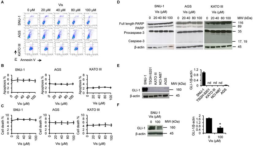
To investigate the effects of vis across gastric cancer cell models with different histological characteristics, we initially considered five gastric cancer cell lines. Among these, SNU-1, AGS, and KATO III cells were selected for subsequent functional experiments, whereas TSGH-9201 and NCI-N87 cells were not included because of their relatively slower growth rates under our experimental culture conditions. Apoptosis and cell death were assessed using annexin V/PI double staining. We found that vis did not induce either apoptosis or non-apoptotic cell death in the three tested gastric cancer cell lines (Figure 1A-C). Consistent with these observations, vis treatment did not induce PARP cleavage or caspase-3 activation (Figure 1D). We next examined baseline GLI-1 protein expression across the five gastric cancer cell lines. GLI-1 expression was relatively high in SNU-1 cells and weak in AGS cells, whereas GLI-1 protein was below the detection limit of our western blot assay in KATO III, TSGH-9201, and NCI-N87 cells (Figure 1E). Thus, the three cell lines selected for functional experiments represented distinct baseline GLI-1 protein expression profiles. Furthermore, vis treatment significantly reduced GLI-1 protein expression in SNU-1 cells (Figure 1F).
Vis does not induce detectable cell death. (A) Cells were incubated with vis at the indicated concentrations for 48 h. Apoptosis (B) and cell death (C) were assessed using annexin V/PI double staining assays. (D) Cells were incubated with vis at the indicated concentrations for 48 h. Cleavage of PARP and activation of caspase-3 were analyzed by western blotting. (E) GLI-1 expression in various gastric cancer cell lines. (F) Effects of vis treatment on GLI-1 protein expression in SNU-1 cells. nd: not detectable. *P < 0.05.
Vis inhibits cell proliferation and alters cell cycle phase distribution
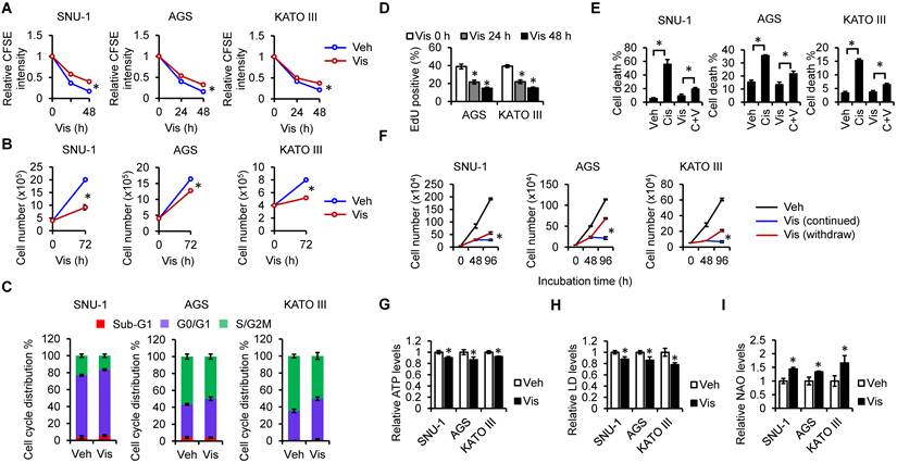
Next, we assessed the effect of vis on cell proliferation according to CFSE fluorescence intensity and cell counting assays. We found that compared to the vehicle group, the decrease in CFSE fluorescence intensity in vis-treated cells was less pronounced (Figure 2A). Furthermore, vis significantly inhibited cell proliferation (Figure 2B) and induced cell cycle arrest at the G0/G1 phase (Figure 2C). Consistent with these findings, vis treatment markedly reduced the proportion of EdU-positive cells, indicating decreased DNA synthesis and further supporting the inhibitory effect of vis on cell proliferation (Figure 2D). We further investigated whether lower concentrations of vis could inhibit gastric cancer cell proliferation. Cells were treated with vis (0-100 μM) for 72 h, and viable and nonviable cell numbers were determined using the trypan blue exclusion assay. A significant reduction in viable cell number was observed only at 100 μM, whereas lower concentrations (0.01-10 μM) showed no significant effect. No significant increase in nonviable cell number was observed at any concentration (Supplementary Figure. S1). We also observed that vis could mitigate the cytotoxic effects of cisplatin (Figure 2E). Importantly, following vis withdrawal, cell proliferation resumed, demonstrating the reversibility of the growth-arrested state and providing further evidence that vis induces a quiescent-like state in gastric cancer cells (Figure 2F) [22]. Since cellular quiescence has been associated with metabolic adaptations, including alterations in mitochondrial ATP production [23-25], mitochondrial mass [26], and lipid metabolism [27], we evaluated the effects of vis treatment on these parameters. As predicted, vis treatment caused all gastric cancer cells to exhibit decreased mitochondrial ATP levels (Figure 2G) and increased mitochondrial mass (Figure 2H). However, vis reduced lipid droplet accumulation in gastric cancer cells (Figure 2I). Taken together, these results further support the idea that vis induces a quiescent-like state in the gastric cancer cell lines.
Vis inhibits cell proliferation. (A) Cells were treated with vis (100 µM) for 24 and 48 h. CFSE intensity was measured using flow cytometry. (B) Cells were treated with vis (100 µM) for 72 h. Cell numbers were determined using the trypan blue exclusion assay. (C) Cells were treated with vis (100 µM) for 48 h. Cell cycle distribution was assessed by PI staining. (D) Cells were treated with vis (100 µM) for 24 and 48 h. Cell proliferation was assessed by EdU incorporation assay. (E) Cells were co-treated with vis (100 µM) and cisplatin (cis; 20 μM) for 48 h. Cell death was assessed by annexin V/PI double staining assay. (F) Reversibility of vis-induced growth arrest following drug withdrawal. Cells were seeded on day 0 and subjected to either continuous vis treatment (Continued) or vis withdrawal (Withdraw). In the Continued group, vis was replenished daily throughout the experimental period. In the Withdraw group, cells were treated with vis for 48 h, washed twice with PBS, and subsequently cultured in fresh drug-free medium until 96 h. Cell numbers were determined at the indicated time points to assess the recovery of cell proliferation following vis withdrawal. Cells were treated with vis (100 µM) for 48 h; mitochondrial ATP (G), mitochondrial mass (H), and lipid droplet (LD) contents (I) were assessed. Veh: vehicle, C+V: cis+vis. *P < 0.05.
Repeated administration of vis effectively inhibits clonogenic growth of gastric cancer cells
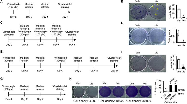
Recent studies suggest that inducing quiescence in cancer cells can be an effective cancer treatment strategy [28, 29]. Since vis induced quiescence-like responses in the tested gastric cancer cell lines, we further investigated whether repeated vis administration could provide sustained inhibition of clonogenic growth. These experiments were performed using AGS cells because their adherent growth characteristics make them suitable for conventional clonogenic assays, whereas SNU-1 cells predominantly grow in suspension and KATO III cells exhibit mixed adherent and suspension growth characteristics. Although a single administration of vis markedly suppressed clonogenic growth in AGS cells (Figure 3A and B), a small number of colonies remained detectable (yellow arrows in Figure 3B). We next examined whether repeated administration of vis at intervals of 2 days (Figure 3C and D) or 7 days (Figure 3E and F) could further enhance its inhibitory effect. We found that administration every 2 days almost completely suppressed clonogenic growth (Figure 3C and D), while administration every 7 days also markedly reduced colony formation (Figure 3E and F). These findings suggest that repeated vis administration may provide more sustained suppression of clonogenic growth in AGS cells. To further investigate whether initial cell density was associated with the inhibitory effect of vis on clonogenic growth, we examined the effects of vis treatment at different initial seeding densities (4,000, 40,000, and 80,000 cells per well; Figure 3G and H). The inhibitory effect of vis appeared less pronounced at higher initial cell densities. However, whether this observation reflects differences in cellular responses or changes in effective drug exposure remains unclear.
Repeated administration of vis enhances inhibition of cell proliferation. AGS cells were treated with vis (100 µM) as described in panels (A, C, E, G). A clonogenic assay was performed as shown in panels (B, D, F, H). Veh: vehicle. Colony area was quantified using ImageJ software. Data were obtained from three independent biological experiments and are presented as the mean ± SEM. Statistical significance was determined using a two-tailed unpaired Student's t-test for panels B, D, and F, and two-way ANOVA followed by Tukey's multiple comparisons test for panel H. *P < 0.05.
Vis increases surface MHC-I and surface PD-L1
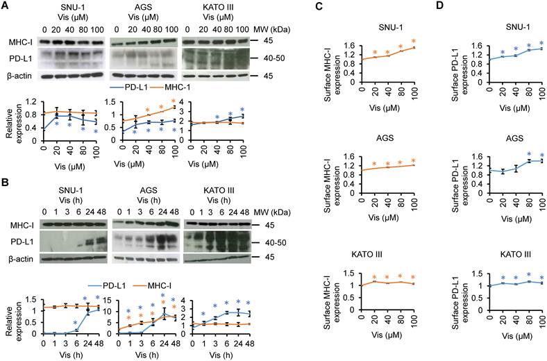
When cancer cells enter a quiescent state, it is common for MHC-I expression to decrease while PD-L1 expression increases. These alterations reduce immunogenicity and enable quiescent cancer cells to evade immune surveillance [22, 27, 30]. Therefore, we further analyzed the effects of vis on MHC-I and PD-L1 expression in gastric cancer cells. Results showed that under different doses (Figure 4A) or durations (Figure 4B) of vis treatment, total MHC-I expression was increased in AGS cells. However, total MHC-I expression was not significantly affected in SNU-1 and KATO III cells. Meanwhile, vis stimulation significantly increased total PD-L1 expression in all gastric cancer cell lines. Next, we analyzed the effects of vis on MHC-I and PD-L1 surface expression, finding that vis significantly enhanced the surface expression levels of both MHC-I (Figure 4C) and PD-L1 (Figure 4D).
Vis modulates MHC-I and PD-L1 expression. (A) Cells were treated with vis at the indicated concentrations for 48 h. Total MHC-I and total PD-L1 expression were assessed by western blot analysis. (B) Cells were treated with vis (100 µM) for the indicated durations. Total MHC-I and total PD-L1 expression were assessed by western blot analysis. Protein band intensities were analyzed using ImageJ software. Blue asterisks indicate significant differences in PD-L1 expression compared with the corresponding baseline control (0 µM for panel A or 0 h for panel B). Orange asterisks indicate significant differences in MHC-I expression compared with the corresponding baseline control. Cells were treated with vis at the indicated concentrations for 48 h. Surface MHC-I (C) and surface PD-L1 (D) expression were assessed by flow cytometry. *P < 0.05.
Involvement of ERK1/2 in vis-mediated increase of surface MHC-I and surface PD-L1
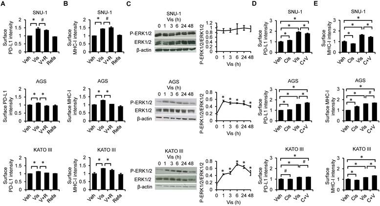
Since ERK1/2 is known to regulate PD-L1 and MHC-I expression in other cancer types [31-33], we next wanted to test whether vis increases surface PD-L1 and surface MHC-I expression via ERK1/2 in gastric cancer cells. Therefore, we investigated whether inhibition of ERK1/2 would impact vis-induced surface PD-L1 and surface MHC-I expression. The results showed that the ERK1/2 inhibitor refametinib suppressed the vis-induced increases of surface PD-L1 (Figure 5A) and surface MHC-I (Figure 5B) in AGS and KATO III cells. However, refametinib did not inhibit the vis-induced increase in surface PD-L1 and MHC-I in SNU-1 cells. Next, we performed western blot analysis to assess the effect of vis on ERK1/2 phosphorylation and found ERK1/2 phosphorylation was increased only in AGS and KATO III cells (Figure 5C). Furthermore, since vis inhibits the cytotoxic effects of cisplatin on gastric cancer cells (Figure 2E), we examined whether vis alters cisplatin-induced increases of surface PD-L1 (Figure 5D) and surface MHC-I expression (Figure 5E). Cisplatin and vis alone each increased surface PD-L1 in SNU-1 and AGS cells, while vis further enhanced cisplatin-induced increases. Interestingly, cisplatin alone decreased surface MHC-I in SNU-1 and KATO III cells but increased the levels in AGS cells. Compared to vehicle controls, combined treatment with vis and cisplatin significantly increased surface MHC-I expression, suggesting that the effect of the combination treatment may vary depending on the cell histological type.
ERK1/2 modulates vis-induced changes in surface PD-L1 and surface MHC-I. Cells were pretreated with refametinib (refa; 50 nM) for 1 h, followed by treatment with vis (100 µM) for 48 h. Levels of surface PD-L1 (A) and surface MHC-I (B) were assessed. (C) Cells were treated with vis (100 µM) for indicated time points. Levels of phospho-ERK1/2 and total ERK1/2 were evaluated by western blot analysis. The relative phosphorylation levels of ERK1/2 were quantified by densitometry and normalized to total ERK1/2. Protein band intensities were analyzed using ImageJ software. Cells were co-treated with vis (100 µM) and cisplatin (cis; 20 μM) for 48 h. Levels of surface PD-L1 (D) and surface MHC-I (E) were assessed. Veh: vehicle, V+R: vis+refa, C+V: cis+vis. *P < 0.05. #P > 0.05.
Involvement of ERK1/2 in vis-mediated cisplatin resistance
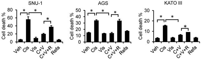
Next, we sought to evaluate whether ERK1/2 is involved in vis-induced cisplatin resistance. Our findings revealed that vis-induced cisplatin resistance could be abolished by refametinib (Figure 6). This result was consistent across all three tested cell lines, suggesting a critical involvement of ERK1/2 in vis-induced cisplatin resistance and quiescence of gastric cancer cells.
ERK1/2 modulates vis-induced cisplatin resistance. Cells were preincubated with refametinib (refa; 50 nM) and vis (100 µM) for 1 h, followed by cisplatin (cis; 20 μM) for an additional 48 h. Cell death was assessed using an annexin V/PI double staining assay. Veh: vehicle, C+V: cis+vis, C+V+R: cis+vis+refa. *P < 0.05. #P > 0.05.
Inhibitory effect of vis on gastric cancer organoids
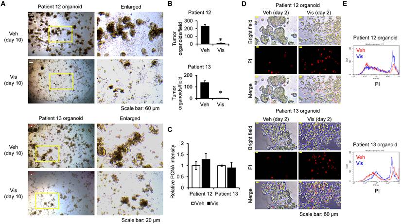
Gastric cancer organoids may be useful to accurately predict the response of a patient's tumor to different drugs [34]. Therefore, we evaluated the effects of vis on gastric cancer patient-derived tumor organoids. Characteristics of each tumor case are shown in Table 1. After 10 days of vis stimulation, growth of tumor organoids was significantly inhibited (Figure 7A), and the number of tumor organoids was markedly lower than in the vehicle group (Figure 7B). Interestingly, vis treatment did not significantly alter PCNA expression at Day 2 (Figure 7C). To further investigate the effects of vis on tumor organoids, we performed a PI uptake assay on Day 2 to assess plasma membrane integrity (Figure 7D and E). Vis treatment significantly increased PI positivity, indicating compromised plasma membrane integrity in patient-derived gastric cancer organoids.
Patient characteristics
| Age | Gender | Histology | Types | TNM stage | MMR | |
|---|---|---|---|---|---|---|
| Patient 12 | 83 | Male | Adenocarcinoma | Intestinal | Stage IA, pT1aN0 | PMS, MLH1 loss |
| Patient 13 | 73 | Male | Adenocarcinoma | Diffuse | Stage IIIA, pT4aN2 | Intact |
Vismodegib inhibits the growth of patient-derived gastric cancer organoids and increases PI positivity. Gastric cancer tumor organoids were incubated with vis (100 µM) for 10 days, and their growth was monitored by light microscopy (A). Scale bars = 60 μm (upper panels, Patient 12) and 20 μm (lower panels, Patient 13). (B) The number of gastric cancer tumor organoids was quantified based on observations of four randomly selected regions of interest. (C) Gastric cancer tumor organoids were incubated with vis (100 µM) for 2 days. Flow cytometry-based analyses were performed on Day 2 because prolonged vismodegib treatment resulted in a substantial reduction in recoverable organoid cells by Day 10, making subsequent flow cytometric analysis impractical. The expression level of PCNA was analyzed by flow cytometry. PI positivity was analyzed by fluorescence microscopy (D) and flow cytometry (E). Scale bar = 60 μm. *P < 0.05.
Discussion
In this study, we explored the potential of using vis as a treatment for gastric cancer. Unlike conventional chemotherapy agents that induce cancer cell death, vis primarily induces quiescence in gastric cancer cells (Figure 2). As a result, cell proliferation decreases, cell cycle arrest occurs, sensitivity to chemotherapy drugs diminishes, and cell surface PD-L1 increases (Figure 4). Furthermore, we identified ERK1/2 as a key mediator of this process, as pharmacological inhibition of ERK1/2 by refametinib attenuated vis-induced effects on chemoresistance-associated readouts (Figure 6). It is noteworthy that although vis does not stimulate ERK1/2 phosphorylation in SNU-1 cells (Figure 5C), inhibition of ERK1/2 in this cell line still prevents vis-induced chemoresistance (Figure 6). This finding suggests that basal ERK1/2 activity is likely necessary for chemoresistance. Additionally, we demonstrated that vis inhibited the growth of patient-derived gastric cancer organoids (Figure 7). Although PCNA expression was not significantly altered at Day 2, vis treatment markedly increased PI positivity, supporting a cytotoxic effect. Notably, PI uptake and intracellular PCNA expression reflect distinct biological parameters and are not necessarily mutually exclusive. Nevertheless, the absence of a significant reduction in PCNA expression at a single time point does not exclude the possibility that vis also suppresses cell proliferation. Collectively, these findings demonstrate that vis induces quiescence-like responses in gastric cancer cells with distinct baseline GLI-1 protein expression profiles, although the contribution of canonical Hedgehog signaling to these effects remains to be determined.
Vis was initially used to treat advanced basal cell carcinoma [14], and it has been recently tested in other cancer types, including gastric cancer [15]. A previous in vitro study demonstrated that vis exhibits significant anti-gastric cancer efficacy. In particular, the study showed that vis induces cytotoxicity and inhibits the proliferation of SGC-7901 (moderately differentiated) gastric cancer cells by suppressing GLI-1 [35]. Nevertheless, a phase II clinical trial showed that although vis demonstrates a favorable safety profile in patients with advanced gastric cancer, the disease control rate was only 5.3% [15]. These findings suggest that the appropriate patient population and optimal mode of vis administration have yet to be identified.
In clinical studies, whole exon-transcriptome analyses suggest that the hedgehog signaling pathway is not robustly activated in gastric cancer patient samples [15]. However, a small subset of patients demonstrated improvement from vis [15]. Among five tested gastric cancer cell lines, only SNU-1 exhibited significant GLI-1 expression (Figure 1E). Nevertheless, we found that vis induced quiescence in cells with varying levels of GLI-1 expression, including SNU-1 (high GLI-1 expression), AGS (low GLI-1 expression), and KATO III (GLI-1 expression below the detection limit) (Figure 2). These findings indicate that the cellular responses to vis were observed across gastric cancer cell lines with different baseline GLI-1 protein expression levels. However, baseline GLI-1 expression alone does not establish functional Hedgehog pathway activity, and further pathway characterization is required to determine the contribution of canonical Hedgehog signaling to these responses.
In recent years, mounting evidence suggests that inducing cancer cells into a quiescent state may help suppress tumor growth [28, 36]. However, reactivation of quiescent tumor cells has also been implicated in cancer recurrence [27], highlighting the importance of maintaining cancer cells in a sustained quiescent state to prevent re-entry into the cell cycle. It is important to distinguish the therapeutic concept proposed in the present study from the therapy-induced quiescence that has been associated with resistance following conventional chemotherapy or radiotherapy. Rather than generating a transient population of dormant cells that survives cytotoxic treatment [37], our strategy aims to pharmacologically maintain gastric cancer cells in a non-proliferative quiescent state through continuous vis administration. Consistent with this concept, our repeated-treatment experiments demonstrated that repeated vis administration resulted in more sustained suppression of clonogenic growth in AGS cells compared with a single administration (Figure 3). These findings suggest that repeated vis administration may provide sustained suppression of clonogenic growth in AGS cells, although whether similar effects occur in other gastric cancer models or in vivo remains to be determined. Nevertheless, several important considerations should be addressed before this strategy can be translated into clinical practice. Although continuous vis administration may provide sustained suppression of gastric cancer cell proliferation, our findings also demonstrated reduced cisplatin sensitivity, increased cell-surface PD-L1 expression, and restoration of cell proliferation following drug withdrawal. These observations raise concerns that vis-induced quiescence may protect tumor cells from conventional chemotherapy and potentially facilitate tumor regrowth after treatment discontinuation. Furthermore, increased PD-L1 expression may have implications for tumor immune evasion, although its functional consequences remain unclear. Therefore, the potential therapeutic application of prolonged vis administration should be considered exploratory, and further preclinical studies are required to determine whether sustained growth suppression can be achieved without promoting treatment resistance or subsequent tumor recurrence.
Further analysis of vis-induced quiescent cells involved measurements of mitochondrial ATP, lipid droplets, and mitochondrial mass. Surprisingly, vis-induced quiescent cells exhibited characteristics that differ slightly from those of other quiescent cells. Studies have shown that quiescent cells generally have low mitochondrial ATP [23], high mitochondrial mass [26], and increased lipid droplet content [27]. However, vis-induced quiescent cells displayed low mitochondrial ATP and increased mitochondrial mass but reduced lipid droplet content (Figure 2G-I). Interestingly, the reduction in lipid droplet accumulation observed in vis-treated gastric cancer cells differs from findings reported in certain other quiescent cell populations. This observation suggests that vis-induced growth arrest may be accompanied by distinct metabolic alterations. However, whether reduced lipid droplet accumulation contributes to the maintenance or reversibility of this growth-arrested state remains unclear and warrants further investigation.
Previous studies have shown that quiescent cancer cells often exhibit increased surface PD-L1 and decreased surface MHC-I, and these characteristics help the cells evade immune surveillance [22, 30, 38]. However, vis-induced quiescent cells had increased surface expression of both PD-L1 and MHC-I (Figure 4C and D), which contrasts with previous findings [22, 30, 38]. Whether the immunogenicity of vis-induced quiescent cells differs from that of other quiescent cancer cells remains unclear. We also found that in SNU-1 and KATO III cells, vis did not increase total MHC-I levels (Figure 4A and B), but it did enhance surface MHC-I expression (Figure 4C and D). This phenomenon may be related to the intracellular trafficking of MHC-I. One possible explanation is that vis treatment alters the intracellular trafficking or surface retention of MHC-I, although this possibility requires further investigation. Further studies will be conducted to evaluate the immunogenicity of vis-induced quiescent gastric cancer cells.
Unlike its activity in gastric cancer cell lines, vis treatment increased PI positivity in patient-derived gastric cancer organoids, supporting a cytotoxic effect (Figure 7). The differential responses observed between conventional gastric cancer cell lines and patient-derived tumor organoids may reflect differences in cellular composition, microenvironmental conditions, or drug exposure. Patient-derived tumor organoids have been reported to preserve cancer stem cell-like populations [39]. Since Hedgehog signaling has been implicated in the maintenance of cancer stem cells [40-42], the potential involvement of these cells may represent one possible explanation for the observed differences. However, our study did not directly assess cancer stem cell markers or stemness-related functions and therefore does not provide direct evidence for selective CSC elimination. Furthermore, potential effects associated with the Matrigel-based culture system, prolonged drug exposure, or non-specific cytotoxicity cannot be excluded. Thus, the mechanisms underlying the differential responses between gastric cancer cell lines and patient-derived tumor organoids remain to be elucidated.
Although vismodegib showed limited clinical efficacy in a previous phase II trial involving patients with advanced gastric cancer [15], the potential influence of treatment schedules and disease characteristics on its antitumor activity remains to be investigated. Our study demonstrated that vis inhibited clonogenic growth in AGS cells, with repeated administration producing more sustained growth suppression under the experimental conditions (Figure 3). These findings suggest that sustained vis administration may represent a potential strategy for maintaining the suppression of gastric cancer cell proliferation, although its therapeutic efficacy remains to be established. Notably, we observed that the inhibitory effect of vis was attenuated under higher cell-density conditions (Figure 3G and H). However, cell density in an in vitro culture system does not directly reflect tumor burden in patients. Furthermore, because drug concentrations were not monitored throughout the treatment period, the possibility that changes in effective drug exposure contributed to the observed density-associated differences cannot be excluded. Therefore, the mechanisms underlying this observation remain unclear, and further studies are required to determine its biological significance and potential clinical relevance.
Despite the limited clinical efficacy reported in a previous trial involving patients with advanced gastric cancer [15], our findings suggest that the potential therapeutic applications of vis in gastric cancer warrant further investigation. One possible direction is to explore its use as an adjuvant treatment following surgical resection, with the aim of suppressing the proliferation of residual tumor cells. However, this proposed application remains hypothetical, as our study did not directly evaluate postoperative residual disease, cancer recurrence, or the selective targeting of cancer stem cells. Moreover, our findings that vis-induced growth inhibition was reversible and accompanied by reduced cisplatin sensitivity and increased cell-surface PD-L1 expression raise important concerns regarding treatment resistance and potential tumor regrowth following drug withdrawal. Although vis has an established clinical safety profile in approved indications, its tolerability and efficacy during prolonged administration in patients with gastric cancer remain uncertain. Therefore, further preclinical studies, particularly those investigating sustained treatment, treatment discontinuation, and interactions with conventional chemotherapy, are necessary before the potential clinical benefits of vis can be established.
Limitations
This study has several limitations. First, all experiments were performed using gastric cancer cell lines and patient-derived tumor organoids. Therefore, the physiological and therapeutic effects of vis could not be fully evaluated. Future studies using appropriate animal models will be necessary to validate the antitumor efficacy and safety of repeated vis administration in vivo. Second, the relatively high concentration of vis (100 μM) used in most functional experiments represents an important pharmacological limitation. This concentration substantially exceeds its reported inhibitory potency against SMO and clinically relevant unbound drug concentrations. Consequently, the possibility of SMO-independent or other off-target effects cannot be excluded. In our additional dose-response experiments, a significant reduction in viable cell number was observed only at 100 μM, whereas lower concentrations (0.01-10 μM) did not significantly affect viable cell numbers following 72 h of treatment. These findings further highlight the limited clinical relevance of the concentrations required to induce growth suppression under our experimental conditions. Although the growth-inhibitory effects of vis were observed only at a relatively high concentration, future investigations may explore whether advanced drug delivery strategies, including liposome-based formulations or tumor-targeted delivery systems, could enhance intratumoral drug accumulation and exposure. However, the feasibility of achieving therapeutically effective drug exposure and the potential efficacy of such approaches remain to be experimentally established. Furthermore, although the additional cell-counting experiments provided information regarding net cell growth, potential effects of lower vis concentrations on cell cycle progression and DNA synthesis were not directly examined. Further investigations are therefore required to determine whether these cellular responses can occur at pharmacologically relevant concentrations. Third, although patient-derived tumor organoids may retain cancer stem cell-like populations, we did not directly evaluate cancer stem cell markers, self-renewal capacity, or tumor-initiating potential. Therefore, the effects of vis on cancer stem cells remain speculative. Furthermore, because non-tumor gastric organoids were not included as controls, the tumor selectivity of vis-induced effects could not be determined. Fourth, Hedgehog pathway activity was not comprehensively characterized in the examined gastric cancer cell lines. Although vis induced similar biological responses across cell lines with different baseline GLI-1 protein expression levels, these findings do not establish independence from canonical Hedgehog signaling. Further studies incorporating multiple pathway readouts and functional interventions are required to clarify the contribution of Hedgehog signaling to vis-induced cellular responses. Finally, the repeated-administration experiments were performed only in AGS cells, and the patient-derived organoid experiments included only two individual cases. Therefore, the generalizability of these findings across different gastric cancer models remains uncertain.
Supplementary Material
Supplementary figures.

Acknowledgements
We thank Marcus J. Calkins, PhD for language editing.
Funding
This research was supported by the National Science and Technology Council (NSTC), NSTC 113-2313-B-038-003, NSTC 114-2320-B-038-057. This research was also funded by the Taipei Medical University-Wan Fang Hospital, 113TMU-WFH-18, 114TMU-WFH-19, 114-wf-eva-11, 115-wf-eva-21. This work was also financially supported by the Higher Education Sprout Project of the Ministry of Education (MOE), Taiwan (DP2-TMU-113-C-02).
Ethics approval
This study complies with the ethical guidelines of the 1975 Helsinki Declaration (modified in 2008). The study protocol was approved by the Taipei Medical University-Joint Institutional Review Board (Approval No: N202405105). Informed consent was obtained from all participants prior to study enrollment. Confidentiality of patient information was strictly maintained in accordance with the Declaration of Helsinki. This study does not include identifiable patient data or images requiring publication consent.
Data availability
The data used in the present study are available from the corresponding author upon reasonable request.
Author contributions
YHY performed experiments, analyzed data, generated figures and wrote the manuscript. XCL performed experiments and analyzed data. CCT performed experiments and analyzed data. KCH performed experiments and analyzed data. JJC performed experiments and analyzed data. CJH performed experiments and analyzed data. HSC performed experiments and analyzed data. TCC performed experiments and analyzed data. YSL performed experiments and analyzed data. CCC performed experiments and analyzed data. HHY supervised the project, designed experiments, formulated the major research plans, reviewed, and revised the manuscript. BCS supervised the project, designed experiments, formulated the major research plans, reviewed, and revised the manuscript.
ORCID
Yi-Hsuan Yin: 0009-0004-9696-0985
Xu-Chen Liu: 0009-0003-8412-2434
Chia-Chi Tsai: 0009-0007-9882-548X
Kai-Chun Hong: 0009-0000-8129-1185
Jen-Jui Chang: 0009-0003-7935-5989
Chian-Ju Hsu: 0009-0008-2087-828X
Heng-Sheng Chiu: 0009-0001-1967-4641
Tzu-Cheng Chien: 0009-0001-9112-2702
You-Syuan Lou: 0009-0000-0181-0943
Chih-Cheng Cheng: 0009-0002-7432-2224
Hsin-Hsien Yu: 0000-0001-9706-8869
Bor-Chyuan Su: 0000-0002-0454-0624
Competing Interests
The authors have declared that no competing interests exist.
References
1. Mousavi SE, Ilaghi M, Elahi Vahed I, Nejadghaderi SA. Epidemiology and socioeconomic correlates of gastric cancer in Asia: results from the GLOBOCAN 2020 data and projections from 2020 to 2040. Sci Rep. 2025;15:6529
2. Thrift AP, Wenker TN, El-Serag HB. Global burden of gastric cancer: epidemiological trends, risk factors, screening and prevention. Nat Rev Clin Oncol. 2023;20:338-49
3. Ramachandran R, Grantham T, Parvataneni S, Budh D, Gollapalli S, Reddy M. et al. Gastric Cancer: Clinical Features, Screening, Diagnosis, Treatment, and Prevention. J Community Hosp Intern Med Perspect. 2024;14:49-57
4. Lou YS, Liu XC, Cheng CC, Yin YH, Chien TC, Hsu PL. et al. Chemotherapy elevates cell surface PD-L1 and MHC-I expression in apoptotic gastric cancer cells. Int J Immunopathol Pharmacol. 2025;39:3946320251338662
5. Su BC, Chen GY, Yang CM, Chuang WT, Lin MC, Hsu PL. et al. Impacts of hyperthermic chemotherapeutic agent on cytotoxicity, chemoresistance-related proteins and PD-L1 expression in human gastric cancer cells. Int J Hyperthermia. 2024;41:2310017
6. Gao JP, Xu W, Liu WT, Yan M, Zhu ZG. Tumor heterogeneity of gastric cancer: From the perspective of tumor-initiating cell. World J Gastroenterol. 2018;24:2567-81
7. Coccolini F, Montori G, Ceresoli M, Cima S, Valli MC, Nita GE. et al. Advanced gastric cancer: what we know and what we still have to learn. World J Gastroenterol. 2016;22:1139-59
8. Jing JJ, Wu ZX, Wang JH, Luo GW, Lin HY, Fan Y. et al. Hedgehog signaling in tissue homeostasis, cancers, and targeted therapies. Signal Transduct Target Ther. 2023 8
9. Zhang JH, Fan JJ, Zeng X, Nie MM, Luan JY, Wang YC. et al. Hedgehog signaling in gastrointestinal carcinogenesis and the gastrointestinal tumor microenvironment. Acta Pharmaceutica Sinica B. 2021;11:609-20
10. Lu L, Wu ML, Zhao FX, Fu WH, Li WD, Li X. et al. Prognostic and clinicopathological value of Gli-1 expression in gastric cancer: A meta-analysis. Oncotarget. 2016;7:69087-96
11. Han ME, Lee YS, Baek SY, Kim BS, Kim JB, Oh SO. Hedgehog Signaling Regulates the Survival of Gastric Cancer Cells by Regulating the Expression of Bcl-2. Int J Mol Sci. 2009;10:3033-43
12. Bai RX, Zhao HC, Zhang X, Du SY. Characterization of sonic hedgehog inhibition in gastric carcinoma cells. Oncol Lett. 2014;7:1381-4
13. Hu Q, Hou YC, Huang J, Fang JY, Xiong H. Itraconazole induces apoptosis and cell cycle arrest via inhibiting Hedgehog signaling in gastric cancer cells. J Exp Clin Cancer Res. 2017;36:50
14. Ayen-Rodriguez A, Linares-Gonzalez L, Llamas-Segura C, Almazan-Fernandez FM, Ruiz-Villaverde R. Retrospective Real-Life Data, Efficacy and Safety of Vismodegib Treatment in Patients with Advanced and Multiple Basal Cell Carcinoma: 3-Year Experience from a Spanish Center. Int J Environ Res Public Health. 2023;20:5824
15. Kim R, Ji JH, Kim JH, Hong JY, Lim HY, Kang WK. et al. Safety and anti-tumor effects of vismodegib in patients with refractory advanced gastric cancer: A single-arm phase-II trial. J Cancer. 2022;13:1097-102
16. Maehara Y, Anai H, Kusumoto H, Sugimachi K. Poorly Differentiated Human Gastric-Carcinoma Is More Sensitive to Antitumor Drugs Than Is Well Differentiated Carcinoma. Eur J Surg Oncol. 1987;13:203-6
17. Mashukov A, Shapochka D, Seleznov O, Kobyliak N, Falalyeyeva T, Kirkilevsky S. et al. Histological differentiation impacts the tumor immune microenvironment in gastric carcinoma: Relation to the immune cycle. World J Gastroenterol. 2021;27:5259-71
18. Lin MC, Chen GY, Yu HH, Hsu PL, Lee CW, Cheng CC. et al. Repurposing the diuretic benzamil as an anti-osteosarcoma agent that acts by suppressing integrin/FAK/STAT3 signalling and compromising mitochondrial function. Bone Joint Res. 2024;13:157-68
19. Chung S, Kim SH, Seo Y, Kim SK, Lee JY. Quantitative Analysis of Cell Proliferation by a Dye Dilution Assay: Application to Cell Lines and Cocultures. Cytom Part A. 2017;91:704-712
20. Kim KH, Sederstrom JM. Assaying Cell Cycle Status Using Flow Cytometry. Curr Protoc Mol Biol. 2015;111:28.6.1-28.6.11
21. Rahme R. Assaying Cell Cycle Status Using Flow Cytometry. Methods Mol Biol. 2021;2267:165-79
22. Hu J, Zhou W, Zhao Y, Li P, Zhao Z, Wang W. et al. Cancer Cell Dormancy and Chemotherapy Resistance. J Cancer. 2026;17:59-73
23. Fiorillo M, Ozsvári B, Sotgia F, Lisanti MP. High ATP Production Fuels Cancer Drug Resistance and Metastasis: Implications for Mitochondrial ATP Depletion Therapy. Front Oncol. 2021;11:740720
24. Tau S, Miller TW. The role of cancer cell bioenergetics in dormancy and drug resistance. Cancer Metastasis Rev. 2023;42:87-98
25. Akkoc Y, Peker N, Akcay A, Gozuacik D. Autophagy and Cancer Dormancy. Front Oncol. 2021;11:627023
26. Takihara Y, Nakamura-Ishizu A, Tan DQ, Fukuda M, Matsumura T, Endoh M. et al. High mitochondrial mass is associated with reconstitution capacity and quiescence of hematopoietic stem cells. Blood Adv. 2019;3:2323-7
27. Jiao YQ, Yu YJ, Zheng MY, Yan M, Wang JP, Zhang Y. et al. Dormant cancer cells and polyploid giant cancer cells: The roots of cancer recurrence and metastasis. Clin Transl Med. 2024;14:e1567
28. Skarkova A, Bizzarri M, Janostiak R, Masek J, Rosel D, Brabek J. Educate, not kill: treating cancer without triggering its defenses. Trends Mol Med. 2024;30:673-85
29. Recasens A, Munoz L. Targeting Cancer Cell Dormancy. Trends Pharmacol Sci. 2019;40:128-41
30. Baldominos P, Barbera-Mourelle A, Barreiro O, Huang Y, Wight A, Cho JW. et al. Quiescent cancer cells resist T cell attack by forming an immunosuppressive niche. Cell. 2022;185:1694-1708.e19
31. Ng HY, Jian L, Tao LH, Lam AKY, Chan KW, Ko JMY. et al. Chemotherapeutic Treatments Increase PD-L1 Expression in Esophageal Squamous Cell Carcinoma through EGFR/ERK Activation. Transl Oncol. 2018;11:1323-33
32. Mimura K, Shiraishi K, Mueller A, Izawa S, Kua LF, So J. et al. The MAPK Pathway Is a Predominant Regulator of HLA-A Expression in Esophageal and Gastric Cancer. J Immunol. 2013;191:6261-72
33. Stutvoet TS, Kol A, de Vries EGE, de Bruyn M, Fehrmann RSN, Terwisscha van Scheltinga AG. et al. MAPK pathway activity plays a key role in PD-L1 expression of lung adenocarcinoma cells. J Pathol. 2019;249:52-64
34. Smabers LP, Wensink GE, Verissimo CS, Doorn M, Yang T, Voskuilen T. et al. Patient-Derived Organoids Predict Treatment Response in Metastatic Colorectal Cancer. Clin Cancer Res. 2025;31:5015-26
35. Wu CQ, Cheng J, Hu SB, Deng R, Muangu YW, Shi L. et al. Reduced proliferation and increased apoptosis of the SGC-7901 gastric cancer cell line on exposure to GDC-0449. Mol Med Rep. 2016;13:1434-40
36. Truskowski K, Amend SR, Pienta KJ. Dormant cancer cells: programmed quiescence, senescence, or both? Cancer Metastasis Rev. 2023;42:37-47
37. Lindell E, Zhong L, Zhang X. Quiescent Cancer Cells-A Potential Therapeutic Target to Overcome Tumor Resistance and Relapse. Int J Mol Sci. 2023;24:3762
38. Wu YA, Sun ZY, Xia L, Tian PP, Jiao LY, Li YZ. et al. MHC-I pathway disruption by viruses: insights into immune evasion and vaccine design for animals. Front Immunol. 2025;16:1540159
39. Azim MMN, Bhajan SK, Park JH, Abass KS, Rahman A, Choi M. et al. Stem Cell-Derived Organoids for Cancer Therapy: Precision Medicine and Drug Selection. Int J Mol Sci. 2026;27:2954
40. Zhao C, Chen A, Jamieson CH, Fereshteh M, Abrahamsson A, Blum J. et al. Hedgehog signalling is essential for maintenance of cancer stem cells in myeloid leukaemia. Nature. 2009;458:776-779
41. Su WX, Meng FK, Huang LF, Zheng M, Liu WL, Sun HY. Sonic hedgehog maintains survival and growth of chronic myeloid leukemia progenitor cells through β-catenin signaling. Exp Hematol. 2012;40:418-27
42. Cochrane CR, Szczepny A, Watkins DN, Cain JE. Hedgehog Signaling in the Maintenance of Cancer Stem Cells. Cancers (Basel). 2015;7:1554-1585
Author contact
Corresponding author: Bor-Chyuan Su, Department of Anatomy and Cell Biology, School of Medicine, College of Medicine, Taipei Medical University, Taipei, Taiwan; Tel+886-2-2736-1661 ext 3253; Email subc8265edu.tw.