Impact Factor ISSN: 1449-1907




Int J Med Sci 2026; 23(12):3693-3709. doi:10.7150/ijms.141625 This issue Cite
Review
The Relationship Between Fatty Acid β-oxidation and Ferroptosis in Kidney Diseases: Mechanisms and Therapeutic Potential
Aijia Cao1#, Xuejiao Li2#, Junhua Li3, Xia Song1, Hui Zhao1, Chang Wang1, Fanxiang Xu1, Chuanxin Liu2  , Yinliang Bai1
1. Department of Pharmacy, The Second Hospital & Clinical Medical School, Lanzhou University, Lanzhou 730030, China.
2. Luoyang Key Laboratory of Clinical Multiomics and Translational Medicine, Key Laboratory of Hereditary Rare Diseases of Health Commission of Henan Province, Henan Key Laboratory of Rare Diseases, Endocrinology and Metabolism Center, The First Affiliated Hospital, and College of Clinical Medicine of Henan University of Science and Technology, Luoyang 471003, China.
3. Department of Pulmonology I, Tianshui Hospital of Traditional Chinese Medicine, Tianshui 741000, China.
# Co-first author: These authors contributed equally to this work.
Received 2026-7-28; Accepted 2026-10-29; Published 2026-11-13
Abstract
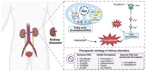
Kidney diseases represent a critical global health challenge, yet no specific or satisfactory therapeutic options are currently available in clinical practice, contributing to rising prevalence and substantial economic burden, and the etiopathogenesis is still elusive. Mounting evidence indicates that the kidneys, particularly proximal tubular cells, rely heavily on mitochondrial fatty acid β-oxidation (FAO) to maintain energy homeostasis and cellular stability. Notably, dysregulation of FAO has been identified as an important driver contributing to diverse kidney diseases, including acute kidney injury (AKI), chronic kidney disease (CKD), and diabetic nephropathy (DN). Concurrently, ferroptosis, an iron-dependent lipid peroxidation process, has attracted increasing attention due to its close association with the pathophysiology of multiple renal conditions. This article elucidates the core regulatory factors and mechanisms of FAO and ferroptosis, highlights their roles, and examines their complex relationship in kidney diseases. Additionally, it also seeks to explore effective interventions by targeting FAO and ferroptosis to alleviate kidney disease progression, particularly by addressing the research gap regarding the intrinsic relationship between aberrant FAO and ferroptosis. By examining these integration mechanisms, disease correlations, and therapeutic potentials, this article offers new perspectives on the management and prevention of kidney disorders.
Keywords: fatty acid β-oxidation, ferroptosis, acute kidney injury, chronic kidney disease, therapeutic targeting
Introduction
Kidney diseases are the tenth most prevalent cause of mortality in the world, which has historically been a threat to human life [1]. According to recent epidemiological investigations, acute kidney injury (AKI) is a serious complication that is widespread, affecting about 10-15% of all hospitalizations, and more than 50% of intensive care unit (ICU) patients [2]. As reported in The Lancet Global Health report, the global prevalence rate of chronic kidney disease (CKD) stands at 9.5% in the meantime [3]. The underlying mechanisms of kidney injury are highly complex and remain incompletely understood, and are proposed to involve the interplay of numerous pivotal factors and pathways. While fatty acid metabolism and cell death among them significantly contribute to the progression of diverse kidney diseases [4-6], their specific roles and the underlying regulatory mechanisms in kidney injury remain to be fully elucidated.
The kidney is a highly energy-demanding organ, and it primarily depends on mitochondria- and peroxisome-mediated fatty acid β-oxidation (FAO) to sustain its functions, growth, and survival under normal conditions [7, 8]. FAO dysfunction is closely associated with the development of AKI, characterized by impaired FAO leading to energy deficiency and lipotoxicity, which manifests as decreased renal adenosine triphosphate (ATP) levels, intracellular lipid buildup, inflammatory reaction, tubular cell damage, and cell death [9, 10]. Similarly, in CKD, disturbed FAO in renal tubular cells is also considered to be a primary mechanism of tubular injury and a promoter of renal interstitial fibrosis [11, 12].
Regulatory forms of cell death, including pyroptosis, apoptosis, autophagy, and necroptosis, play an important role in maintaining normal physiological homeostasis [13]. With advancements in research, several types of cell death have been known to contribute to the pathogenesis of kidney diseases [14]. Notably, ferroptosis is a form of oxidative cell death characterized by distinctive mitochondrial morphological changes [15], and it has been gradually discovered to be related to nephropathy [16]. Emerging evidence reveals that ferroptosis plays a pivotal role in driving the pathological progression of AKI, and its presence has been observed in multiple AKI models [17]. Interestingly, it was observed that ferroptosis not only participates in AKI but also contributes to the pathogenesis of CKD, particularly in chronic progressive nephropathies such as diabetic nephropathy (DN) and renal fibrosis [18, 19].
Lipid metabolism pathways, especially mitochondrial FAO, are involved in the regulation of ferroptosis [20-22]. In a physiological environment, FAO uses a large majority of intracellular fatty acids (FAs), so as to reduce lipid peroxidation levels and suppress ferroptosis [23]. On the contrary, impaired FAO function brings about the accumulation of intracellular lipids, giving plenty of substrates to trigger ferroptosis [24, 25]. In harmony with this, previous investigations have shown that the sensitivity of renal cancer cells to ferroptosis can be enhanced by inhibiting FAO [26]. Hence, the connection between FAO regulation and ferroptosis emerges as a critical mechanism underlying kidney disease progression.
This paper reviews the significant roles of FAO and ferroptosis in kidney diseases and their underlying mechanisms. It also highlights how their interaction influences renal function, providing a basis for developing novel and promising therapeutic approaches.
Regulatory mechanisms of FAO in kidney diseases
The physiological function of FAO
The kidneys are the second-largest metabolic organ in the human body, and their mitochondrial density ranks second only to that of the heart [27]. In most cases, different areas of the kidney prefer different types of energy substrates to match their particular energy requirements: glomeruli prefer glucose, and tubules use FAs as their primary energy source [28]. Tubular epithelial cells provide most of the mass of the kidney, which is enriched with mitochondria, serving as a source of energy, and perform functions such as active reabsorption of the filtered sodium, glucose, ions, and other metabolites [29]. FAO is a common and complex metabolic process in living organisms that produces ATP more efficiently than glycolysis for use by the body. Therefore, healthy renal tubular epithelial cells mainly rely on FAO to meet their functional needs and have a preference for FAs as an energy source [11, 30].
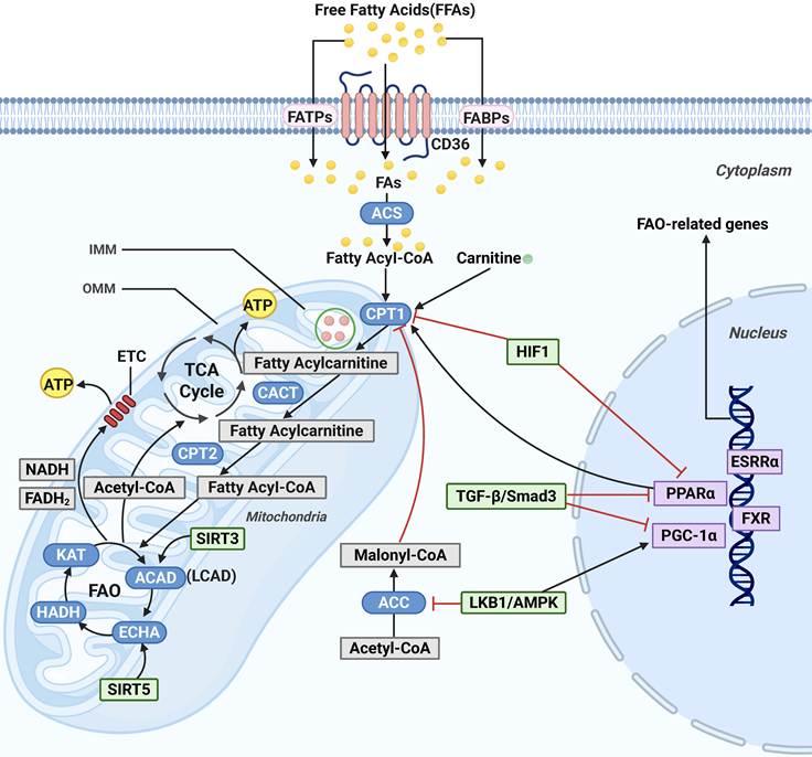
In the renal tubules, FAO primarily occurs in the mitochondria, but also in the peroxisomes [31]. In this article, we specifically focus on mitochondrial FAO (Figure 1). Free fatty acids (FFAs) are transported across the plasma membrane into the cytoplasm by specialized transport proteins, such as fatty acid transport proteins (FATPs), fatty acid binding proteins (FABPs), and the scavenger receptor CD36 [32-35]. After being taken up, FFAs can be used as a substrate in anabolic and catabolic metabolic pathways. After entering cells, FFAs are not directly delivered to mitochondria. Rather, they are enzymatically activated to fatty acyl-coenzyme A (CoA) in response to acyl-CoA synthetase (ACS), a mandatory step preceding further β-oxidation [36, 37]. Long-chain fatty acyl-CoA is not freely able to enter the mitochondrial matrix because of the impermeability of the inner mitochondrial membrane (IMM). The solution to this limitation involves the carnitine palmitoyl transferase 1 (CPT1), an enzyme located on the outer mitochondrial membrane (OMM) that catalyzes the conversion of acyl-CoA into fatty acylcarnitine. This response is a rate-limiting process that contributes to its transportation via the carnitine shuttle system [38]. After that, when the action of the carnitine/acylcarnitine translocase (CACT) on the IMM occurs [39], acylcarnitine is transported into the matrix of the mitochondrion and converted back to acyl-CoA by CPT2 on the matrix side of the IMM [40]. The β-oxidation cycle in mitochondria sequentially catalyzes fatty acyl-CoA degradation through four enzymatic reactions: (1) oxidation by acyl-CoA dehydrogenase (ACAD), (2) hydration by enoyl-CoA hydratase (ECH), (3) secondary oxidation by (S)-3-hydroxyacyl-CoA dehydrogenase (HADH), and (4) thiolytic cleavage by 3-ketoacyl-CoA thiolase (KAT), to generate acetyl-CoA, reduced nicotinamide adenine dinucleotide (NADH), and reduced flavin adenine dinucleotide (FADH2). The breakdown product acetyl-CoA enters the tricarboxylic acid (TCA) cycle to be fully oxidized to yield ATP, while by-products NADH and FADH2 enter the electron transport chain (ETC) reaction, donating electrons to generate an electrochemical gradient, which in turn produces ATP [41-43]. Physiologically, these processes produce large quantities of ATP per fatty acid molecule, which is necessary to support the energy-intensive transport and reabsorption processes of TECs [36].
The regulatory pathways of mitochondrial FAO in kidney diseases. FAs enter the cytoplasm through CD36, FABPs, or FATPs. Once in the cytoplasm, FAs are activated by ACS to form fatty acyl-CoA, which is subsequently transported to the mitochondrial matrix via CPT1, CACT, and CPT2. In the mitochondria, acyl-CoA is converted into acetyl-CoA through FAO and enters the TCA for further oxidation, ultimately leading to ATP production. The enhanced FAO occurs through the upregulation of CPT1 and PPAR-related signaling pathways. FAO, fatty acid β-oxidation; FAs, fatty acids; FABPs, FA-binding proteins; FATPs, FA-transport proteins; ACS, acyl-CoA synthetase; CPT1, carnitine palmitoyl transferase 1; CACT, carnitine-acylcarnitine translocase; CPT2, carnitine palmitoyl transferase 2; TCA, tricarboxylic acid cycle; HIF1, hypoxia-inducible factor 1; SIRT3, sirtuin 3; SIRT5, sirtuin 5; ACAD, acyl-CoA dehydrogenase; LCAD, long-chain acyl-CoA dehydrogenase; ECHA, enoyl-CoA hydratase; HADH, (S)-3-hydroxyacyl-CoA dehydrogenase; KAT, 3-ketoacyl-CoA thiolase; NADH, reduced nicotinamide adenine dinucleotide; FADH2, reduced flavin adenine dinucleotide; ETC, electron transport chain; ACC, acetyl-CoA carboxylase; LKB1, Liver kinase B1; AMPK, adenosine monophosphate-activated protein kinase; PGC-1α, peroxisome proliferator-activated receptor gamma coactivator 1alpha; PPARα, peroxisome proliferator-activated receptor alpha; FXR, farnesoid X receptor; ESRRα, estrogen-related receptor alpha; TGF-β, transforming growth factor-β; Smad3, mothers against decapentaplegic homolog 3.
Predisposing factors of FAO
Nuclear receptors
Most of the transcription factors involved in regulating FAO gene expression are nuclear receptors (NRs), including peroxisome proliferator-activated receptor alpha (PPARα, estrogen-related receptor alpha (ERRα), and farnesoid X receptor (FXR), all of which can directly manipulate FAO genes [30]. Here, we focus on reviewing the roles of these three key renal NRs in maintaining normal renal homeostasis.
The PPAR family, comprising the subtypes α, β/δ, and γ, is a group of ligand-activated transcription factors within the type II nuclear hormone receptor superfamily [44]. PPARα is primarily expressed in renal tubular cells with high metabolic activity and plays a key role in facilitating FAO [45]. After being activated by circulating FFAs, PPARα transcriptionally upregulates key enzymes involved in both mitochondrial and peroxisomal FAO pathways, promoting the clearance of superfluous lipid from kidneys [46]. Targeted modulation of PPARα activity through regulation of its expression, ligand interactions, coactivators or corepressors, cofactor recruitment, and post-translational modifications offers a potential therapeutic approach for managing renal lipid disorders [47]. Blanquart et al. first established that synthetic PPARα ligands (e.g., Wy14,643, GW7647, fibrates) enhance PPARα transcriptional activity by prolonging its half-life and increasing protein stability, consequently upregulating downstream target gene expression [48]. Furthermore, PPARγ coactivator 1α (PGC-1α), a transcriptional coactivator of PPARα, further regulates FAO by modulating mitochondrial biogenesis [49].
ERR family, orphan members of the nuclear receptor superfamily, have emerged as crucial regulators of renal homeostasis and pathophysiology. ERRs (ERRα, β, and γ in mammals) are widely expressed throughout the body, with the highest levels observed in tissues with high oxidative activity, such as kidneys, skeletal muscle, and liver [50]. Among them, ERRα functions as a nuclear receptor with fundamental metabolic regulatory properties [51, 52]. Growing evidence suggests that ERRα may represent a novel therapeutic target for modulating renal FAO and oxidative phosphorylation (OXPHOS) [53]. PGC-1α, a coactivator of ERRα, not only triggers the expression of ERRα but also creates a viable complex with ERRα. They collaborate to upregulate the transcription of downstream targets, such as enzymes and regulatory proteins of FAO [54]. According to the emerging evidence, ERRα, a transcriptional regulator of human peroxisomal acyl-coenzyme oxidases (ACOXs) genes, was observed to reduce lipid accumulation in myocytes upon activation by increasing the expression of genes involved in FAO and OXPHOS pathways [55].
FXR is a nuclear receptor with high expression in the kidneys, liver, vasculature, intestines, and adrenal glands [50]. FXR is also a key mediator of bile acid metabolism, that is, regulating its synthesis, transport, and reabsorption, while regulating the homeostasis of carbohydrates and lipids [56]. To be more precise, FXR triggers a metabolic repositioning of FA synthesis to FAO and lipid catabolism, which is marked by the downregulation of FA synthase gene expression (e.g., fatty acid synthase (FASN), acetyl-CoA carboxylase (ACC), sterol regulatory element-binding protein 1c (SREBP-1c), and stearoyl-CoA desaturase 1 (SCD1)) and the upregulation of FAO and lipid catabolism-related factors (e.g., PPARα, uncoupling protein 2 (UCP2), CPT1α, lipoprotein lipase, and PGC-1α) [57]. As demonstrated by Xu et al., FXR potentiates FAO and attenuates lipid accumulation through a PPARγ-dependent mechanism in proximal tubule epithelial cells of the kidney [9].
Adenosine monophosphate-activated protein kinase
Adenosine monophosphate-activated protein kinase (AMPK) is a master metabolic sensor that controls cellular energy homeostasis, ion transport, inflammation, autophagy, cell growth, and survival function [58]. AMPK maintains cellular energy balance by facilitating catabolic pathways (e.g., FAO, glycolysis, and autophagy) while suppressing energy-consuming anabolic processes, including protein and lipid biosynthesis [59]. In response to nutrient deprivation and cellular stress, AMPK is activated through the phosphorylation of the α-catalytic subunit at threonine-172 (Thr (172)) mediated by its upstream kinase, liver kinase B1 (LKB1) [60, 61]. Activated AMPK phosphorylates and inactivates acetyl-CoA carboxylase (ACC), thereby inhibiting the conversion of acetyl-CoA to malonyl-CoA. The result of this cascade is the activation of FAO because malonyl-CoA is a potent allosteric inhibitor of CPT1 [62]. Moreover, PGC-1α is directly phosphorylated at Thr177 and Ser538 by AMPK, and the post-translational modification increases the activity of the transcriptional coactivator of PGC-1α. This control system is very important in skeletal muscle mitochondrial biogenesis, especially during exercise-induced metabolic adaptation [63].
Sirtuins
Mitochondrial FAO is dynamically regulated by reversible post-translational modifications [64], especially lysine acylation, which is a process modulated by NAD⁺-dependent class III histone deacetylases family, sirtuins (SIRTs). SIRT3, localized to the mitochondrial matrix, upregulates FAO by regulating long-chain acyl-CoA dehydrogenase (LCAD) and the LKB1/AMPK pathway, and protects the kidney by improving mitochondrial morphology and performance, increasing ATP generation, and lowering reactive oxygen species (ROS), as well as lipid peroxidation [65, 66]. However, in contrast, SIRT5, which is found in mitochondria and in peroxisomes, inhibits peroxisomal FAO by suppressing the ACOX1 activity and improves mitochondrial FAO by activating enoyl-CoA hydratase (ECHA) [67, 68].
TGF-β/Smad3
Transforming growth factor-β (TGF-β), an important upstream regulator related to FA metabolism, induces lipid accumulation in the kidney by downregulating FAO [11]. When bound to the dual serine/threonine kinase receptor, TGF-β stimulates and phosphorylates Smad3 [69], which is translocated to the nucleus and inhibits the expression of PPARα and PGC-1α. This lowers the activity of CPT1 and ACOX1, downstream FAO-related enzymes, and leads to metabolism inhibition, cellular ATP reduction, and lipid droplet deposition [11, 70]. Moreover, Sekiguchi et al. indicated that the TGF-β/Smad3 signaling pathway can suppress directly the activity of PPARα, such that the expression of its target genes is inhibited, leading to reduced FAO in cardiomyocytes [71].
Hypoxia-inducible factors
Hypoxia-inducible factors (HIFs), crucial transcriptional regulators, may play a central role in controlling the cellular response to ensure oxygen homeostasis. HIF1α and HIF2α are mostly transcriptional activators that have broad-ranging regulation of biological processes, including glucose, cholesterol, and FA metabolism [72]. As opposed to the well-known effect of HIFs on carbohydrate metabolism, the impacts of HIFs on lipid metabolism have only recently captured interest [73]. HIF reduces the expression of PGC-1α and CPT1A and increases the expression of FA synthase, which boosts de novo lipogenesis in the presence of hypoxia. These adaptations reduce the opportunity of FA transporting into the mitochondria and impair FAO [74, 75], thus increasing lipid accumulation [76].
Impaired FAO in kidney damage
Acute kidney injury
Research has proven that FAO is suppressed in many types of AKI, and FAO deficiency is very important in ischemia/reperfusion (I/R)-induced and cisplatin-induced AKI [77, 78]. Mechanistically, the transcription of genes encoding key FAO enzymes, such as CPT1 and medium-chain specific acyl-CoA dehydrogenase (MCAD), is suppressed through downregulation of the expression of PPARα and PGC-1α, and further results in decreasing FA entry into the mitochondrial matrix and impairing FAO [38, 79, 80]. Further research has demonstrated that cyclophilin D (CypD) in the proximal tubules can bind and block PPARα in mitochondria to suppress the transcriptional regulation of FAO-related genes by PPARα, leading to FAO dysfunction, intracellular lipid accretion, later lipotoxicity, and cellular necrosis in cisplatin-induced AKI [77]. Recent research has found that FXR is essential in the pathogenesis of cisplatin-induced AKI. It's observed that overexpression or activation of FXR enhanced FAO and decreased lipid accumulation primarily through regulation of PPARγ expression in renal tubular epithelial cells (RTECs) [9]. Juszczak et al. found that Sirt3 regulates FAO by deacetylating LKB1 and activating AMPK in renal cells in cisplatin-induced AKI [81]. Interestingly, Chiba et al. discovered that loss of SIRT5 stimulates a metabolic shift of FAO from mitochondria to peroxisomes, contributing to preserving and even improving renal function following cisplatin treatment or I/R injury in the kidneys [82].
Chronic kidney disease
In CKD, sustained suppression of FAO in RTECs is closely associated with the progression of fibrosis, a typical pathological hallmark of various CKD [83]. This coincided with hypertension-induced and folate-induced CKD models. Specifically, it was observed that the core transcriptional regulators and metabolic enzymes involved in FAO were persistently downregulated, causing insufficient ATP production, accelerating tubular injury, and the progression of renal fibrosis [11, 84]. Moreover, Kang et al. and Jao et al. found that TGF-β/Smad3 and ATF6αpathways can directly inhibit the PPARα/PGC-1α axis, leading to dysregulation of FAO, mitochondrial impairment, lipid deposition, and accelerating the generation of the fibrotic phenotype [11, 85]. Ke et al. found that increased mitochondrial UCP2 induces the HIF-1α stabilization, diminishes the expression of PPARα and CPT1A, and further exacerbates lipid deposition and tubulointerstitial fibrosis [86]. At the epigenetic level, PPARα, PGC-1α, and the mitochondrial transcription factor TFAM are negatively regulated by miR-21, miR-150, and miR-495 in fibrotic kidneys, thereby reducing FAO and further promoting renal fibrotic progression. miR-21 directly targets and silences PPARα [87]. In experimental renal fibrosis, miR-150-5p and miR-495-3p are upregulated; overexpression of these two miRNAs in renal tubular epithelial cells reduces PGC-1α and TFAM mRNA levels by approximately 50%[88]. miR-33, a different microRNA, is also involved in the pathophysiological mechanism of lipid accumulation and fibrosis of the kidney by regulating FAO in both folic acid nephropathy and the unilateral ureteral obstruction (UUO) model [89].
DN is characterized by podocyte damage and intracellular lipid buildup that ends up causing kidney dysfunction. In the progression of DN, the expression of the co-activator Lipin-1 in renal tissues initially is elevated and then reduced. The downregulation of Lipin-1 suppresses the PGC-1α/PPARα signaling pathway, resulting in the defective utilization of FA in RTECs and aggravated fibrosis [90]. Chen et al. found that the elevated expression of the E3 ubiquitin ligase WWP2 further limits the process of FAO and OXPHOS by inhibiting PGC-1α, thereby stimulating lipid deposition in fibroblasts [91]. The activation of HIF-1α by hypoxia in the DN microenvironment has a direct suppressive effect on CPT1A, which disrupts mitochondrial FA transport and FAO [75, 92, 93]. Li et al. demonstrated that AMPK, a major regulator of FAO, is decreased in hypoxia, and this further weakens FA catabolism and increases lipotoxicity [94]. Emerging evidence indicates that downregulation of ERRα in podocytes suppresses the ACOX1-mediated FAO pathway and aggravates podocyte injury in DN [95].
Ferroptosis in renal diseases: biological mechanisms and evidence
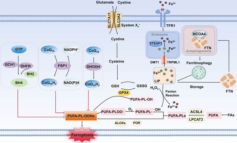
Ferroptosis is a recently identified type of regulated cell death, recognized for its unique mitochondrial morphology, featured membrane contraction, elevated membrane density, diminished mitochondrial cristae, and outer membrane disruption associated with lipid peroxide [96]. Mechanistically, this cell death process is directly or indirectly regulated by interconnected pathways of iron metabolism, antioxidant defense, and lipid metabolism [22]. An overview of these related regulatory pathways involved in ferroptosis is presented in Figure 2.
Regulatory pathways involved in ferroptosis. The major regulatory mechanisms include the iron metabolism pathway, the lipid peroxidation-related pathway, and the antioxidant defense system (e.g., System Xc-/GSH/GPX4 axis, NAD(P)H/FSP1/CoQ10 axis, GCH1/BH4 axis, DHODH/CoQ10 axis). TF, transferrin; TFR1, transferrin receptor 1; STEAP3, six-transmembrane epithelial antigen of the prostate 3; DMT1, divalent metal transporter 1; TRPML1, transient receptor potential mucolipin 1; LIP, labile iron pool; FTN, ferritin; NCOA4, nuclear receptor coactivator 4; SLC3A2, solute carrier family 3 member 2; SLC7A11, solute carrier family 7 member 11; System Xc-, cystine/glutamate antiporter; GPX4, glutathione peroxidase 4; GSH, glutathione; GSSG, oxidized glutathione; ACSL4, acyl-CoA synthetase long-chain family member 4; LPCAT3, lysophosphatidylcholine acyltransferase 3; ALOXs, arachidonate lipoxygenases; POR, cytochrome P450 reductase; GTP, guanosine triphosphate; GCH1, GTP cyclohydrolase 1; BH4, tetrahydrobiopterin; BH2, 7,8-dihydrobiopterin; DHFR, dihydrofolate reductase; CoQ10, coenzyme Q10; CoQ10H2, reduced coenzyme Q10; FSP1, ferroptosis suppressor protein 1; DHODH, dihydroorotate dehydrogenase; NAD(P)H, nicotinamide adenine dinucleotide (phosphate); FAs, fatty acids; PUFA, polyunsaturated fatty acid; PUFA-PLs, polyunsaturated fatty acid-phospholipids; PUFA-PL-OH, polyunsaturated fatty acid-phospholipid alcohol; PUFA-PL-OOHs, polyunsaturated fatty acid-phospholipid hydroperoxides.
A brief overview of the core engines of ferroptosis
The accumulation of labile iron, lethal ROS and membrane lipid peroxides constitutes key predisposing factors for ferroptosis, which may be induced by any factor affecting the imbalance between oxidative damage and antioxidant defenses [97].
Fe3+ is taken up by binding to transferrin (TF) first, following binding to transferrin receptor 1 (TFR1) on the cell membrane to produce a complex, which was subsequently internalized by endocytosis. Within the endosome, six-transmembrane epithelial antigen of the prostate 3 (STEAP3) mediates the reduction of Fe3+ to Fe2+, which is subsequently exported to the cytoplasmic labile iron pool (LIP) via transient receptor potential mucolipin 1 (TRPML1) or divalent metal transporter 1 (DMT1) [98-100]. Ferritin (FTN), composed of ferritin light-chain and ferritin heavy-chain, possesses ferric oxidase activity that converts Fe2+ to Fe3+, which plays a crucial role in maintaining iron homeostasis by storing excess intracellular iron ions [101]. When the intracellular iron levels decrease, nuclear receptor coactivator 4 (NCOA4) facilitates ferritinophagy in autophagosomes, consequently, causing the autophagic degradation of FTN, resulting in the release of Fe2+. The surplus of Fe2+ is used as a catalyst and leads to the Fenton reaction that promotes ferroptosis [102].
Intracellular ROS can be indirectly generated from free iron ions via the Fenton reaction, in which Fe2+ catalyzes the decomposition of H2O2 into highly reactive hydroxyl radicals (·OH) and hydroxide ions (OH⁻) [103, 104]. Oxidative stress is a state characterized by an excess of ROS that is beyond the cell's capacity to eliminate them [105]. Oxidative stress ultimately induces lipid peroxidation, a key component of ferroptosis, which is counteracted by the antioxidant defense system. The System Xc-/GSH/GPX4 axis is an important antioxidant system that protects cells from lipid oxidation [22]. System Xc-, which functions as the cystine/glutamate antiporter, is a heterodimer complex consisting of two specific subunits, namely solute carrier family 7 member 11 (SLC7A11) and solute carrier family 3 member 2 (SLC3A2). In this pathway, system Xc- can exchange glutamate for cystine and subsequently reduce intracellular cystine to cysteine, which is further used for glutathione (GSH) synthesis [106]. Glutathione peroxidase 4 (GPX4) is a phospholipid peroxidase that utilizes its essential cofactor GSH to protect membranes from oxidative damage of lipid peroxidation by converting cytotoxic phospholipid hydroperoxides (PLOOH) into harmless corresponding phospholipid alcohols (PLOH) and simultaneously transforming GSH into glutathione disulfide (GSSG) [107, 108]. Additionally, a growing number of GPX4-independent axes have also been found involved in impeding ferroptosis by reducing the deposition of lipid hydroperoxides, such as GTP cyclohydrolase 1-tetrahydrobiopterin (GCH1/BH4) axis, nicotinamide adenine dinucleotide (phosphate)-ferroptosis suppressor protein 1-ubiquinol (NAD(P)H/FSP1/CoQ10) axis, and mitochondrial dihydroorotate dehydrogenase (DHODH) / CoQ10 axis [109].
Lipid peroxidation constitutes an integral process in ferroptosis, initiated by either enzymatic or non-enzymatic means [110]. Especially, the peroxidation of polyunsaturated fatty acids (PUFAs) within membrane lipid settings is known to be the key final event driving ferroptosis [111]. This process begins with the incorporation of PUFAs into membrane phospholipids (PUFA-PLs), a key step orchestrated by lysophosphatidylcholine acyltransferase 3 (LPCAT3) and acyl-CoA synthetase long-chain family member 4 (ACSL4) [112]. Due to their bisallylic moieties, PUFA-PLs are highly susceptible to peroxidation, a process that involves two different pathways: iron-dependent non-enzymatic autoxidation and enzymatic oxidation [113]. Non-enzymatic peroxidation, which depends on by the Fenton reaction, is triggered by hydroxyl radical (·OH)-mediated abstraction of a bisallylic hydrogen atom from PUFA-PLs embedded within lipid bilayers, thereby generating a phospholipid radical (PL·). Subsequently, reaction of this radical with molecular oxygen generates a PUFA peroxide radical (PUFA-PL-OO·), which then abstracts a hydrogen atom from an adjacent PUFA, producing phospholipid hydroperoxides (PUFA-PL-OOHs) [113, 114]. In addition to non-enzymatic mechanisms, enzymatic lipid peroxidation plays a critical role in ferroptosis. Several enzymes, such as the arachidonate lipoxygenase (ALOX) family and the cytochrome P450 reductase (POR), can directly catalyze the oxidation of PUFA-PLs into toxic PUFA-PL-OOHs through their catalytic activity. The excessive accumulation of these peroxidized lipids disrupts membrane integrity and permeability, ultimately leading to ferroptosis [115].
Ferroptosis and kidney damage
Numerous studies have demonstrated that ferroptosis is closely associated with the pathogenesis and progression of diverse renal diseases, including AKI, CKD, and DN. It is further believed that ferroptosis may be associated with RTEC damage, glomerular mesangial cell damage, and podocyte damage in the kidney.
In RTECs, various pathogenic insults lead to the aberrant accumulation of Fe2+ or H2O2. This, in turn, drives Fenton-like reactions that catalyze the oxidation of membrane lipids into peroxides, culminating in ferroptosis and AKI [116]. Renal ischemia/reperfusion (I/R), a serious illness, is the predominant cause of AKI. Emerging evidence has identified ferroptosis as a pivotal mechanism underlying I/R-induced renal damage [117]. The development of AKI following cardiac surgery, a clinical condition largely driven by renal I/R injury, involves a multifaceted pathogenesis. Notably, elevated free iron levels during this process can initiate ferroptosis, which in turn causes renal tubular necrosis [118, 119]. Furthermore, I/R-induced AKI is also related to the GSH/GPX system [120, 121]. Previous research has indicated cell ferroptosis rather than necroptosis as one of the main factors of drug-induced AKI, as exemplified by the folic acid (FA) model [122, 123]. In particular, the occurrence of ferroptosis in FA-induced AKI is driven by the suppression of GPX4, SLC7A11, and FSP1, concurrent with an increase in the levels of iron ions and malondialdehyde (MDA), which is a major product of lipid peroxidation [124].
In CKD, the interplay of micronutrient imbalance, ectopic lipid deposition, immune activation due to inflammation, and dysregulation of the antioxidant system collectively drives lipid peroxidation, leading to ferroptosis in renal parenchymal cells and thereby propelling disease progression [12]. This is supported by the characteristic indicators of ferroptosis, including depleted GSH, reduced GPX activity, and increased concentrations of ROS in CKD animals and patients [125, 126]. DN is a leading cause of mortality and morbidity in CKD patients [127], whose pathogenesis involves a variety of factors, with ferroptosis emerging as a key pathological mechanism [18]. More specifically, significant downregulation of the key ferroptosis-related molecules SLC7A11 and GPX4 leads to exacerbated lipid peroxidation, and then initiating ferroptosis has been observed in the renal tubules of DN patients compared to non-DN patients, as demonstrated in vivo and in vitro experiments [128]. These insights offer a potential therapeutic avenue for attenuating the chronic progression of kidney disease.
The dual role of FAO in Ferroptosis
Recently, a form of oxidative cell death known as ferroptosis has been associated with the presence of PUFAs on the cell membrane that undergoes irreversible lipid peroxidation, which leads to the disruption of cell membrane integrity and subsequent cell death. FA metabolism plays a central role in regulating ferroptosis and involves the synthesis and catabolism of FA, including FAO [22]. Given the significance of FAO in energy supply and the regulation of lipid homeostasis, the effect of FAO in ferroptosis regulation has garnered increasing interest.
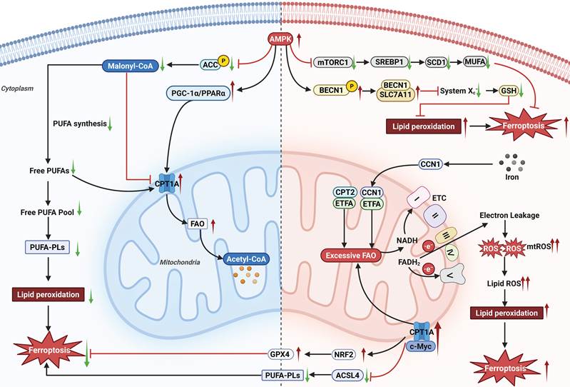
Accumulating evidence reveals that mitochondrial FAO exhibits a complex and context-dependent dual role in modulating ferroptosis (Figure 3). It serves as a suppressor or a promoter, even a crucial pathway for cellular defense against ferroptosis in particular circumstances [129]. Furthermore, FAO can alter the lipid composition of cell membranes by modulating the balance between fatty acid synthesis and degradation, thereby influencing cellular sensitivity to ferroptosis [20].
The dual role of mitochondrial FAO in ferroptosis regulation. AMPK, adenosine monophosphate-activated protein kinase; ACC, acetyl-CoA carboxylase; PUFA, polyunsaturated fatty acid; PUFA-PLs, polyunsaturated fatty acid-phospholipids; PGC-1α, peroxisome proliferator-activated receptor gamma coactivator 1-alpha; PPARα, peroxisome proliferator-activated receptor alpha; CPT1A, carnitine palmitoyltransferase 1A; FAO, fatty acid β-oxidation; mTORC1, mechanistic target of rapamycin complex 1; SREBP1, sterol regulatory element-binding protein 1; SCD1, stearoyl-CoA desaturase 1; MUFA, monounsaturated fatty acid; BECN1, Beclin 1; SLC7A11, solute carrier family 7 member 11; System Xc-, cystine/glutamate antiporter; GSH, glutathione; CCN1, cellular communication network factor 1; ETFA, electron transfer flavoprotein subunit alpha; CPT2, carnitine palmitoyltransferase 2; ETC, electron transport chain; NADH, reduced nicotinamide adenine dinucleotide; FADH2, reduced flavin adenine dinucleotide; ROS, reactive oxygen species; mtROS, mitochondrial reactive oxygen species; c-Myc, cellular myelocytomatosis oncogene; NRF2, nuclear factor erythroid 2-related factor 2; GPX4, glutathione peroxidase 4; ACSL4, acyl-CoA synthetase long-chain family member 4.
FAO as an inhibitor for ferroptosis
Here, we discuss the inhibitory role of FAO in ferroptosis, primarily through the reduction of lipid peroxidation substrates and the modulation of membrane lipid composition.
Enhanced FAO can reduce the levels of incorporation of peroxidation-susceptible FAs into membrane phospholipids, thereby attenuating lipid peroxidation and suppressing ferroptosis. This mechanism has been experimentally validated in tumor and immune cell models. For instance, IL-9/STAT3 axis-mediated FAO signaling is significantly upregulated in tumor-infiltrating Tc9 cells, resulting in reduced lipid peroxidation and ferroptosis sensitivity [130]. This finding suggests that the FAO pathway is an endogenous ferroptotic inhibitor. According to the most direct theory, FAO is suggested to impede ferroptosis by utilizing intracellular FFAs and especially unesterified PUFAs [131, 132]. PUFAs are the primary substrates of lipid peroxidation whose abundance in the cell membrane directly affects cellular sensitivity to ferroptosis [133]. Mechanistically, FAO diminishes the substrate pool available for oxidation and modification by decomposing these fatty acids into acetyl-CoA, reducing the risk of ferroptosis at its source [41, 134]. Conversely, ferroptosis may be enhanced by FAO deficiency, which increases the free PUFAs and stimulates lipid peroxidation, which is evident in related research [24, 25]. Hence, FAO is regarded as a metabolic defense system that preserves the lipid homeostasis of cellular membranes by breaking the fuel supply line and preventing oxidative damage in lipid peroxidation-driven diseases. This mechanism may be crucial for maintaining cell survival under physiological conditions.
Additionally, increased FAO activity promotes the breakdown of excess free PUFAs into acetyl-CoA and other metabolites, thereby reducing both the storage of peroxidation-prone FAs and the precursor pool for membrane phospholipid synthesis. Subsequently, the clearance of PUFAs alters the dynamic composition of membrane lipids, which increases the relative proportion of MUFAs incorporated into membrane phospholipids instead of PUFAs. This shift results in decreasing the membrane lipids' susceptibility to oxidative attack, thereby reducing the generation of toxic lipid peroxides and ultimately suppressing ferroptosis [135].
Taken together, FAO impedes ferroptosis through two main ways that regulate lipid peroxidation. First, it reduces the supply of easily peroxidized PUFAs, which are the materials needed for peroxidation. Second, it changes the composition of cellular membrane lipids. FAO curbs the initiation and execution of ferroptosis by reducing lipid peroxidation through these direct and indirect methods. These mechanisms expand our understanding of how ferroptosis is regulated and provide promising therapies for diseases linked to ferroptosis, such as cardiovascular conditions, neurodegenerative diseases, and cancer.
FAO as a promoter or resistance pathway for ferroptosis
Unlike the protective effects described above, emerging evidence indicates that under specific pathological conditions, particularly in cancer, the intrinsic relation between FAO and ferroptosis is more complex.
Firstly, the ongoing FAO process generates substantial amounts of reduced coenzymes NADH and FADH2, which subsequently transfer electrons to the mitochondrial respiratory chain. However, the partial electrons may inadvertently leak and interact with oxygen, resulting in the overproduction of superoxides such as mitochondrial reactive oxygen species (mtROS) [136-139]. Excessive mtROS, a potent oxidant, can directly assault PUFAs within cellular membranes, initiate pronounced lipid peroxidation, and thereby drive ferroptosis [140]. This finding is further substantiated in recent research. As previously mentioned, FAs should be conjugated with CoA to form acyl-CoA before undergoing FAO, then converted into acylcarnitine and transported into the mitochondria by CPT1 and CACT. Within the mitochondrion, CPT2 reconverts it back to acyl-CoA, the direct substrate for FAO. Electron transfer flavoprotein subunit alpha (ETFA) promotes the initial steps of FAO by transferring electrons to the ETC, thereby driving mtROS production and sensitizing cells to ferroptosis [139, 141]. Notably, a pivotal study demonstrated that following induction, cellular communication network factor 1 (CCN1) translocates to the mitochondria in response to elevated iron levels, where it forms a complex with ETFA and collaborates with CPT2 to provide adequate substrates for ETFA-driven mtROS generation, thereby exacerbating ferroptosis by facilitating excessive FAO [129]. Consequently, this mechanism increased the metabolic flux mediated by CPT2, indicating that overactive FAO may serve as an intrinsic oxidative stressor, thereby escalating the cellular risk of undergoing ferroptosis [142-144]. Stimulating CCN1-mediated FAO and mtROS generation could enhance the efficacy of ferroptosis-targeted cancer treatments. Furthermore, recent advancements have been made in exploring the delicate relationship between FAO and ferroptosis in cancer therapy. A recent study reported that modulating FAO activity can trigger selective, iron-independent ferroptosis in cancer cells, revealing a potential novel treatment strategy [142]. This was achieved by combining mild tetrakis (4-carboxyphenyl) porphyrin (TCPP)-based photodynamic therapy with xanthohumol (Xan) to inhibit the conversion of FAs to triglycerides, concurrently circumventing the suppressive signaling mediated by AMPK in malignant cells. This approach enabled sustained activation of AMPK and FAO. The excessive and prolonged ROS generated by FAO, along with the increased levels of PUFAs and PUFA-PLs induced by Xan, were identified as key factors driving the robust ferroptosis response. Overall, this study further highlights the critical role of FAO in the induction of ferroptosis.
Secondly, it's interesting that active FAO has been identified as an essential survival mechanism for cellular resistance to ferroptosis in certain cancer cells, in contrast to the previous situation. For instance, Miess et al. demonstrated that FAO inhibition by drugs such as the CPT1 inhibitor Etomoxir can reverse the resistance of renal cancer cells against ferroptosis, rendering them sensitive to ferroptosis [26]. This observation indicates that such cancer cells are highly dependent on FAO to sustain sufficient energy production and metabolic homeostasis, and alleviate cellular oxidative stress generated by ferroptosis inducers. In this regard, though FAO does not directly cause ferroptosis, its persistent activity acts as a shelter that allows cells to survive and resist ferroptosis.
Metabolic tipping points determining the dual role of FAO in ferroptosis
The dual role of FAO in ferroptosis is not absolute but is governed by several metabolic tipping points. First, the balance between free PUFA supply and FAO-mediated clearance is critical. When intracellular free PUFAs remain within physiological limits and mitochondrial FAO flux is moderate, FAO primarily converts unesterified PUFAs into acetyl-CoA, thereby depleting the substrate pool available for phospholipid synthesis and lipid peroxidation, and thus acting as an anti-ferroptotic defense [41, 134]. However, when the rate of free PUFA input or release exceeds the clearance capacity of FAO, excess PUFAs are esterified and incorporated into membrane phospholipids, providing substrates for lipid peroxidation [113]. Therefore, the ratio of free PUFA supply to FAO clearance flux represents the first critical tipping point.
Second, the activation status of the CCN1-ETFA pathway determines whether FAO-derived electrons are safely transferred or leak to generate mtROS. Recent studies have shown that CCN1 translocates to mitochondria under ferroptosis-inducing conditions and forms a complex with ETFA, thereby supplying additional substrates for mtROS production. Compared with the conventional CPT2-ETFA pathway, the CCN1-ETFA axis markedly increases the probability of electron leakage between FAO and the electron transport chain, leading to excessive mtROS generation [129]. Thus, the level of mitochondrial CCN1 translocation and the intensity of ETFA-dependent FAO flux constitute the second tipping point: when the CCN1-ETFA pathway is activated and FAO flux exceeds the mitochondrial antioxidant buffering capacity, FAO-derived mtROS overwhelm its PUFA clearance effect, converting FAO into a pro-ferroptotic driver.
Third, cell type and metabolic context further shape the direction of FAO function. In cells highly dependent on FAO for energy supply, such as renal tubular epithelial cells, basal FAO flux is relatively high and antioxidant reserves are sufficient, favoring the protective role of FAO in maintaining lipid homeostasis [145]. In contrast, in certain tumor cells, high CCN1 expression or upregulated CPT1A activity sustains elevated FAO flux, causing mtROS production to exceed the cellular antioxidant threshold [129, 146]. Moreover, under pathological conditions such as diabetic kidney disease, NOX4-mediated oxidative stress can suppress FAO and accelerate ferroptosis, suggesting that the redox state of the pathological microenvironment is also a critical variable [147].
In summary, the role of FAO in ferroptosis is not fixed but is shaped by a dynamic balance determined by the PUFA supply/FAO clearance ratio, the degree of CCN1-ETFA pathway activation, and cellular antioxidant reserves. When FAO flux is sufficient to clear free PUFAs without causing net mtROS accumulation, FAO functions as an anti-ferroptotic defense; when FAO flux exceeds the electron-accepting capacity of the mitochondrial ETC, causing net mtROS production to surpass the cellular antioxidant threshold, FAO shifts to a pro-ferroptotic driver. This framework not only reconciles the seemingly contradictory roles of FAO in ferroptosis but also provides a theoretical basis for precision therapeutic strategies targeting the FAO-ferroptosis axis.
Key molecular mechanisms between FAO and ferroptosis
In the ferroptosis process, the main molecules involved in FAO play a central role in directly or indirectly affecting the initiation and progression of ferroptosis by regulating fatty acid metabolism [12].
CPT1A, a rate-limiting enzyme of FAO, facilitates the translocation of long-chain fatty acids into the mitochondrial matrix to be subjected to FAO. Numerous experimental studies have established that CPT1A is an important connection between FAO and ferroptosis. Chen et al. discovered that interventions such as Etomoxir treatment or siRNA-mediated inhibition of CPT1A activity or expression directly led to ferroptosis in human HK-2 cells, which was manifested by elevated lipid peroxidation and a decrease in cell viability [25], while Gan et al. noted that ferroptosis was effectively alleviated and renal function was maintained in a diabetic mouse model by the enhancement of CPT1A expression [148]. It has recently been shown that lung cancer stem cells rely on the highly expressed CPT1A to promote FAO and result in the activation of an NRF2/GPX4 antioxidant pathway regulated by CPT1A/c-Myc positive feedback pathway. This process also suppresses the production of PUFA-PLs by down-regulating ACSL4 to create a defensive barrier against ferroptosis. In a further study, Ma et al. found that this protective shield can be effectively dismantled by targeted blocking of CPT1A [149]. These findings not only define CPT1A as a hub of interaction between FAO and ferroptosis but also provide a direct foundation for innovative anticancer approaches aimed at inducing ferroptosis.
AMPK, a central metabolic sensor, modulates ferroptosis through a series of downstream effectors. Its stimulation usually facilitates FAO by several mechanisms, such as ACC phosphorylation and inactivation, which lessens the inhibition of CPT1A [150], and the enhancement of PGC-1α and PPARα [151]. The process enhances the use of potential lipid peroxidation substrates and energy generation. At the same time, AMPK inhibits the ACC activity to weaken the process of de novo production of PUFAs. This cascade reaction subsequently reduces the content of PUFA-PLs in the cell membrane that are prone to oxidative reactions, thereby exerting a negative regulatory effect on ferroptosis [152]. The synergistic action of these two mechanisms helps to maintain bioenergetic homeostasis in the cells under conditions of energy stress or metabolic reprogramming by upregulating FAO. Moreover, the lipid buildup alleviation can indirectly reduce cell exposure to ferroptosis. Notably, recent findings have revealed a paradoxical role for AMPK in promoting ferroptosis in particular conditions, and this is the opposite of the repressive actions presented above. The relevant study has shown that AMPK inhibits the mechanistic target of rapamycin complex 1 (mTORC1) - SREBP1 axis, and down-regulates SCD1, which is the enzyme of restricting the rate of synthesis of the monounsaturated fatty acids (MUFAs). This suppression disturbs homeostatic PUFA/MUFA ratio in the membrane lipid environment, which contributes to the relative increase in the accumulation of oxidation-prone PUFAs and an increase in cell susceptibility to ferroptosis [21, 153]. Additionally, activated AMPK coordinates the phosphorylation of Beclin 1 (BECN1) at the Ser90/93/96 residues to promote the formation of the BECN1-SLC7A11 complex. The ensuing complex directly suppresses the activity of the system Xc- transporter, curtails intracellular GSH biosynthesis, exacerbates lipid peroxidation, and leads cells towards ferroptotic destiny [154, 155].
The relationship between FAO and ferroptosis is complicated. Under different cellular environments and metabolic states, FAO may either promote or inhibit ferroptosis. Understanding this interaction is crucial, as it facilitates the development of cancer therapies targeting both FAO and ferroptosis, while also aiding in the prevention of oxidative stress-induced kidney damage.
Evidence in different kidney disease scenarios
The link between FAO and ferroptosis in renal pathology has gained increasing attention, and recent studies have identified their roles in renal injury and disease progression. It has been observed that AKI is associated with suppression of FAO-related gene expression and mitochondrial dysfunction, which is accompanied by critical molecular alterations leading to ferroptosis. In an AKI model caused by cisplatin, FXR expression, as well as several important FAO genes, were significantly suppressed, resulting in lipid deposition in the kidneys and aggravated ferroptosis. Conversely, the upregulation of FXR expression may enhance FAO by elevating PPARγ levels, reducing lipid accumulation, and preventing the increase of ROS in RTECs [9, 156]. Also, FXR can directly regulate the expression of ferroptosis-related genes, including Aifm2, Ggt6, and Gsta4, suppress ferroptosis via various pathways, and finally, mitigate kidney damage [157]. In cadmium-induced AKI, Guo et al. found that the downregulation of the deacetylase Sirt3 triggers renal cell ferroptosis through the acetylation of GPX4, thus exacerbating renal injury [158]. Conversely, Li et al. discovered that enhancing Sirt3 activity can improve mitochondrial function by deacetylating liver kinase B1 and activating AMPK to promote FAO, reduce ROS generation, and lessen lipid peroxidation, which ultimately attenuates cisplatin-induced AKI in mice [65]. Normally, I/R injury impairs mitochondrial function and exacerbates oxidative stress on one hand, while shifting renal tubular cell metabolism from FAO to glycolysis on the other, leading to PUFAs accumulation. Both processes collectively promote lipid peroxidation, ultimately triggering ferroptosis in RTECs [159]. Luseogliflozin inhibits renal tubular cellular ferroptosis by enhancing AMPK phosphorylation and SIRT3 expression, promoting FAO and reducing PUFAs buildup, and simultaneously strengthening antioxidant defense effect through the Nrf2-GPX4 pathway, ultimately delaying the transformation progression of AKI to CKD after I/R injury [160].
Main regulators of FAO are also significant modulators of ferroptosis in CKD. Cai et al. reported that FAO dysfunction in diabetes positively influences the ferroptosis-prone condition via oxidative stress and energy depletion [161]. Chen et al. demonstrated that fatty acid binding protein 4 (FABP4) has been proposed to be a major pathological inducer that suppresses FAO by down-regulating the level of CPT1A, resulting in the accumulation and peroxidation of PUFAs that trigger ferroptosis [25]. Therefore, targeted improvement of FAO shows therapeutic potential for addressing the process of ferroptosis in kidney disease. Notably, it's reported that canagliflozin is considered to enhance FAO in RTECs by increasing CPT1A expression via the transcription factor FOXA1, which alleviates lipid overload and ferroptosis [148]. Zhang et al. found that augmenter of liver regeneration (ALR) exerts its protective effect by directly binding to CPT1A, stimulating the FAO pathway, maintaining lipid droplet-mitochondria coupling, and suppressing lethal lipid peroxidation [162]. Moreover, Lu et al. utilized an AMPK agonist to sufficiently prevent kidney tubular ferroptosis through coactivation of AMPK and NRF2 signaling in DN [163]. Duan et al. found that PGC-1α exerts a protective effect by recruiting NRF2 to co-activate GPX4 transcription, thereby attenuating cell ferroptosis in calcium oxalate-induced renal injury [164]. In summary, these results demonstrate a new regulatory mechanism of ferroptosis that revolves around FAO and the molecular nodes it is linked to, which reveal potential therapeutic targets for CKD.
Potential strategies for clinical treatment
Increasing evidence indicates that FAO and ferroptosis are closely implicated in the pathogenesis and progression of kidney diseases. Meanwhile, the relationship between impaired FAO function and triggered ferroptosis offers multiple intervention targets for protecting the kidneys (Table 1).
Targeting therapeutic agents and mechanisms for FAO dysfunction and ferroptosis in kidney disorders.
| Types | Agents | Targets | Mechanisms | Injury Models | References |
|---|---|---|---|---|---|
| PPARα agonist | Fenofibrate | PPARα | Enhance FAO | AKI and CKD | [11, 165] |
| PPARα/β activator | MHY2013 | PPARα/β | Enhance FAO | CKD | [166] |
| PGC-1α agonist | ZLN005 | PGC-1α | Enhance FAO | Kidney I/R injury | [167] |
| AMPK agonist | A769662 | AMPK | Enhance FAO | CKD | [168] |
| AMPK activator | AICAR | AMPK | Enhance FAO | Kidney I/R injury | [169] |
| Ferroptosis inhibitors | Ferrostatin-1 | PUFA-PL-OO· (chain-carrying lipid peroxyl radicals) | Inhibit lipid peroxidation | AKI and CKD | [170-173] |
| Ferroptosis inhibitors | Liproxstatin-1 | PUFA-PL-OO· (chain-carrying lipid peroxyl radicals) | Inhibit lipid peroxidation | AKI and CKD | [19, 174] |
| Iron chelator | Deferoxamine | Intracellular iron | Inhibit iron overload | AKI and CKD | [175, 176] |
| Enzymatic lipid peroxidation | Fisetin | ACSL4 | Reduce phospholipid fatty acids | CKD | [177] |
| Enzymatic lipid peroxidation | AS-252424 | ACSL4 | Reduce phospholipid fatty acids | Kidney I/R injury | [178] |
| SGLT2 inhibitors | Canagliflozin | CPT1A | Improve FAO and ameliorate ferroptosis through FOXA1-CPT1A axis | DN | [148] |
| SGLT2 inhibitors | Dapagliflozin | PGC-1α | Improve FAO and ameliorate ferroptosis through SIRT3/PGC-1α axis | Diabetic AKI | [179] |
FAO, fatty acid oxidation; AKI, acute kidney injury; CKD, chronic kidney disease; I/R, ischemia/reperfusion; DN, diabetic nephropathy.
Impaired FAO is a major factor involved in kidney disorders, and restoring FAO is a potential treatment approach. Therefore, considering the identified renoprotective properties of PPARα, the use of PPARα agonists has also become a central treatment alternative to repair the defective FAO [46]. Zeng et al. discovered that the PPARα agonist fenofibrate upregulated the expression of FAO-related enzymes, improved mitochondrial function, and attenuated renal injury in sepsis-associated AKI [165], which is consistent with its verified effectiveness in lessening renal damage and fibrosis in folic acid and UUO models [11]. In parallel, Chung et al. noted that PPARα/β activator MHY2013 also markedly increased the expression of FAO-related genes, diminished lipid deposition in renal tissue, and inhibited age-related renal fibrosis [166]. Furthermore, augmenting the level of PGC-1α in RTECs is another effective approach that can be used to correct energy imbalance and alleviate different nephropathies. Wang et al. observed that PGC-1α agonist ZLN005 can protect against I/R-induced renal damage by restoring mitochondrial FAO and alleviating endoplasmic reticulum stress [167]. Activation of AMPK has also been successful in restoring FAO. Han et al. found that AMPK agonist A769662 can up-regulate the expression of CPT1, CPT2, and ACOX1, promote FAO, leading to inhibition of apoptosis and dedifferentiation in cells of LKB1 deficiency [168]. In addition, combined administration of 5-aminoimidazole-4-carboxylamide nucleotide (AICAR) and carnitine, where AICAR is used to activate CPT1 by AMPK, may synergistically increase CPT1 expression and ATP contents and alleviate renal I/R injury [169].
Nowadays, the further study of the pathophysiology of ferroptosis in kidney diseases has stimulated the creation of therapeutic drugs targeting its fundamental pathways, mainly containing ferroptosis inhibitors, labile iron modulators, and lipid peroxidation pathway inhibitors. First, small-molecule ferroptosis inhibitors, such as ferrostatin-1 and liproxstatin-1, have been consistently shown to attenuate renal injury in both AKI and CKD models by scavenging oxygen radicals, reducing lipid peroxidation, and consequently suppressing cell death [19, 170-174]. Moreover, deferoxamine (DFO), an iron chelator, can alleviate kidney injury in AKI and CKD models [175, 176]. Mechanistically, DFO chelates circulating iron to form a DFO-iron complex (ferrioxamine), which is excreted via urine and feces [180], thereby mitigating iron-dependent lipid ROS accumulation and lipid peroxidation, suppressing ferroptosis [181], and ultimately alleviating renal dysfunction and tissue damage. Recent studies have shown that modulating the pivotal regulatory factor NCOA4 of ferritinophagy can effectively diminish the labile iron pool levels. The Chinese herbal formula Kunkui Baoshen decoction (KKBS) was found to ameliorate renal fibrosis in diabetic nephropathy by promoting HERC2-mediated ubiquitination and degradation of NCOA4, and then suppressing autophagy-dependent ferroptosis [182]. In terms of targeting crucial enzymes in lipid metabolism, suppressing the activity of the lipid synthase ACSL4 can decrease the enrichment of PUFAs in membrane phospholipids, thereby reducing lipid peroxidation susceptibility at its origin. Research has validated that the inhibitors Fisetin and AS-252424 notably ameliorated the progression of renal injury by impeding ACSL4-mediated ferroptosis in RTECs [177, 178].
Given the complex link between FAO dysfunction and ferroptosis, the combined therapeutic strategy exhibits promising potential in the treatment of kidney diseases. Theoretically, enhancing FAO while suppressing ferroptosis may produce an additive effect, simultaneously improving energy metabolic homeostasis and antioxidant defense, thereby more effectively delaying the progression of kidney disease. This dual-pronged strategy is expected to achieve stronger renal protection with lower drug doses and may overcome the drug resistance caused by single-drug treatment. The multi-target therapeutic approach derived from this provides a new direction for intervening in kidney diseases. To date, in the treatment of kidney diseases, although there are still few clinical reports on such combination therapy mentioned above, existing research has provided a solid theoretical framework and encouraging experimental evidence for this synergistic approach. Gan et al. discovered that the hypoglycemic drug canagliflozin plays a potential renal protective role in DN. The underlying mechanism is that canagliflozin restores FAO and alleviates ferroptosis by regulating the FOXA1-CPT1A axis [148]. Dapagliflozin, another sodium-glucose cotransporter 2 (SGLT2) inhibitor, also reduces renal injury through dual effects of improving FAO and ferroptosis in RTECs in DN [179, 183].
Perspectives and Conclusions
FAO and ferroptosis play vital roles in the development of kidney disorders. On one hand, disturbed FAO makes cells more sensitive to ferroptosis by elevating lipid peroxidation substrates and promoting oxidative stress. On the other hand, the process of ferroptosis and the resulting membrane damage subsequently further disrupt cellular metabolic homeostasis and accelerate renal injury. Therefore, multi-target intervention treatments combine FAO promotion and ferroptosis inhibition, holding significant therapeutic potential in theory. However, further studies in vivo and in vitro, as well as clinical validation trials, are still needed to elucidate the comprehensive mechanisms and translate these potentials into clinical reality. In this article, we have reviewed the pathophysiological roles of FAO and ferroptosis in kidney diseases, the correlations between them, and the recent research advances in the pathological mechanisms of the kidneys, thereby providing a theoretical basis for the novel treatment of kidney diseases. However, several critical gaps remain, particularly in the discovery of clinical biomarkers for early diagnosis and the development of targeted nanoscale drug delivery systems for renal therapeutics. Future studies should further elucidate the specific mechanisms of renal disorders, identify and validate early metabolic biomarkers, clinically translate safe and effective combination regimens, and develop precise therapeutic drugs, such as nanoscale targeted drugs for renal damage. Currently, elucidating the interconnected roles of FAO and ferroptosis in damaged kidneys in depth, as well as the underlying mechanisms, will offer novel therapeutic opportunities for the prevention and treatment of various kidney disorders.
Acknowledgements
There is no financial conflict of interest between the authors. We acknowledge the support of the funding and the online drawing software Biorender for assisting in drawing graphics.
Funding
This work was supported by the Natural Science Foundation of Gansu Province (No. 23JRRA0983) and the Henan Provincial Natural Science Foundation General Project (No. 262300421558).
Author contributions
AC: Conceptualization, Visualization, Modification, Writing—original draft. XL: Conceptualization, Writing—review and editing. JL: Writing—review and editing. XS: Conceptualization, Visualization, Supervision. HZ: Conceptualization, Supervision, Funding acquisition. CW: Designing the content of the article, Visualization. FX: Literature research. CL and YB: Conceptualization, Supervision, Guiding this article. All authors have read, critically reviewed, and approved the final manuscript for submission.
Competing Interests
The authors have declared that no competing interest exists.
References
1. Lin Y, Xu W, Hou Y, Wang S, Zhang H, Ran M. et al. The multifaceted role of ferroptosis in kidney diseases. Chem Biol Interact. 2022;365:110107
2. Al-Jaghbeer M, Dealmeida D, Bilderback A, Ambrosino R, Kellum JA. Clinical decision support for in-hospital AKI. J Am Soc Nephrol. 2018;29(2):654-60
3. Jadoul M, Aoun M, Masimango Imani M. The major global burden of chronic kidney disease. Lancet Glob Health. 2024;12(3):e342-e3
4. Priante G, Gianesello L, Ceol M, Del Prete D, Anglani F. Cell Death in the Kidney. Int J Mol Sci. 2019;20(14):3598
5. Liu L, Ning X, Wei L, Zhou Y, Zhao L, Ma F. et al. Twist1 downregulation of PGC-1α decreases fatty acid oxidation in tubular epithelial cells, leading to kidney fibrosis. Theranostics. 2022;12(8):3758-75
6. Chung KW, Lee EK, Lee MK, Oh GT, Yu BP, Chung HY. Impairment of PPARα and the fatty acid oxidation pathway aggravates renal fibrosis during aging. J Am Soc Nephrol. 2018;29(4):1223-37
7. Gu M, Tan M, Zhou L, Sun X, Lu Q, Wang M. et al. Protein phosphatase 2Acα modulates fatty acid oxidation and glycolysis to determine tubular cell fate and kidney injury. Kidney Int. 2022;102(2):321-36
8. Minami S, Yamamoto T, Takabatake Y, Takahashi A, Namba T, Matsuda J. et al. Lipophagy maintains energy homeostasis in the kidney proximal tubule during prolonged starvation. Autophagy. 2017;13(10):1629-47
9. Xu S, Jia P, Fang Y, Jin J, Sun Z, Zhou W. et al. Nuclear farnesoid X receptor attenuates acute kidney injury through fatty acid oxidation. Kidney Int. 2022;101(5):987-1002
10. Zhao L, Hao Y, Tang S, Han X, Li R, Zhou X. Energy metabolic reprogramming regulates programmed cell death of renal tubular epithelial cells and might serve as a new therapeutic target for acute kidney injury. Front Cell Dev Biol. 2023;11:1276217
11. Kang HM, Ahn SH, Choi P, Ko Y-A, Han SH, Chinga F. et al. Defective fatty acid oxidation in renal tubular epithelial cells has a key role in kidney fibrosis development. Nat Med. 2015;21(1):37-46
12. Jin R, Dai Y, Wang Z, Hu Q, Zhang C, Gao H. et al. Unraveling ferroptosis: a new frontier in combating renal fibrosis and CKD progression. Biology. 2025;14(1):12
13. Uysal E, Dokur M, Kucukdurmaz F, Altınay S, Polat S, Batcıoglu K. et al. Targeting the PANoptosome with 3,4-methylenedioxy-β-nitrostyrene, reduces PANoptosis and protects the kidney against renal İschemia-reperfusion injury. J Invest Surg. 2022;35(11-12):1824-35
14. Garg JP, Vucic D. Targeting cell death pathways for therapeutic intervention in kidney diseases. Semin Nephrol. 2016;36(3):153-61
15. Jiang Y, Liu X, Sun M. The mist of ferroptosis: The orpheus journey of mitochondria - exploring the symphony of cell fate. Int J Biol Macromol. 2025;319:145472
16. Li J, Cao F, Yin H, Huang Z, Lin Z, Mao N. et al. Ferroptosis: past, present and future. Cell Death Dis. 2020;11(2):88
17. Ni L, Yuan C, Wu X. Targeting ferroptosis in acute kidney injury. Cell Death Dis. 2022;13(2):182
18. Wang Y, Bi R, Quan F, Cao Q, Lin Y, Yue C. et al. Ferroptosis involves in renal tubular cell death in diabetic nephropathy. Eur J Pharmacol. 2020;888:173574
19. Zhang B, Chen X, Ru F, Gan Y, Li B, Xia W. et al. Liproxstatin-1 attenuates unilateral ureteral obstruction-induced renal fibrosis by inhibiting renal tubular epithelial cells ferroptosis. Cell Death Dis. 2021;12(9):843
20. Lin Z, Liu J, Kang R, Yang M, Tang D. Lipid metabolism in ferroptosis. Adv Biol (Weinh). 2021;5(8):e2100396
21. Pope LE, Dixon SJ. Regulation of ferroptosis by lipid metabolism. Trends Cell Biol. 2023;33(12):1077-87
22. Sun D, Wang L, Wu Y, Yu Y, Yao Y, Yang H. et al. Lipid metabolism in ferroptosis: mechanistic insights and therapeutic potential. Front Immunol. 2025;16:1545339
23. Chen X, Li J, Kang R, Klionsky DJ, Tang D. Ferroptosis: machinery and regulation. Autophagy. 2020;17(9):2054-81
24. Niu B, Lei X, Xu Q, Ju Y, Xu D, Mao L. et al. Protecting mitochondria via inhibiting VDAC1 oligomerization alleviates ferroptosis in acetaminophen-induced acute liver injury. Cell Biol Toxicol. 2021;38(3):505-30
25. Chen J, Wu K, Lei Y, Huang M, Cheng L, Guan H. et al. Inhibition of fatty acid β-oxidation by fatty acid binding protein 4 induces ferroptosis in HK2 cells under high glucose conditions. Endocrinol Metab (Seoul). 2023;38(2):226-44
26. Miess H, Dankworth B, Gouw AM, Rosenfeldt M, Schmitz W, Jiang M. et al. The glutathione redox system is essential to prevent ferroptosis caused by impaired lipid metabolism in clear cell renal cell carcinoma. Oncogene. 2018;37(40):5435-50
27. Pagliarini DJ, Calvo SE, Chang B, Sheth SA, Vafai SB, Ong SE. et al. A mitochondrial protein compendium elucidates complex I disease biology. Cell. 2008;134(1):112-23
28. Forbes JM, Thorburn DR. Mitochondrial dysfunction in diabetic kidney disease. Nat Rev Nephrol. 2018;14(5):291-312
29. Tian Z, Liang M. Renal metabolism and hypertension. Nat Commun. 2021;12(1):963
30. Lee LE, Doke T, Mukhi D, Susztak K. The key role of altered tubule cell lipid metabolism in kidney disease development. Kidney Int. 2024;106(1):24-34
31. Fransen M, Lismont C, Walton P. The peroxisome-mitochondria connection: how and why? Int J Mol Sci. 2017;18(6):1126
32. Baek J, He C, Afshinnia F, Michailidis G, Pennathur S. Lipidomic approaches to dissect dysregulated lipid metabolism in kidney disease. Nat Rev Nephrol. 2022;18(1):38-55
33. Khan S, Cabral PD, Schilling WP, Schmidt ZW, Uddin AN, Gingras A. et al. Kidney proximal tubule lipoapoptosis is regulated by fatty acid transporter-2 (FATP2). J Am Soc Nephrol. 2018;29(1):81-91
34. Storch J, McDermott L. Structural and functional analysis of fatty acid-binding proteins. J Lipid Res. 2009;50:S126-S31
35. Scerbo D, Son NH, Sirwi A, Zeng L, Sas KM, Cifarelli V. et al. Kidney triglyceride accumulation in the fasted mouse is dependent upon serum free fatty acids. J Lipid Res. 2017;58(6):1132-42
36. Jang HS, Noh MR, Kim J, Padanilam BJ. Defective mitochondrial fatty acid oxidation and lipotoxicity in kidney diseases. Front Med (Lausanne). 2020;7:65
37. Grevengoed TJ, Klett EL, Coleman RA. Acyl-CoA metabolism and partitioning. Annu Rev Nutr. 2014;34:1-30
38. Console L, Scalise M, Giangregorio N, Tonazzi A, Barile M, Indiveri C. The link between the mitochondrial fatty acid oxidation derangement and kidney injury. Front Physiol. 2020;11:794
39. Almannai M, Alfadhel M, El-Hattab AW. Carnitine inborn errors of metabolism. Molecules. 2019;24(18):3251
40. Bonnefont JP, Djouadi F, Prip-Buus C, Gobin S, Munnich A, Bastin J. Carnitine palmitoyltransferases 1 and 2: biochemical, molecular and medical aspects. Mol Aspects Med. 2004;25(5):495-520
41. Houten SM, Violante S, Ventura FV, Wanders RJA. The biochemistry and physiology of mitochondrial fatty acid β-oxidation and its genetic disorders. Annu Rev Physiol. 2016;78:23-44
42. Gewin LS. Sugar or fat? Renal tubular metabolism reviewed in health and disease. Nutrients. 2021;13(5):1580
43. Carracedo A, Cantley LC, Pandolfi PP. Cancer metabolism: fatty acid oxidation in the limelight. Nat Rev Cancer. 2013;13(4):227-32
44. Wagner N, Wagner KD. The Role of PPARs in Disease. Cells. 2020;9(11):2367
45. Simon N, Hertig A. Alteration of fatty acid oxidation in tubular epithelial cells: from acute kidney injury to renal fibrogenesis. Front Med (Lausanne). 2015;2:52
46. Cheng CF, Chen HH, Lin H. Role of PPARα and Its Agonist in Renal Diseases. PPAR Research. 2010;2010:345098
47. Bougarne N, Weyers B, Desmet SJ, Deckers J, Ray DW, Staels B. et al. Molecular actions of PPARα in lipid metabolism and inflammation. Endocr Rev. 2018;39(5):760-802
48. Blanquart C, Barbier O, Fruchart JC, Staels B, Glineur C. Peroxisome proliferator-activated receptor alpha (PPARalpha ) turnover by the ubiquitin-proteasome system controls the ligand-induced expression level of its target genes. J Biol Chem. 2002;277(40):37254-9
49. Scarpulla RC, Vega RB, Kelly DP. Transcriptional integration of mitochondrial biogenesis. Trends Endocrinol Metab. 2012;23(9):459-66
50. Libby AE, Jones B, Lopez-Santiago I, Rowland E, Levi M. Nuclear receptors in the kidney during health and disease. Mol Aspects Med. 2021;78:100935
51. Billon C, Schoepke E, Avdagic A, Chatterjee A, Butler AA, Elgendy B. et al. A synthetic ERR agonist alleviates metabolic syndrome. J Pharmacol ExpTher. 2024;388(2):232-40
52. Cerutti C, Shi JR, Vanacker JM. Multifaceted transcriptional network of estrogen-related receptor alpha in health and disease. Int J Mol Sci. 2023;24(5):4265
53. Tripathi M, Yen PM, Singh BK. Estrogen-related receptor alpha: an under-appreciated potential target for the treatment of metabolic diseases. Int J Mol Sci. 2020;21(5):1645
54. Spinelli S, Bruschi M, Passalacqua M, Guida L, Magnone M, Sturla L. et al. Estrogen-related receptor α: a Key transcription factor in the regulation of energy metabolism at an organismic level and a target of the ABA/LANCL hormone receptor system. Int J Mol Sci. 2024;25(9):4796
55. Kitamura K, Erlangga JS, Tsukamoto S, Sakamoto Y, Mabashi-Asazuma H, Iida K. Daidzein promotes the expression of oxidative phosphorylation- and fatty acid oxidation-related genes via an estrogen-related receptor α pathway to decrease lipid accumulation in muscle cells. J Nutr Biochem. 2020;77:108315
56. Jiang L, Zhang H, Xiao D, Wei H, Chen Y. Farnesoid X receptor (FXR): structures and ligands. Comput Struct Biotechnol J. 2021;19:2148-59
57. Wang XX, Jiang T, Shen Y, Adorini L, Pruzanski M, Gonzalez FJ. et al. The farnesoid X receptor modulates renal lipid metabolism and diet-induced renal inflammation, fibrosis, and proteinuria. Am J Physiol Renal Physiol. 2009;297(6):F1587-F96
58. Juszczak F, Caron N, Mathew AV, Declèves AE. Critical role for AMPK in metabolic disease-induced chronic kidney disease. Int J Mol Sci. 2020;21(21):7994
59. Gómez H. Reprogramming metabolism to enhance kidney tolerance during sepsis: the role of fatty acid oxidation, aerobic glycolysis, and epithelial de-differentiation. Nephron. 2023;147(1):31-4
60. Lin SC, Hardie DG. AMPK: sensing glucose as well as cellular energy status. Cell Metab. 2018;27(2):299-313
61. Oakhill JS, Steel R, Chen Z-P, Scott JW, Ling N, Tam S. et al. AMPK is a direct adenylate charge-regulated protein kinase. Science. 2011;332(6036):1433-5
62. Jeon SM, Chandel NS, Hay N. AMPK regulates NADPH homeostasis to promote tumour cell survival during energy stress. Nature. 2012;485(7400):661-5
63. Herzig S, Shaw RJ. AMPK: guardian of metabolism and mitochondrial homeostasis. Nat Rev Mol Cell Biol. 2018;19(2):121-35
64. Rardin Matthew J, He W, Nishida Y, Newman John C, Carrico C, Danielson Steven R. et al. SIRT5 regulates the mitochondrial lysine succinylome and metabolic networks. Cell Metab. 2013;18(6):920-33
65. Li M, Li CM, Ye ZC, Huang J, Li Y, Lai W. et al. Sirt3 modulates fatty acid oxidation and attenuates cisplatin-induced AKI in mice. J Cell Mol Med. 2020;24(9):5109-21
66. Hirschey MD, Shimazu T, Goetzman E, Jing E, Schwer B, Lombard DB. et al. SIRT3 regulates mitochondrial fatty-acid oxidation by reversible enzyme deacetylation. Nature. 2010;464(7285):121-5
67. Sadhukhan S, Liu X, Ryu D, Nelson OD, Stupinski JA, Li Z. et al. Metabolomics-assisted proteomics identifies succinylation and SIRT5 as important regulators of cardiac function. Proc Natl Acad Sci U S A. 2016;113(16):4320-5
68. Chen XF, Tian MX, Sun RQ, Zhang ML, Zhou LS, Jin L. et al. SIRT5 inhibits peroxisomal ACOX1 to prevent oxidative damage and is downregulated in liver cancer. EMBO Rep. 2018;19(5):e45124
69. Feng XH, Derynck R. Specificity and versatility in tgf-beta signaling through Smads. Annu Rev Cell Dev Biol. 2005;21:659-93
70. Cheng L, Ge M, Lan Z, Ma Z, Chi W, Kuang W. et al. Zoledronate dysregulates fatty acid metabolism in renal tubular epithelial cells to induce nephrotoxicity. Arch Toxicol. 2018;92(1):469-85
71. Sekiguchi K, Tian Q, Ishiyama M, Burchfield J, Gao F, Mann DL. et al. Inhibition of PPAR-α activity in mice with cardiac-restricted expression of tumor necrosis factor: potential role of TGF-β/Smad3. Am J Physiol Heart Circ Physiol. 2007;292(3):H1443-H51
72. Gaspar JM, Velloso LA. Hypoxia inducible factor as a central regulator of metabolism -implications for the development of obesity. Front Neurosci. 2018;12:813
73. Mylonis I, Simos G, Paraskeva E. Hypoxia-inducible factors and the regulation of lipid metabolism. Cells. 2019;8(3):214
74. Liu Y, Ma Z, Zhao C, Wang Y, Wu G, Xiao J. et al. HIF-1α and HIF-2α are critically involved in hypoxia-induced lipid accumulation in hepatocytes through reducing PGC-1α-mediated fatty acid β-oxidation. Toxicol Lett. 2014;226(2):117-23
75. Du W, Zhang L, Brett-Morris A, Aguila B, Kerner J, Hoppel CL. et al. HIF drives lipid deposition and cancer in ccRCC via repression of fatty acid metabolism. Nat Commun. 2017;8(1):1769
76. Hu Z, Zhu Q, Wang Y, Deng X, Yang H, Zhou M. et al. Lipid nephrotoxicity mediated by HIF-1α activation accelerates tubular injury in diabetic nephropathy. Ren Fail. 2024;46(1):2347446
77. Jang HS, Noh MR, Jung EM, Kim WY, Southekal S, Guda C. et al. Proximal tubule cyclophilin D regulates fatty acid oxidation in cisplatin-induced acute kidney injury. Kidney Int. 2020;97(2):327-39
78. Zhao L, Zhang T, Zhao Z, Ren X, Huang X, Hao Y. et al. Irisin improves acute kidney injury induced by ischemia-reperfusion through targeting energy metabolism reprogramming. Int J Biol Macromol. 2025;323:147073
79. Tang WB, Wei QQ. The metabolic pathway regulation in kidney injury and repair. Front Physiol. 2024;14:1344271
80. Portilla D, Dai G, McClure T, Bates L, Kurten R, Megyesi J. et al. Alterations of PPARalpha and its coactivator PGC-1 in cisplatin-induced acute renal failure. Kidney Int. 2002;62(4):1208-18
81. Juszczak F, Arnould T, Declèves AE. The role of mitochondrial sirtuins (SIRT3, SIRT4 and SIRT5) in renal cell metabolism: implication for kidney diseases. Int J Mol Sci. 2024;25(13):6936
82. Chiba T, Peasley KD, Cargill KR, Maringer KV, Bharathi SS, Mukherjee E. et al. Sirtuin 5 regulates proximal tubule fatty acid oxidation to protect against AKI. J Am Soc Nephrol. 2019;30(12):2384-98
83. Zhou S, Ling X, Zhu J, Liang Y, Feng Q, Xie C. et al. MAGL protects against renal fibrosis through inhibiting tubular cell lipotoxicity. Theranostics. 2024;14(4):1583-601
84. Kim HJ, Moradi H, Yuan J, Norris K, Vaziri ND. Renal mass reduction results in accumulation of lipids and dysregulation of lipid regulatory proteins in the remnant kidney. Am J Physiol Renal Physiol. 2009;296(6):F1297-F306
85. Jao TM, Nangaku M, Wu CH, Sugahara M, Saito H, Maekawa H. et al. ATF6α downregulation of PPARα promotes lipotoxicity-induced tubulointerstitial fibrosis. Kidney Int. 2019;95(3):577-89
86. Ke Q, Yuan Q, Qin N, Shi C, Luo J, Fang Y. et al. UCP2-induced hypoxia promotes lipid accumulation and tubulointerstitial fibrosis during ischemic kidney injury. Cell Death Dis. 2020;11(1):26
87. Chau BN, Xin C, Hartner J, Ren S, Castano AP, Linn G. et al. MicroRNA-21 promotes fibrosis of the kidney by silencing metabolic pathways. Sci Transl Med. 2012;4(121):121ra18
88. Miguel V, Ramos R, García-Bermejo L, Rodríguez-Puyol D, Lamas S. The program of renal fibrogenesis is controlled by microRNAs regulating oxidative metabolism. Redox Biol. 2021;40:101851
89. Price NL, Miguel V, Ding W, Singh AK, Malik S, Rotllan N. et al. Genetic deficiency or pharmacological inhibition of miR-33 protects from kidney fibrosis. JCI insight. 2019;4(22):e131102
90. Lin S, Wang L, Jia Y, Sun Y, Qiao P, Quan Y. et al. Lipin-1 deficiency deteriorates defect of fatty acid β-oxidation and lipid-related kidney damage in diabetic kidney disease. Transl Res. 2024;266:1-15
91. Chen H, You R, Guo J, Zhou W, Chew G, Devapragash N. et al. WWP2 regulates renal fibrosis and the metabolic reprogramming of profibrotic myofibroblasts. J Am Soc Nephrol. 2024;35(6):696-718
92. Semenza Gregg L. Hypoxia-inducible factors in physiology and medicine. Cell. 2012;148(3):399-408
93. Yu W, Haoyu Y, Ling Z, Xing H, Pengfei X, Anzhu W. et al. Targeting lipid metabolic reprogramming to alleviate diabetic kidney disease: molecular insights and therapeutic strategies. Front Immunol. 2025;16:1549484
94. Li H, Satriano J, Thomas JL, Miyamoto S, Sharma K, Pastor-Soler NM. et al. Interactions between HIF-1α and AMPK in the regulation of cellular hypoxia adaptation in chronic kidney disease. Am J Physiol Renal Physiol. 2015;309(5):F414-F28
95. Hu H, Wang J, Peng Z, Fan Y, Yang Q, Hu J. Dapagliflozin attenuates diabetes-induced podocyte lipotoxicity via ERRα-Mediated lipid metabolism. Free Radical Biol Med. 2025;234:178-91
96. Xu X, Xu XD, Ma MQ, Liang Y, Cai YB, Zhu ZX. et al. The mechanisms of ferroptosis and its role in atherosclerosis. Biomed Pharmacother. 2024;171:116112
97. Ru Q, Li Y, Chen L, Wu Y, Min J, Wang F. Iron homeostasis and ferroptosis in human diseases: mechanisms and therapeutic prospects. Signal Transduct Target Ther. 2024;9(1):271
98. Fang X, Ardehali H, Min J, Wang F. The molecular and metabolic landscape of iron and ferroptosis in cardiovascular disease. Nat Rev Cardiol. 2022;20(1):7-23
99. Tang D, Chen X, Kang R, Kroemer G. Ferroptosis: molecular mechanisms and health implications. Cell Res. 2020;31(2):107-25
100. Dong XP, Cheng X, Mills E, Delling M, Wang F, Kurz T. et al. The type IV mucolipidosis-associated protein TRPML1 is an endolysosomal iron release channel. Nature. 2008;455(7215):992-6
101. Latunde-Dada GO. Ferroptosis: Role of lipid peroxidation, iron and ferritinophagy. Biochim Biophys Acta Gen Subj. 2017;1861(8):1893-900
102. He J, Li Z, Xia P, Shi A, FuChen X, Zhang J. et al. Ferroptosis and ferritinophagy in diabetes complications. Mol Metab. 2022;60:101470
103. Bresgen N, Eckl PM. Oxidative stress and the homeodynamics of iron metabolism. Biomolecules. 2015;5(2):808-47
104. Muckenthaler MU, Galy B, Hentze MW. Systemic iron homeostasis and the iron-responsive element/iron-regulatory protein (IRE/IRP) regulatory network. Annu Rev Nutr. 2008;28:197-213
105. Hensel A, Váraljai R, Knauer SK. Raising the iron curtain: Lactate's secret role in oxidative stress defense. Redox Biol. 2025;85:103754
106. Dixon SJ, Lemberg KM, Lamprecht MR, Skouta R, Zaitsev EM, Gleason CE. et al. Ferroptosis: an iron-dependent form of nonapoptotic cell death. Cell. 2012;149(5):1060-72
107. Seiler A, Schneider M, Förster H, Roth S, Wirth EK, Culmsee C. et al. Glutathione peroxidase 4 senses and translates oxidative stress into 12/15-lipoxygenase dependent- and AIF-mediated cell death. Cell Metab. 2008;8(3):237-48
108. Yang WS, SriRamaratnam R, Welsch ME, Shimada K, Skouta R, Viswanathan VS. et al. Regulation of ferroptotic cancer cell death by GPX4. Cell. 2014;156(1-2):317-31
109. Ma T, Du J, Zhang Y, Wang Y, Wang B, Zhang T. GPX4-independent ferroptosis-a new strategy in disease's therapy. Cell Death Discov. 2022;8(1):434
110. Yang WS, Kim KJ, Gaschler MM, Patel M, Shchepinov MS, Stockwell BR. Peroxidation of polyunsaturated fatty acids by lipoxygenases drives ferroptosis. Proc Natl Acad Sci U S A. 2016;113(34):E4966-E75
111. Lei G, Zhuang L, Gan B. mTORC1 and ferroptosis: Regulatory mechanisms and therapeutic potential. Bioessays. 2021;43(8):2100093
112. Dixon SJ, Winter GE, Musavi LS, Lee ED, Snijder B, Rebsamen M. et al. Human haploid cell genetics reveals roles for lipid metabolism genes in nonapoptotic cell death. ACS Chem Biol. 2015;10(7):1604-9
113. Jiang X, Stockwell BR, Conrad M. Ferroptosis: mechanisms, biology and role in disease. Nat Rev Mol Cell Biol. 2021;22(4):266-82
114. Zhong W, Xie Z, Wei S, Zhong T, Jiang T. Regulatory mechanism of ferroptosis in acute kidney injury and potential therapeutic strategies. Life Sci. 2025;378:123828
115. Bayır H, Dixon SJ, Tyurina YY, Kellum JA, Kagan VE. Ferroptotic mechanisms and therapeutic targeting of iron metabolism and lipid peroxidation in the kidney. Nat Rev Nephrol. 2023;19(5):315-36
116. Hu Z, Zhang H, Yang S-k, Wu X, He D, Cao K. et al. Emerging role of ferroptosis in acute kidney injury. Oxid Med Cell Longev. 2019;2019(1):8010614
117. Thapa K, Singh TG, Kaur A. Targeting ferroptosis in ischemia/reperfusion renal injury. Naunyn-Schmiedeberg's Arch Pharmacol. 2022;395(11):1331-41
118. Han H, Wen Z, Wang J, Zhang P, Gong Q, Ge S. et al. Prediction of short-term mortality with renal replacement therapy in patients with cardiac surgery-associated acute kidney injury. Front Cardiovasc Med. 2021;8:738947
119. Choi N, Whitlock R, Klassen J, Zappitelli M, Arora RC, Rigatto C. et al. Early intraoperative iron-binding proteins are associated with acute kidney injury after cardiac surgery. J Thorac Cardiovasc Surg. 2019;157(1):287-97.e2
120. Ding C, Ding X, Zheng J, Wang B, Li Y, Xiang H. et al. miR-182-5p and miR-378a-3p regulate ferroptosis in I/R-induced renal injury. Cell Death Dis. 2020;11(10):929
121. Chen Ca, Wang D, Yu Y, Zhao T, Min N, Wu Y. et al. Legumain promotes tubular ferroptosis by facilitating chaperone-mediated autophagy of GPX4 in AKI. Cell Death Dis. 2021;12(1):65
122. Martin-Sanchez D, Ruiz-Andres O, Poveda J, Carrasco S, Cannata-Ortiz P, Sanchez-Niño MD. et al. Ferroptosis, but not necroptosis, is important in nephrotoxic folic acid-induced AKI. J Am Soc Nephrol. 2017;28(1):218-29
123. Yan LJ. Folic acid-induced animal model of kidney disease. Animal Model Exp Med. 2021;4(4):329-42
124. Li D, Liu B, Fan Y, Liu M, Han B, Meng Y. et al. Nuciferine protects against folic acid-induced acute kidney injury by inhibiting ferroptosis. Br J Pharmacol. 2021;178(5):1182-99
125. Zhang Y, Mou Y, Zhang J, Suo C, Zhou H, Gu M. et al. Therapeutic implications of ferroptosis in renal fibrosis. Front Mol Biosci. 2022;9:890766
126. Zhou J, Tan Y, Wang R, Li X. Role of ferroptosis in fibrotic diseases. J Inflammation Res. 2022;15:3689-708
127. Zhang K, Kan C, Han F, Fan Q, Wang Y, Li X. et al. Global Trends and Projections in Chronic Kidney Disease Burden from Diabetes, Hypertension, and Glomerulonephritis: A Population-Based Study. Kidney Blood Press Res. 2026;51(1):72-90
128. Kim S, Kang SW, Joo J, Han SH, Shin H, Nam BY. et al. Characterization of ferroptosis in kidney tubular cell death under diabetic conditions. Cell Death Dis. 2021;12(2):160
129. Guo W, Zhang C, Zhou Q, Chen T, Xu X, Zhang J. et al. Mitochondrial CCN1 drives ferroptosis via fatty acid β-oxidation. Dev Cell. 2025;60(17):2294-312.e11
130. Xiao L, Ma X, Ye L, Su P, Xiong W, Bi E. et al. IL-9/STAT3/fatty acid oxidation-mediated lipid peroxidation contributes to Tc9 cell longevity and enhanced antitumor activity. J Clin Invest. 2022;132(7):e153247
131. Blomme A, Ford CA, Mui E, Patel R, Ntala C, Jamieson LE. et al. 2,4-dienoyl-CoA reductase regulates lipid homeostasis in treatment-resistant prostate cancer. Nat Commun. 2020;11(1):2508
132. Xin S, Schick JA. PUFAs dictate the balance of power in ferroptosis. Cell Calcium. 2023;110:102703
133. Agmon E, Solon J, Bassereau P, Stockwell BR. Modeling the effects of lipid peroxidation during ferroptosis on membrane properties. Sci Rep. 2018;8(1):5155
134. Fu C, Cao N, Zeng S, Zhu W, Fu X, Liu W. et al. Role of mitochondria in the regulation of ferroptosis and disease. Front Med (Lausanne). 2023;10:1301822
135. Li Z, Lange M, Dixon SJ, Olzmann JA. Lipid quality control and ferroptosis: from concept to mechanism. Annu Rev Biochem. 2024;93:499-528
136. Ma ZA. The role of peroxidation of mitochondrial membrane phospholipids in pancreatic β-cell failure. Curr Diabetes Rev. 2012;8(1):69-75
137. Murphy MP. How mitochondria produce reactive oxygen species. Biochem J. 2009;417(1):1-13
138. Rodrigues JV, Gomes CM. Mechanism of superoxide and hydrogen peroxide generation by human electron-transfer flavoprotein and pathological variants. Free Radical Biol Med. 2012;53(1):12-9
139. Perevoshchikova IV, Quinlan CL, Orr AL, Gerencser AA, Brand MD. Sites of superoxide and hydrogen peroxide production during fatty acid oxidation in rat skeletal muscle mitochondria. Free Radical Biol Med. 2013;61:298-309
140. Battaglia AM, Chirillo R, Aversa I, Sacco A, Costanzo F, Biamonte F. Ferroptosis and cancer: mitochondria meet the "Iron Maiden" cell death. Cells. 2020;9(6):1505
141. Zhang X, Zhang Y, Qiu G, Pian L, Guo L, Cao H. et al. Hepatic neddylation targets and stabilizes electron transfer flavoproteins to facilitate fatty acid β-oxidation. Proc Natl Acad Sci U S A. 2020;117(5):2473-83
142. Gao Y, Song Z, Gan W, Zou X, Bai Y, Zhao X. et al. Selective and iron-independent ferroptosis in cancer cells induced by manipulation of mitochondrial fatty acid oxidation. Biomaterials. 2025;320:123259
143. Rosca MG, Vazquez EJ, Chen Q, Kerner J, Kern TS, Hoppel CL. Oxidation of fatty acids is the source of increased mitochondrial reactive oxygen species production in kidney cortical tubules in early diabetes. Diabetes. 2012;61(8):2074-83
144. Song X, Liu J, Kuang F, Chen X, Zeh HJ, Kang R. et al. PDK4 dictates metabolic resistance to ferroptosis by suppressing pyruvate oxidation and fatty acid synthesis. Cell Rep. 2021;34(8):108767
145. Chiba T, Oda A, Zhang Y, Pfister K, Bons J, Bharathi SS. et al. Loss of long-chain acyl-CoA dehydrogenase protects against acute kidney injury. JCI insight. 2025;10(6):e186073
146. Zeng F, Chen XH, Long KG, Yu BJ, Cai YH, Cai YL. et al. CPT1A as a metabolic gatekeeper linking fatty acid oxidation, ferroptosis resistance, and breast cancer stemness. Front Oncol. 2026;16:1959579
147. Han YZ, Zhu XY, Wang YZY, Du BX, Zhou YQ, Zhang XQ. et al. Understanding the Mechanisms of Lipid Metabolism Reprogramming and Ferroptosis in Diabetic Kidney Disease Progression. J Food Biochem. 2025;2025(1):8388326
148. Gan T, Wang Q, Song Y, Shao M, Zhao Y, Guo F. et al. Canagliflozin improves fatty acid oxidation and ferroptosis of renal tubular epithelial cells via FOXA1-CPT1A axis in diabetic kidney disease. Mol Cell Endocrinol. 2023;582:112139
149. Ma L, Chen C, Zhao C, Li T, Ma L, Jiang J. et al. Targeting carnitine palmitoyl transferase 1A (CPT1A) induces ferroptosis and synergizes with immunotherapy in lung cancer. Signal Transduct Target Ther. 2024;9(1):64
150. Hardie DG, Pan DA. Regulation of fatty acid synthesis and oxidation by the AMP-activated protein kinase. Biochem Soc Trans. 2002;30(6):1064-70
151. Lee WJ, Kim M, Park H-S, Kim HS, Jeon MJ, Oh KS. et al. AMPK activation increases fatty acid oxidation in skeletal muscle by activating PPARα and PGC-1. Biochem Biophys Res Commun. 2006;340(1):291-5
152. Lee H, Zandkarimi F, Zhang Y, Meena JK, Kim J, Zhuang L. et al. Energy-stress-mediated AMPK activation inhibits ferroptosis. Nat Cell Biol. 2020;22(2):225-34
153. Su J, Zhao Q, Zheng Z, Wang H, Bian C, Meng L. et al. Prospective application of ferroptosis in hypoxic cells for tumor radiotherapy. Antioxidants (Basel). 2022;11(5):921
154. Xu W, Huang Y. Regulation of inflammatory cell death by phosphorylation. Front Immunol. 2022;13:851169
155. Song X, Zhu S, Chen P, Hou W, Wen Q, Liu J. et al. AMPK-mediated BECN1 phosphorylation promotes ferroptosis by directly blocking system Xc- activity. Curr Biol. 2018;28(15):2388-99.e5
156. Kim DH, Park JS, Choi HI, Kim CS, Bae EH, Ma SK. et al. The critical role of FXR is associated with the regulation of autophagy and apoptosis in the progression of AKI to CKD. Cell Death Dis. 2021;12(4):320
157. Kim DH, Choi HI, Park JS, Kim CS, Bae EH, Ma SK. et al. Farnesoid X receptor protects against cisplatin-induced acute kidney injury by regulating the transcription of ferroptosis-related genes. Redox Biol. 2022;54:102382
158. Guo YY, Liang NN, Zhang XY, Ren YH, Wu WZ, Liu ZB. et al. Mitochondrial GPX4 acetylation is involved in cadmium-induced renal cell ferroptosis. Redox Biol. 2024;73:103179
159. Guo R, Duan J, Pan S, Cheng F, Qiao Y, Feng Q. et al. The road from AKI to CKD: molecular mechanisms and therapeutic targets of ferroptosis. Cell Death Dis. 2023;14(7):426
160. Hirashima Y, Nakano T, Torisu K, Aihara S, Wakisaka M, Kitazono T. SGLT2 inhibition mitigates transition from acute kidney injury to chronic kidney disease by suppressing ferroptosis. Sci Rep. 2024;14(1):20386
161. Cai T, Ke Q, Fang Y, Wen P, Chen H, Yuan Q. et al. Sodium-glucose cotransporter 2 inhibition suppresses HIF-1α-mediated metabolic switch from lipid oxidation to glycolysis in kidney tubule cells of diabetic mice. Cell Death Dis. 2020;11(5):390
162. Zhang Y, Zhang Z, Huang L, Wang C, Yang P, Zhang L. et al. Augmenter of liver regeneration knockout aggravates tubular ferroptosis and macrophage activation by regulating carnitine palmitoyltransferase-1A-induced lipid metabolism in diabetic nephropathy. Acta Physiol (Oxf). 2024;240(7):e14159
163. Lu QY, Yang LJ, Xiao JJ, Liu Q, Ni LH, Hu JW. et al. Empagliflozin attenuates the renal tubular ferroptosis in diabetic kidney disease through AMPK/NRF2 pathway. Free Radic Biol Med. 2022;195:89-102
164. Duan C, Li B, Liu H, Zhang Y, Yao X, Liu K. et al. Sirtuin1 suppresses calcium oxalate nephropathy via inhibition of renal proximal tubular cell ferroptosis through PGC-1α-mediated transcriptional coactivation. Adv Sci (Weinh). 2024;11(48):e2408945
165. Zeng S, Wang J, Guan C, Yao Z, Zhang Y, Li Y. et al. Fenofibrate alleviates sepsis-associated acute kidney injury by enhancing renal fatty acid oxidation. BMC Pharmacol Toxicol. 2025;26(1):160
166. Chung KW, Ha S, Kim SM, Kim DH, An HJ, Lee EK. et al. PPARα/β activation alleviates age-associated renal fibrosis in sprague dawley rats. J Gerontol A Biol Sci Med Sci. 2020;75(3):452-8
167. Wang Z, Fu Z, Wang C, Xu J, Ma H, Jiang M. et al. ZLN005 protects against ischemia-reperfusion-induced kidney injury by mitigating oxidative stress through the restoration of mitochondrial fatty acid oxidation. Am J Transl Res. 2021;13(9):10014-37
168. Han SH, Malaga-Dieguez L, Chinga F, Kang HM, Tao J, Reidy K. et al. Deletion of lkb1 in renal tubular epithelial cells leads to CKD by altering metabolism. J Am Soc Nephrol. 2015;27(2):439-53
169. Idrovo JP, Yang WL, Matsuda A, Nicastro J, Coppa GF, Wang P. Post-treatment with the combination of 5-aminoimidazole-4-carboxyamide ribonucleoside and carnitine improves renal function after ischemia/reperfusion injury. Shock. 2012;37(1):39-46
170. Deng F, Sharma I, Dai Y, Yang M, Kanwar YS. Myo-inositol oxygenase expression profile modulates pathogenic ferroptosis in the renal proximal tubule. J Clin Invest. 2019;129(11):5033-49
171. Martín-Saiz L, Guerrero-Mauvecin J, Martín-Sanchez D, Fresnedo O, Gómez MJ, Carrasco S. et al. Ferrostatin-1 modulates dysregulated kidney lipids in acute kidney injury. J Pathol. 2022;257(3):285-99
172. Linkermann A, Skouta R, Himmerkus N, Mulay SR, Dewitz C, De Zen F. et al. Synchronized renal tubular cell death involves ferroptosis. Proc Natl Acad Sci U S A. 2014;111(47):16836-41
173. Feng X, Wang S, Sun Z, Dong H, Yu H, Huang M. et al. Ferroptosis Enhanced Diabetic Renal Tubular Injury via HIF-1α/HO-1 Pathway in db/db Mice. Front Endocrinol (Lausanne). 2021;12:626390
174. Ma D, Li C, Jiang P, Jiang Y, Wang J, Zhang D. Inhibition of ferroptosis attenuates acute kidney injury in rats with severe acute pancreatitis. Dig Dis Sci. 2020;66(2):483-92
175. Ikeda Y, Hamano H, Horinouchi Y, Miyamoto L, Hirayama T, Nagasawa H. et al. Role of ferroptosis in cisplatin-induced acute nephrotoxicity in mice. J Trace Elem Med Biol. 2021;67:126798
176. Wang J, Wang Y, Liu Y, Cai X, Huang X, Fu W. et al. Ferroptosis, a new target for treatment of renal injury and fibrosis in a 5/6 nephrectomy-induced CKD rat model. Cell Death Discov. 2022;8(1):127
177. Wang B, Yang LN, Yang LT, Liang Y, Guo F, Fu P. et al. Fisetin ameliorates fibrotic kidney disease in mice via inhibiting ACSL4-mediated tubular ferroptosis. Acta Pharmacol Sin. 2023;45(1):150-65
178. Huang Q, Ru Y, Luo Y, Luo X, Liu D, Ma Y. et al. Identification of a targeted ACSL4 inhibitor to treat ferroptosis-related diseases. Sci Adv. 2024;10(13):eadk1200
179. Li H, Xia Y, Zha H, Zhang Y, Shi L, Wang J. et al. Dapagliflozin attenuates AKI to CKD transition in diabetes by activating SIRT3/PGC1-α signaling and alleviating aberrant metabolic reprogramming. Biochim Biophys Acta Mol Basis Dis. 2024;1870(7):167433
180. Sharma S, Leaf DE. Iron Chelation as a Potential Therapeutic Strategy for AKI Prevention. J Am Soc Nephrol. 2019;30(11):2060-71
181. Cao JY, Dixon SJ. Mechanisms of ferroptosis. Cell Mol Life Sci. 2016;73(11):2195-209
182. Song SY, Shan CC, Zhou PP, Xu WL, Tan Y, Zhou XQ. et al. Nephroprotective mechanism of Kunkui Baoshen decoction in diabetic kidney disease: Targeting the HERC2/NCOA4-mediated autophagy-dependent ferroptosis pathway. World J Diabetes. 2025;16(10):109568
183. Zhang Z, Li L, Dai Y, Lian Y, Song H, Dai X. et al. Dapagliflozin inhibits ferroptosis and ameliorates renal fibrosis in diabetic C57BL/6J mice. Sci Rep. 2025;15(1):7117
Author contact
Corresponding authors: Dr Chuanxin Liu, E-mail address: 15222003775com (Chuanxin Liu) Professor Yinliang Bai, E-mail address: ylbedu.cn (Yinliang Bai).