Impact Factor ISSN: 1449-1907




Int J Med Sci 2026; 23(12):3667-3677. doi:10.7150/ijms.140932 This issue Cite
Research Paper
DPP-4 Inhibition Differentially Reprograms Inflammatory and Nociceptive Responses in Human Osteoarthritic Chondrocytes and Synovial Fibroblasts
Yu-Hsiu Chen1,2, Zi-Ling Lin3,4, Chih-Chien Wang5, Yi-Jen Peng4 
1. Division of Rheumatology, Immunology and Allergy, Department of Internal Medicine, Tri-Service General Hospital, National Defense Medical University, Taipei, Taiwan.
2. Division of Rheumatology, Immunology and Allergy, Department of Medicine, School of Medicine, College of Medicine, National Defense Medical University, Taipei, Taiwan.
3. Program for Precision Health and Intelligent Medicine, Graduate School of Advanced Technology, National Taiwan University, Taipei, Taiwan
4. Department of Pathology, Tri-Service General Hospital, National Defense Medical University, Taipei, Taiwan.
5. Department of Orthopedics, Tri-Service General Hospital, National Defense Medical University, Taipei, Taiwan.
Received 2026-7-15; Accepted 2026-9-16; Published 2026-10-25
Abstract
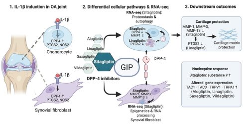
Introduction: Dipeptidyl peptidase-4 (DPP-4/CD26) has emerged as an important regulator of osteoarthritis (OA) and cellular senescence. Although increasing evidence suggests that DPP-4 inhibitors (DPP-4i) attenuate joint inflammation, post-marketing surveillance and FDA safety warnings have linked DPP-4i therapy to severe arthralgia. The biological mechanisms underlying these seemingly paradoxical effects remain poorly understood. This study investigated the effects of DPP-4 inhibition on inflammatory and pain-related responses in human OA chondrocytes and synovial fibroblasts and explored the underlying molecular mechanisms using transcriptomic analysis.
Methods: Primary human OA chondrocytes and synovial fibroblasts isolated from patients undergoing total knee arthroplasty were stimulated with IL-1β and treated with five clinically available DPP-4 inhibitors (sitagliptin, saxagliptin, vildagliptin, linagliptin, and alogliptin). Expression of inflammatory mediators, matrix-degrading enzymes, senescence markers, pain-related neuropeptides, and nociceptive ion channels was quantified by quantitative RT-qPCR. Substance P secretion was measured by ELISA. RNA sequencing was performed in sitagliptin-treated cells, followed by Gene Ontology pathway enrichment analysis to characterize the biological pathways regulated by DPP-4 inhibition.
Results: IL-1β markedly induced DPP4 expression together with inflammatory mediators (PTGS2, NOS2), matrix metalloproteinases, and pain-related genes in both chondrocytes and synovial fibroblasts. Individual DPP-4 inhibitors exerted differential effects on inflammatory and catabolic gene expression, with significant reductions in selected inflammatory and matrix-degrading markers observed for specific inhibitors. Individual DPP-4 inhibitors also differentially modulated pain-associated neuropeptides and nociceptive ion channels, indicating heterogeneous responses across the DPP-4 inhibitor class. Sitagliptin further increased substance P secretion in IL-1β-stimulated chondrocytes. Transcriptomic analysis demonstrated that IL-1β activated conserved inflammatory pathways in both cell types, whereas sitagliptin treatment elicited cell type-specific responses, predominantly regulating proteostasis- and autophagy-related pathways in chondrocytes and epigenetic and RNA-processing pathways in synovial fibroblasts.
Conclusion: These findings demonstrate heterogeneous effects among individual DPP-4 inhibitors on inflammatory, catabolic, and pain-associated molecular responses in osteoarthritic joint cells. In-depth analysis of sitagliptin further revealed cell type-specific transcriptomic responses and increased substance P secretion, highlighting the complex biological effects of DPP-4 inhibition beyond inflammatory suppression. These findings provide a potential molecular basis for DPP-4 inhibitor-associated arthralgia.
Keywords: osteoarthritis, dipeptidyl peptidase-4 inhibitors, chondrocytes, synovial fibroblasts, substance P, nociception
Introduction
Dipeptidyl peptidase-4 (DPP-4) inhibitors have emerged as an important class of oral antidiabetic agents due to their ability to enhance the action of incretin hormones, thereby promoting insulin secretion[1]. These agents offer advantages such as a lower risk of hypoglycemia and neutral effect on body weight, making them a preferred choice for many patients[2]. DPP-4, also known as CD26, is a transmembrane serine peptidase that is widely expressed in various tissues, including the liver, kidneys, intestines, immune cells, and cardiovascular system [3]. Besides glucose metabolism, DPP-4 is involved in immune modulation and regulation of inflammatory responses [4].
DPP-4 exists not only as a membrane-bound protein but also in a soluble form (sDPP-4) that circulates in blood and other body fluids [3]. Emerging evidence suggests that sDPP-4 contributes to the pathophysiology of various metabolic and inflammatory diseases, including type 2 diabetes mellitus (T2DM), cardiovascular disorders, and osteoarthritis (OA) [5-7]. Inflammation plays a crucial role in OA [8, 9]. Pro-inflammatory cytokines such as interleukin-1 beta (IL-1β) and tumor necrosis factor-alpha (TNF-α) have been implicated in cartilage degradation and pain sensitization in OA [10] [11]. Additionally, DPP-4 has been linked to the regulation of neuropeptides such as substance P, which plays a role in pain signaling and inflammatory responses[12]. Studies have shown that DPP-4 expression is elevated in OA cartilage, suggesting its involvement in cartilage degeneration and joint inflammation [7, 13]. Furthermore, soluble DPP-4 has been associated with increased levels of inflammatory markers in OA patients, indicating a potential role in disease progression [7]. Preclinical findings in rat models of OA have demonstrated that DPP-4 inhibitors can attenuate OA-like changes, suggesting a possible disease-modifying role [14].
Despite these promising findings, post-marketing surveillance and FDA safety warnings have raised concerns regarding potential adverse effects associated with DPP-4 inhibitors, including joint pain and arthritis[15-19]. Despite their widespread use, the mechanisms underlying these adverse effects remain unclear.
The interplay between DPP-4, diabetes, and OA raises important clinical questions regarding the long-term impact of DPP-4 inhibition on joint health. While DPP-4 inhibitors effectively regulate glucose levels, their influence on inflammatory pathways and cartilage homeostasis requires further investigation. Some studies suggest that DPP-4 inhibitors may reduce systemic inflammation [3], while others highlight their potential to alter local joint environments in ways that could exacerbate OA-related pathology.
This study aims to explore the dual role of DPP-4 inhibitors in osteoarthritic inflammation and pain. Currently, five DPP-4 inhibitors are approved for clinical use in Taiwan, including sitagliptin, saxagliptin, vildagliptin, linagliptin, and alogliptin. Using an in vitro OA chondrocyte model and a synovial cell model, we investigated the effects of DPP-4 inhibition on inflammatory markers, pain-related neuropeptides, and ion channels involved in joint pain signaling.
Materials and Methods
Human tissue collection
Human articular cartilage and synovial tissues were obtained from patients with osteoarthritis undergoing total knee replacement surgery. The study was approved by the Institutional Review Board of Tri-Service General Hospital (TSGHIRB C202405133), and written informed consent was obtained from all participants prior to tissue collection. A total of 16 independent surgical donors were included in this study. Cells from six donors were used for the IL-1β stimulation experiments, of which three were also used for the CCK-8 assay. Cells from an additional six donors were used for the DPP-4 inhibitor treatment experiments, including RT-qPCR and substance P ELISA analyses. Cells from four additional independent donors were used for RNA-seq analysis. Each donor was considered an independent biological replicate.
Primary chondrocyte isolation and culture
Non-lesioned articular cartilage from the tibial plateau and femoral condyle was finely diced and initially incubated in an antibiotic solution (100 µg/mL penicillin, 100 µg/mL streptomycin, 5 mg/mL Fungizone, 200 mM L-Glutamate) for 2 hours at room temperature. This was followed by digestion with 0.25% (w/v) trypsin (Thermo Fisher, 15050057) for 30 minutes, and subsequently with a mixture of 0.1% (w/v) type II collagenase (Sigma-Aldrich, C6885) with 0.1% type H collagenase (Sigma-Aldrich, C8015) in chondrocyte culture media for 16-18 hours at 37 °C [20]. Chondrocyte media contained DMEM/F-12 (Gibco, 11039) with 10% heat-inactivated fetal bovine serum (FBS, Gibco, 10437028), and 1× penicillin-streptomycin (Gibco, 15140122). Following digestion, isolated chondrocytes were filtered, centrifuged, and seeded into 75 cm² culture flasks (Corning, 430641U) and maintained in a humidified incubator at 37°C with 5% CO₂. The culture medium was replaced every 3-5 days. Cells were passaged at approximately 80% confluence using 0.05% trypsin-EDTA (Gibco, 25300054). To preserve their primary phenotype, all experiments were conducted using chondrocytes within passage 4.
Synovial fibroblast isolation and culture
Synovial fibroblasts were isolated as previously described by our group [21]. Synovial tissues were collected as surgical waste and processed immediately. Tissues were finely minced into ~1 mm³ fragments under sterile conditions and washed thoroughly with phosphate-buffered saline (PBS) containing 100 µg/mL penicillin and 100 µg/mL streptomycin to remove blood and debris. The minced tissue was then digested in 0.1% (w/v) type I collagenase (Sigma-Aldrich, C0130) dissolved in DMEM/F-12 supplemented with 10% FBS for 4-6 hours at 37°C in a shaking water bath.
After enzymatic digestion, the cell suspension was filtered through a 70 µm nylon mesh to remove undigested tissue fragments and centrifuged at 300 × g for 10 minutes. The resulting cell pellet was resuspended in DMEM supplemented with 10% FBS and 1× penicillin-streptomycin, and seeded into 75 cm² culture flasks. Cultures were maintained at 37 °C in a humidified incubator with 5% CO₂. The medium was changed every 2-3 days.
Non-adherent cells were removed during the first medium change. Synovial fibroblasts were expanded and passaged using 0.05% trypsin-EDTA upon reaching 80-90% confluence. Cells from passages 3 to 6 were used for experiments.
Inflammatory stimulation
Human primary chondrocytes and synovial fibroblasts were seeded in 6-cm culture dishes at a density of 3 × 10⁵ cells per dish. After reaching 80% confluence, cells were serum-starved for 12 hours and subsequently treated with the pro-inflammatory cytokine IL-1β at concentrations of 0.1, 1, or 10 ng/mL for 24 hours to mimic inflammatory conditions. Based on the results of these dose-response experiments, 1 ng/mL IL-1β was selected as the stimulation concentration for subsequent experiments evaluating the effects of DPP-4 inhibitors.
CCK-8 assay
Cell viability was assessed using the CCK-8 assay (Abbkine, KTA1020). Each biological sample was analyzed in duplicate as technical replicates. Chondrocytes and synovial fibroblasts were seeded in 96-well plates (5 × 10³ cells/well) and treated with increasing concentrations (0-10 µM) of sitagliptin (MedChemExpress, HY-13749), saxagliptin (MedChemExpress, HY-10285), vildagliptin (MedChemExpress, HY-14291), linagliptin (MedChemExpress, HY-10284), or alogliptin (MedChemExpress, HY-A0023) for 12, 24, and 48 hours. After treatment, 10 µL of CCK-8 reagent was added to each well and incubated for 2 hours at 37 °C. Absorbance was measured at 450 nm, and cell viability was expressed as a percentage relative to untreated controls. Results indicated minimal cytotoxicity at ≤ 1 µM, which was therefore selected as the working concentration for subsequent experiments (Supplementary Fig. S1-2).
DPP-4 inhibitor treatment
Human primary chondrocytes and synovial fibroblasts were seeded in 6-cm culture dishes at a density of 3 × 10⁵ cells per dish. After reaching 80% confluence, cells were serum-starved for 12 hours and subsequently stimulated with IL-1β (1 ng/mL) and concurrently treated with one of five DPP-4 inhibitors—sitagliptin, saxagliptin, vildagliptin, linagliptin, or alogliptin—at a final concentration of 1 μM for 24 hours. After treatment, cells were washed twice with cold PBS and harvested for downstream RT-qPCR analysis. For RNA-seq and substance P ELISA experiments, sitagliptin was selected as the representative DPP-4 inhibitor. Cells were treated with IL-1β (1 ng/mL) with or without sitagliptin (1 μM) for 24 hours. Following treatment, cells were harvested for RNA-seq analysis, while conditioned media were collected for measurement of substance P by ELISA.
Quantitative real-time polymerase chain reaction (RT-qPCR)
RNA was isolated from treated primary chondrocytes and synovial fibroblasts using TRIZOL Reagent (Invitrogen, 15596026). cDNA was synthesized using the SensiFAST cDNA Synthesis kits (BIOSCIENCE, BIO-65054) with 2 µg of RNA. Subsequently, RT-qPCR was performed using SyGreen BlueMix Lo-ROX (PCRBIO, PB20.15-01) with a QuantStudio™ 5 Real-Time PCR System (Applied Biosystems). Gene expression was expressed as ΔCt, calculated as Ct(target gene) - Ct(GAPDH). Senescent and inflammatory responses were evaluated by assessing DPP4, PTGS2, NOS2, MMP1, MMP3, and MMP13 expression. Pain-related neuropeptides (TAC1, TAC3) and nociceptive ion channels (TRPV1, TRPA1, SCN9A) were analyzed to determine the effects of inflammatory cytokines on pain-associated molecular pathways in OA chondrocytes and synovial fibroblasts. Primers used for RT-qPCR are listed in Table 1. RT-qPCR measurements for each biological sample were performed in duplicate as technical replicates, and the mean of the duplicate measurements was used for statistical analysis.
Primers used for RT-qPCR detection of target genes.
| MMP1 | F:ATGAAGCAGCCCAGATGTGGAG |
| R:TGGTCCACATCTGCTCTTGGCA | |
| MMP3 | F:CACTCACAGACCTGACTCGGTT |
| R:AAGCAGGATCACAGTTGGCTGG | |
| MMP13 | F:CCTTGATGCCATTACCAGTCTCC |
| R:AAACAGCTCCGCATCAACCTGC | |
| PTGS2 | F:CGGTGAAACTCTGGCTAGACAG |
| R:GCAAACCGTAGATGCTCAGGGA | |
| NOS2 | F:GCTCTACACCTCCAATGTGACC |
| R:CTGCCGAGATTTGAGCCTCATG | |
| DPP4 | F:AAAGGCACCTGGGAAGTCATCG |
| R:CAGCTCACAACTGAGGCATGTC | |
| TAC1 | F:TTACTGGTCCGACTGGTACGAC |
| R:CAAAGAACTGCTGAGGCTTGGG | |
| TAC3 | F:CAAAAGCCACTCATCTCTGGAGG |
| R:GCTCCTCTTGCCCATAAGTCCC | |
| TRPV1 | F:GTGGACAGCTACAGTGAGATGC |
| R:GGAAGCCACATACTCCTTGAGG | |
| TRPA1 | F:TCCTCTCCATCTGGCAGCAAAG |
| R:GGACCGCATGATGCAAAGCTGTC | |
| SCN9A | F:GTGGAAGGATTGTCAGTTCTGCG |
| R:GCCAACACTAAGGTGAGGTTACC | |
| CDKN2A | F:CTCGTGCTGATGCTACTGAGGA |
| R:GGTCGGCGCAGTTGGGCTCC | |
| GAPDH | F:GTCTCCTCTGACTTCAACAGCG |
| R:ACCACCCTGTTGCTGTAGCCAA |
Measurement of substance P by ELISA
Substance P levels in conditioned media were measured using a commercially available Human Substance P ELISA Kit (ELK1039; ELK Biotechnology) according to the manufacturer's instructions. Conditioned media were collected from primary human OA chondrocytes and synovial fibroblasts following 24 hours of treatment with IL-1β (1 ng/mL), with or without sitagliptin (1 μM). Substance P ELISA was performed using conditioned media. Each biological sample was assayed in duplicate as technical replicates, and the mean of the duplicate measurements was used for statistical analysis.
RNA-seq analysis
Total RNA was extracted from primary human osteoarthritic chondrocytes and synovial fibroblasts using TRIzol reagent (Invitrogen, Carlsbad, CA, USA) according to the manufacturer's instructions. RNA concentration and integrity were assessed prior to library preparation, and samples containing at least 300 ng of total RNA with an RNA integrity number (RIN) greater than 6.3 were subjected to RNA sequencing. The mRNA libraries were prepared using the Illumina Stranded mRNA Prep kit (Illumina, San Diego, CA, USA) and sequenced on the Illumina NovaSeq 6000 platform to generate 150-bp paired-end reads.
RNA sequencing was performed using four independent biological replicates per experimental condition for each cell type (n = 4 per group), with each biological replicate derived from an independent donor. Gene-level raw count data were analyzed using DESeq2 in R. Differential expression analysis was performed separately for each pairwise comparison within each cell type, and log2 fold changes were subjected to shrinkage using the lfcShrink() function with the adaptive shrinkage estimator (type = "ashr"). Principal component analysis was performed using variance-stabilizing transformed counts to assess global transcriptional variation among samples.
No predefined adjusted P-value or fold-change cutoff was applied before gene ranking. Instead, genes were ranked according to a π-value integrating effect size and statistical significance, calculated as π = |log2 fold change| × [-log10 (P value + 1 × 10⁻¹⁰)] [22]. The top 2,000 π-value-ranked genes from each comparison were subjected to Gene Ontology (GO) Biological Process enrichment analysis using clusterProfiler, with multiple-testing correction performed using the Benjamini-Hochberg method.
Statistical analysis
Data are presented as mean ± SEM. Analyses were performed using Prism 9 (GraphPad software). Repeated measures one-way ANOVA with Dunnett's post-hoc test were performed to compare IL-1β-stimulated conditions at different concentrations and to evaluate the effects of individual DPP-4 inhibitors under IL-1β stimulation. P < 0.05 was considered statistically significant.
Results
IL-1β induces DPP4 expression and inflammatory responses in osteoarthritic chondrocytes and synovial fibroblasts
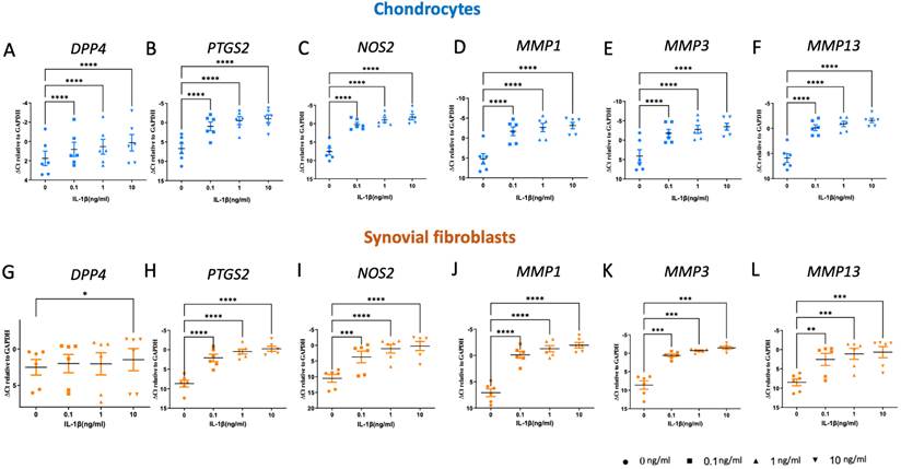
IL-1β stimulation with 0.1, 1, and 10 ng/mL led to a robust and dose-dependent induction of DPP4, reaching mean increases of 1.91-, 2.31-, and 3.07-fold, respectively in chondrocytes (p < 0.01; Fig. 1A). In contrast, in synovial fibroblasts, a significant increase in DPP4 expression was observed only at the highest IL-1β concentration (10 ng/mL), reaching a 2.07-fold increase compared with untreated controls (p < 0.05; Fig. 1G). In parallel, IL-1β stimulation with 0.1, 1, and 10 ng/mL markedly upregulated PTGS2 with a mean increase of 53.19-, 149.29-, and 209.24-fold, respectively, in chondrocytes and 94.68-, 291.02-, and 477.38-fold in synovial fibroblasts (Fig.1B, H). IL-1β stimulation also strongly induced NOS2, with significant increases observed in both chondrocytes and synovial fibroblasts (Fig. 1C and I). Furthermore, matrix-degrading enzymes including MMP1, MMP3, and MMP13 were significantly induced in both cell types (Fig. 1D-F, 1J-L).
IL-1β induces DPP4 expression and inflammatory responses in primary human osteoarthritic chondrocytes and synovial fibroblasts. (A-F) Primary human OA chondrocytes were stimulated with IL-1β (0.1, 1, or 10 ng/mL) for 24 hours. mRNA expression of DPP4, PTGS2, NOS2, MMP1, MMP3, and MMP13 was quantified by quantitative real-time PCR. (G-L) Primary human OA synovial fibroblasts were treated under identical conditions and analyzed for the same inflammatory and catabolic markers. Data are presented as mean ± SEM from six independent biological replicates (n=6) and normalized to GAPDH. Statistical analyses were performed using one-way ANOVA followed by Dunnett's multiple comparisons test. The Y-axis is reversed, with lower ΔCt values indicating higher gene expression. * P < 0.05, ** P < 0.01, *** P < 0.001, **** P < 0.0001.
IL-1β induces pain-related neuropeptides and nociceptive ion channels in osteoarthritic chondrocytes and synovial fibroblasts
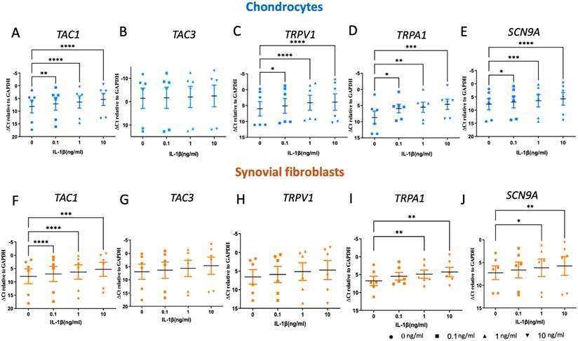
Stimulation with IL-1β (0.1, 1, and 10 ng/mL) significantly upregulated several pain-related genes in OA chondrocytes. TAC1, TRPV1, TRPA1, and SCN9A showed significant increases compared with untreated controls at one or more IL-1β concentrations, whereas TAC3 expression was not significantly altered (Fig. 2A-E). In synovial fibroblasts, IL-1β also significantly upregulated TAC1, TRPA1, and SCN9A, albeit with relatively lower fold-changes than in chondrocytes (Fig. 2F-J).
IL-1β promotes pain-associated neuropeptide and nociceptive ion channel expression in primary human osteoarthritic joint cells. (A-E) Expression of TAC1, TAC3, TRPV1, TRPA1, and SCN9A was determined in OA chondrocytes following IL-1β stimulation. (F-J) Expression of the same pain-related genes was examined in OA synovial fibroblasts after IL-1β treatment. Data are presented as mean ± SEM from six independent biological replicates (n = 6) and normalized to GAPDH. Statistical analyses were performed using one-way ANOVA with Dunnett's multiple comparisons test. The Y-axis is reversed, with lower ΔCt values indicating higher gene expression. * P < 0.05, ** P < 0.01, *** P < 0.001, **** P < 0.0001.
Differential effects of individual DPP-4 inhibitors on IL-1β-induced inflammatory and catabolic genes
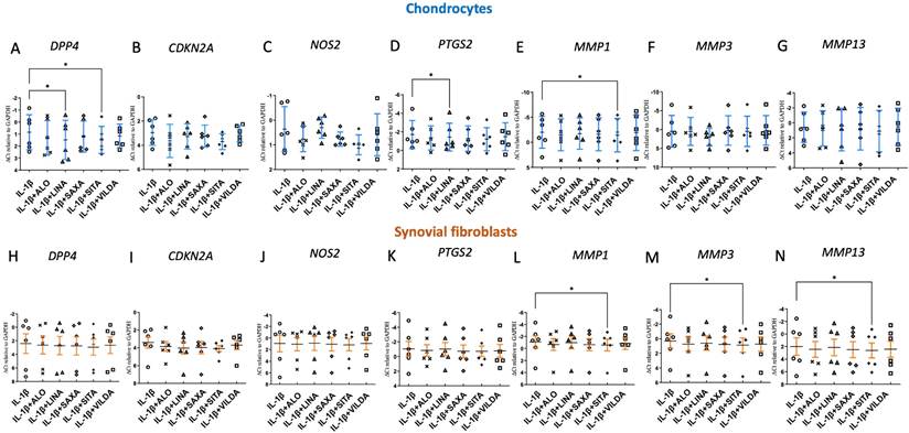
We next investigated the effects of individual DPP-4 inhibitors on IL-1β-induced inflammatory and catabolic responses in OA joint cells. In chondrocytes, sitagliptin significantly suppressed IL-1β-induced expression of both DPP4 and MMP1 (Figure 3A and E). In addition, linagliptin reduced DPP4 and PTGS2 expression (Figure 3A and D). Saxagliptin, vildagliptin, and alogliptin did not produce statistically significant changes in these inflammatory or catabolic targets under the tested conditions. No significant changes in CDKN2A expression were observed following DPP-4 inhibitor treatment in IL-1β-stimulated chondrocytes or synovial fibroblasts (Figure 3B and I). In synovial fibroblasts, sitagliptin consistently inhibited IL-1β-driven MMP1, MMP3 and MMP13 expression, whereas the other DPP-4 inhibitors did not reach statistical significance for these targets (Figure 3L-N). These findings demonstrate differential effects among individual DPP-4 inhibitors rather than a uniform class response.
DPP-4 inhibition modulates IL-1β-induced inflammatory and catabolic gene expression in primary human osteoarthritic joint cells. Primary OA chondrocytes (A-G) and synovial fibroblasts (H-N) were co-treated with IL-1β (1 ng/mL) and one of five clinically available DPP-4 inhibitors (sitagliptin, saxagliptin, vildagliptin, linagliptin, or alogliptin; 1 μM) for 24 hours. Expression of DPP4, CDKN2A, PTGS2, NOS2, MMP1, MMP3, and MMP13 was quantified by quantitative real-time PCR. Data are expressed as mean ± SEM from six independent biological replicates (n = 6) and normalized to GAPDH. Statistical significance was determined using one-way ANOVA with Dunnett's multiple comparisons test. The Y-axis is reversed, with lower ΔCt values indicating higher gene expression. * P < 0.05.
Differential effects of individual DPP-4 inhibitors on pain-related neuropeptides and nociceptive ion channels
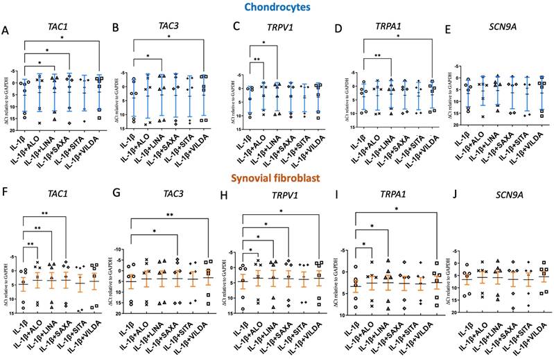
Compared with IL-1β stimulation alone, individual DPP-4 inhibitors produced distinct changes in pain-associated gene expression. In chondrocytes, alogliptin significantly increased TRPV1 expression; linagliptin significantly increased TAC1, TAC3, TRPV1, and TRPA1 expression; saxagliptin significantly increased TAC1 expression; sitagliptin did not significantly alter these targets; and vildagliptin significantly increased TAC1, TAC3, and TRPA1 expression (Figure 4A-D). In synovial fibroblasts, alogliptin significantly increased TAC1, TRPV1, and TRPA1 expression; linagliptin significantly increased TAC1, TRPV1, and TRPA1 expression; saxagliptin significantly increased TAC1, TAC3, and TRPV1 expression; sitagliptin did not significantly alter these targets; and vildagliptin significantly increased TAC3, TRPV1, and TRPA1 expression (Figure 4F-I). SCN9A expression was not significantly altered by any DPP-4 inhibitor in either cell type (Figure 4E and J). Collectively, these findings indicate that modulation of pain-associated neuropeptides and nociceptive ion channels varies among individual DPP-4 inhibitors rather than representing a uniform class effect.
DPP-4 inhibition modulates pain-associated neuropeptides and nociceptive ion channels in primary human osteoarthritic chondrocytes and synovial fibroblasts. (A-E) Expression of TAC1, TAC3, TRPV1, TRPA1, and SCN9A was quantified in OA chondrocytes following co-treatment with IL-1β and DPP-4 inhibitors. (F-J) The same pain-associated genes were analyzed in OA synovial fibroblasts. Data are presented as mean ± SEM from six independent biological replicates (n = 6) and normalized to GAPDH. Statistical analyses were performed using one-way ANOVA followed by Dunnett's multiple comparisons test. The Y-axis is reversed, with lower ΔCt values indicating higher gene expression. * P < 0.05, ** P < 0.01.
Sitagliptin enhances substance P levels and induces cell type-specific transcriptomic responses
Based on the previous results, sitagliptin was selected for further investigation because of its relatively prominent anti-inflammatory and anti-catabolic effects and limited effects on pain-associated gene expression. To further characterize the effects of sitagliptin on pain-associated molecular responses, substance P levels were measured in conditioned media from primary human OA chondrocytes and synovial fibroblasts. In chondrocytes, IL-1β stimulation significantly increased substance P levels compared with untreated controls, and co-treatment with sitagliptin further increased substance P levels compared with IL-1β stimulation alone (Figure 5A). In synovial fibroblasts, substance P levels were significantly higher following IL-1β and sitagliptin co-treatment than in untreated controls, whereas IL-1β stimulation alone did not result in a significant increase (Figure 5D).
Effects of sitagliptin on substance P levels and transcriptomic responses in primary human osteoarthritic chondrocytes and synovial fibroblasts. (A, D) Substance P levels in conditioned media from primary human OA chondrocytes (A) and synovial fibroblasts (D) under control conditions, IL-1β stimulation, or IL-1β stimulation with sitagliptin. Substance P concentrations were measured by ELISA. Each symbol represents an independent donor (n = 6). Data are presented as mean ± SEM. **P < 0.01 and ***P < 0.001. (B) Gene Ontology (GO) Biological Process enrichment analysis of genes associated with IL-1β stimulation in primary OA chondrocytes. (C) GO enrichment analysis of genes altered by sitagliptin treatment in IL-1β-stimulated OA chondrocytes. (E) GO enrichment analysis of genes associated with IL-1β stimulation in primary OA synovial fibroblasts. (F) GO enrichment analysis of genes altered by sitagliptin treatment in IL-1β-stimulated OA synovial fibroblasts. Genes were ranked according to π-values (|log₂ fold change| × -log₁₀ P value), and the top 2,000 ranked genes from each comparison were subjected to GO Biological Process enrichment analysis. Dot size represents the number of genes associated with each GO term, while dot color indicates the adjusted P value (n = 4 independent biological replicates per cell type).
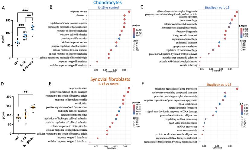
At the transcriptomic level, RNA sequencing was performed to characterize the molecular responses to sitagliptin in IL-1β-stimulated primary human OA chondrocytes and synovial fibroblasts. Gene Ontology enrichment analysis demonstrated that IL-1β induced a robust inflammatory transcriptional program in both cell types (Figure 5B and E). The significantly enriched biological processes included innate immune response, leukocyte cell-cell adhesion, chemotaxis, response to bacterial molecules, response to virus, and interferon-related signaling, indicating broad activation of inflammatory pathways following cytokine stimulation (Figure 5B and E). These transcriptional alterations were largely conserved between chondrocytes and synovial fibroblasts, suggesting that IL-1β drives a common inflammatory phenotype in osteoarthritic joint cells.
Interestingly, sitagliptin treatment resulted in distinct pathway alterations in the two cell populations. In chondrocytes, differentially expressed genes were primarily enriched in pathways associated with ribonucleoprotein complex biogenesis, ribosome biogenesis, proteasome-mediated protein catabolism, macroautophagy (Figure 5C), Golgi vesicle transport, endosomal transport, and translational regulation, suggesting that sitagliptin treatment predominantly modulates cellular proteostasis and stress adaptation rather than directly suppressing inflammatory signaling.
In contrast, synovial fibroblasts displayed enrichment of pathways related to chromatin organization, epigenetic regulation of gene expression, heterochromatin formation, RNA localization, miRNA-mediated gene silencing, and DNA damage checkpoint signaling following sitagliptin treatment (Figure 5F). These findings suggest that sitagliptin treatment induces transcriptional and epigenetic remodeling in synovial fibroblasts, highlighting a cell type-specific response to DPP-4 blockade.
Discussion
In the present study, we comprehensively characterized the biological effects of DPP-4 inhibition in primary human osteoarthritic chondrocytes and synovial fibroblasts under inflammatory conditions. Consistent with previous reports, IL-1β markedly induced DPP4 expression together with inflammatory mediators, matrix-degrading enzymes, and pain-associated molecules in both cell types. Individual DPP-4 inhibitors exerted differential effects on inflammatory and catabolic responses, with significant anti-inflammatory and anti-catabolic effects observed for selected inhibitors. Similarly, individual DPP-4 inhibitors differentially modulated the expression of pain-related neuropeptides and nociceptive ion channels, indicating that these responses were not uniform across the DPP-4 inhibitor class. These findings suggest a potential molecular link between DPP-4 inhibition and the arthralgia reported in patients receiving these agents. Furthermore, transcriptomic analysis of sitagliptin demonstrated that although IL-1β induced a common inflammatory transcriptional program in both chondrocytes and synovial fibroblasts, sitagliptin elicited distinct downstream biological responses in each cell type. Together, these findings suggest that DPP-4 inhibition exerts complex, inhibitor- and cell type-specific effects that extend beyond suppression of inflammatory signaling.
Our findings further support the growing evidence that DPP-4 contributes to osteoarthritis pathogenesis. Previous experimental studies have demonstrated increased DPP4 expression in osteoarthritic cartilage and suggested that pharmacological or genetic inhibition of DPP-4 attenuates cartilage degeneration and inflammatory responses. Consistent with these experimental findings, our previous nationwide population-based cohort study demonstrated that the use of DPP-4 inhibitors was associated with a significantly lower risk of developing osteoarthritis and a reduced incidence of total knee and hip replacement among patients with type 2 diabetes mellitus, supporting a potential disease-modifying role of DPP-4 inhibition in OA [23]. We found IL-1β markedly induced DPP4 expression in our primary chondrocytes and, to a lesser extent, in synovial fibroblasts, indicating that inflammatory cytokines directly regulate DPP4 expression within the osteoarthritic joint. Importantly, individual DPP-4 inhibitors differentially modulated IL-1β-induced inflammatory and catabolic responses, with significant reductions in selected inflammatory and matrix-degrading markers observed for specific inhibitors. These findings reinforce the concept that DPP-4 is not merely a metabolic enzyme but also an important regulator of local inflammatory responses in joint tissues.
One of the most intriguing findings of this study is that individual DPP-4 inhibitors differentially affected inflammatory, catabolic, and pain-associated molecular responses. Interestingly, despite the relatively limited effects of sitagliptin on pain-associated gene expression in the initial RT-qPCR screening, sitagliptin significantly increased substance P secretion in IL-1β-stimulated chondrocytes. This discrepancy suggests that transcriptional changes in pain-associated genes may not directly reflect neuropeptide secretion. This apparent paradox may provide biological insight into the association between DPP-4 inhibitors and arthralgia reported in post-marketing surveillance and FDA safety communications [15]. Although DPP-4 inhibitors effectively reduce inflammatory activation, inhibition of DPP-4 may also alter the metabolism of neuropeptides or activate compensatory pathways involved in nociceptive signaling [24]. Increased expression of nociceptive ion channels may sensitize peripheral joint tissues and contribute to pain perception independently of inflammation [25]. However, functional nociceptive responses, such as TRPV1/TRPA1-mediated calcium influx, were not directly assessed in the present study. Therefore, whether these molecular changes translate into enhanced nociceptive signaling remains to be determined.
Transcriptomic profiling further provided mechanistic insight into these dual biological effects. As expected, IL-1β activated conserved inflammatory pathways in both chondrocytes and synovial fibroblasts, including innate immune responses, leukocyte adhesion, chemotaxis, and responses to microbial stimuli. However, sitagliptin treatment did not simply reverse these inflammatory pathways. Instead, chondrocytes preferentially exhibited enrichment of pathways involved in ribosome biogenesis, ubiquitin-mediated protein degradation, macroautophagy, vesicular trafficking, and translational regulation, suggesting that sitagliptin treatment may promote cellular adaptation through regulation of proteostasis and intracellular stress responses. This specific survival response supports findings where targeting DPP-4 in senescent chondrocytes reverses catabolic stress and prevents joint cartilage degradation [14]. In contrast, synovial fibroblasts predominantly demonstrated enrichment of pathways associated with chromatin organization, epigenetic regulation, RNA processing, and DNA damage checkpoints, indicating that sitagliptin treatment was associated with transcriptional reprogramming rather than directly targeting inflammatory signaling. These observations emphasize that sitagliptin elicits distinct adaptive responses in different joint cell populations and may explain why inflammatory suppression and pain-related signaling coexist following DPP-4 inhibition. Indeed, while clinical cohorts suggest a long-term protective trend for joint structures, clinical evidence has also suggested an association between DPP-4 inhibitor use and an increased risk of arthralgia, emphasizing a complex molecular disconnect between molecular inflammation and nociceptive pathways [26].
This study has several strengths. We employed primary human osteoarthritic chondrocytes and synovial fibroblasts, evaluated all five clinically available DPP-4 inhibitors, integrated molecular and protein-level analyses with transcriptomic profiling, and investigated both inflammatory and nociceptive responses within the same experimental framework. Nevertheless, several limitations should be acknowledged. Chondrocytes were expanded up to P4, and although key chondrogenic markers remained detectable, some degree of passage-associated dedifferentiation cannot be excluded, particularly given the known phenotypic alterations of OA chondrocytes and inter-patient variability. Although the increased substance P levels provide additional evidence supporting modulation of pain-associated pathways, functional nociception was not directly assessed, which limits the interpretation of these molecular findings. Therefore, whether these molecular changes translate into enhanced nociceptive signaling remains to be determined. Moreover, RNA sequencing was primarily used for pathway enrichment and was not followed by functional validation of newly identified signaling pathways. In addition, the present study was performed using in vitro cytokine stimulation models that cannot fully reproduce the complexity of the osteoarthritic joint microenvironment, including immune cell interactions, mechanical loading, and systemic metabolic influences. Future studies using animal models and human clinical samples will be required to validate the transcriptomic findings and determine whether the differential regulation of proteostasis and epigenetic remodeling contributes directly to the beneficial and adverse effects of DPP-4 inhibition in vivo. Despite these limitations, our findings demonstrate that individual DPP-4 inhibitors exert heterogeneous effects in osteoarthritic joint cells, with differential modulation of inflammatory, catabolic, and pain-associated molecular responses. In-depth analysis of sitagliptin further revealed cell type-specific transcriptomic responses and increased substance P secretion in IL-1β-stimulated chondrocytes.
Supplementary Material
Supplementary figures.

Acknowledgements
The authors acknowledge the technical support provided by the Instrument Center of the National Defense Medical University. The graphical abstract was created using BioRender.com. ChatGPT (OpenAI) was used solely for language editing and improvement of manuscript clarity.
Funding
This study was supported in part by grants from the National Science and Technology Council, Taiwan (NSTC 113-2314-B-016-049-MY2 to YHC and NSTC 112-2314-B-016-029 to YJP), and Tri-Service General Hospital (TSGH_C04_115042 to YJP).
Availability of data and material
The data generated or analyzed during this study are included in this article and its supplementary materials. Additional data are available from the corresponding author upon reasonable request.
Ethics approval and consent to participate
All samples were obtained with IRB approval from Tri-service General Hospital TSGHIRB No C202405133.
Authors contributions
YHC conceived and designed the study, analyzed and interpreted the data, and drafted and revised the manuscript. ZLL performed the experiments. CCW provided human tissue specimens and contributed to the study resources. YJP conceived the original study concept, acquired funding, supervised the project, and contributed to data interpretation. All authors critically reviewed, revised, and approved the final version of the manuscript.
Competing Interests
The authors have declared that no competing interest exists.
References
1. Vella A. Mechanism of action of DPP-4 inhibitors-new insights. J Clin Endocrinol Metab. 2012;97:2626-8
2. Gomez-Peralta F, Abreu C, Gomez-Rodriguez S, Barranco RJ, Umpierrez GE. Safety and Efficacy of DPP4 Inhibitor and Basal Insulin in Type 2 Diabetes: An Updated Review and Challenging Clinical Scenarios. Diabetes Ther. 2018;9:1775-89
3. Shao S, Xu Q, Yu X, Pan R, Chen Y. Dipeptidyl peptidase 4 inhibitors and their potential immune modulatory functions. Pharmacol Ther. 2020;209:107503
4. Zhao Y, Yang L, Wang X, Zhou Z. The new insights from DPP-4 inhibitors: their potential immune modulatory function in autoimmune diabetes. Diabetes Metab Res Rev. 2014;30:646-53
5. Cho EH, Kim SW. Soluble Dipeptidyl Peptidase-4 Levels Are Associated with Decreased Renal Function in Patients with Type 2 Diabetes Mellitus. Diabetes Metab J. 2019;43:97-104
6. Chen SY, Kong XQ, Zhang KF, Luo S, Wang F, Zhang JJ. DPP4 as a Potential Candidate in Cardiovascular Disease. J Inflamm Res. 2022;15:5457-69
7. Chen YH, Zhang X, Chou CH, Hsueh MF, Attarian D, Li YJ. et al. Association of Dipeptidylpeptidase 4 (CD26) With Chondrocyte Senescence and Radiographic Progression in Knee Osteoarthritis. Arthritis Rheumatol. 2023;75:1120-31
8. Sokolove J, Lepus CM. Role of inflammation in the pathogenesis of osteoarthritis: latest findings and interpretations. Ther Adv Musculoskelet Dis. 2013;5:77-94
9. De Roover A, Escribano-Núñez A, Monteagudo S, Lories R. Fundamentals of osteoarthritis: Inflammatory mediators in osteoarthritis. Osteoarthritis and Cartilage. 2023;31:1303-11
10. Jenei-Lanzl Z, Meurer A, Zaucke F. Interleukin-1β signaling in osteoarthritis - chondrocytes in focus. Cellular Signalling. 2019;53:212-23
11. Chow YY, Chin K-Y. The Role of Inflammation in the Pathogenesis of Osteoarthritis. Mediators of Inflammation. 2020;2020:8293921
12. Shirakawa Y, Nakasa T, Kanemitsu M, Nekomoto A, Ishikawa M, Yimiti D. et al. Therapeutic effect of targeting Substance P on the progression of osteoarthritis. Mod Rheumatol. 2022;32:1175-85
13. Li X, Zhang Z, Jiang W, Ju Y, Guo W, Huang Z. Dipeptidyl Peptidase 4 (DPP4) Exacerbates Osteoarthritis Progression in an Enzyme-Independent Manner. Advanced Science. 2025;12:2410525
14. Ro DH, Cho GH, Kim JY, Min SK, Yang HR, Park HJ. et al. Selective targeting of dipeptidyl-peptidase 4 (DPP-4) positive senescent chondrocyte ameliorates osteoarthritis progression. Aging Cell. 2024;23:e14161
15. Rai P, Dwibedi N, Rowneki M, Helmer DA, Sambamoorthi U. Dipeptidyl Peptidase-4 Inhibitors and Joint Pain: A Retrospective Cohort Study of Older Veterans with Type 2 Diabetes Mellitus. Am Health Drug Benefits. 2019;12:223-31
16. Mascolo A, Rafaniello C, Sportiello L, Sessa M, Cimmaruta D, Rossi F. et al. Dipeptidyl Peptidase (DPP)-4 Inhibitor-Induced Arthritis/Arthralgia: A Review of Clinical Cases. Drug Saf. 2016;39:401-7
17. Yokota K, Igaki N. Sitagliptin (DPP-4 inhibitor)-induced rheumatoid arthritis in type 2 diabetes mellitus: a case report. Intern Med. 2012;51:2041-4
18. Chaicha-Brom T, Yasmeen T. DPP-IV inhibitor-associated arthralgias. Endocr Pract. 2013;19:377
19. Wang CY, Fu SH, Yang RS, Hsiao FY. Use of dipeptidyl peptidase-4 inhibitors and the risk of arthralgia: Population-based cohort and nested case-control studies. Pharmacoepidemiol Drug Saf. 2019;28:500-6
20. Chen YT, Wang CC, Cheng CP, Liu FC, Lee CH, Lee HS. et al. Interleukin-26 Has Synergistic Catabolic Effects with Palmitate in Human Articular Chondrocytes via the TLR4-ERK1/2-c-Jun Signaling Pathway. Cells. 2021 10
21. Hsieh T-H, Ho J-Y, Wang C-C, Liu F-C, Lee C-H, Lee H-S. et al. Fucoidan Modulates Osteoarthritis Progression Through miR-22/HO-1 Pathway. Cells. 2025 p. 1208
22. Xiao Y, Hsiao TH, Suresh U, Chen HI, Wu X, Wolf SE. et al. A novel significance score for gene selection and ranking. Bioinformatics. 2014;30:801-7
23. Chen YH, Byers Kraus V, Chang YS, Lin TM, Lee HY, Chang CK. et al. Dipeptidyl peptidase-4 inhibitors are associated with a lower risk of osteoarthritis in patients with type 2 diabetes. Ther Adv Musculoskelet Dis. 2026;18:1759720x261434279
24. Guieu R, Fenouillet E, Devaux C, Fajloun Z, Carrega L, Sabatier J-M. et al. CD26 modulates nociception in mice via its dipeptidyl-peptidase IV activity. Behavioural Brain Research. 2006;166:230-5
25. Matta C, Takács R, Ducza L, Ebeid RA, Choi H, Mobasheri A. Ion channels involved in inflammation and pain in osteoarthritis and related musculoskeletal disorders. American Journal of Physiology-Cell Physiology. 2023;325:C257-C71
26. Men P, He N, Song C, Zhai S. Dipeptidyl peptidase-4 inhibitors and risk of arthralgia: A systematic review and meta-analysis. Diabetes & Metabolism. 2017;43:493-500
Author contact
Corresponding author: Yi-Jen Peng, Address: No 325, Sec 2, Chenggong Rd, Neihu, Taipei, Taiwan; Telephone: +886-2-87927135; Fax: +886-2-87927136, Email: yijen0426com.