Impact Factor ISSN: 1449-1907




Int J Med Sci 2026; 23(12):3644-3659. doi:10.7150/ijms.129536 This issue Cite
Review
The Dual Role of Hypoxia-Inducible Factors in Oral and Maxillofacial Bone: From Pathological Destruction to Regenerative Potential
Diwen Shi1*, Wenxuan Huang1*, Yurong Kou2  , Lin Wu1
1. Department of Stomatology, Shengjing Hospital of China Medical University, Shenyang, China.
2. Department of Periodontology, School of Stomatology, China Medical University, Shenyang, China.
* These authors contributed equally to this work.
Received 2025-12-6; Accepted 2026-6-1; Published 2026-10-21
Abstract
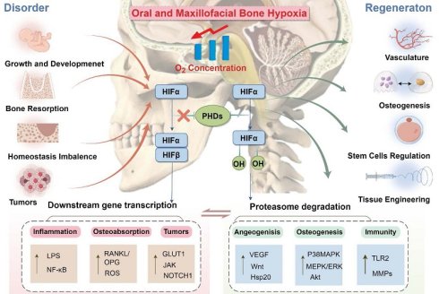
The maxillofacial skeleton forms the foundation of craniofacial structural integrity, regulating both function and morphological stability. The etiology of maxillofacial bone lesions (including trauma, tumors, and infections) exhibits multifactorial complexity, with hypoxia serving as a unifying pathophysiological pivot that drives disease progression and impedes healing. Hypoxia-inducible factors (HIFs), particularly the oxygen-sensitive HIF-1α, have emerged as the primary regulatory factors for cellular adaptation to hypoxic microenvironments, exerting dual roles in pathological responses and regenerative processes. This review summarizes the latest advances in HIF biology in maxillofacial bone diseases, elucidating their dual roles in bone resorption and tissue repair. We first introduce hypoxia-related drivers in maxillofacial pathophysiology, focusing on the regulatory role of HIF-mediated transcriptional networks in angiogenesis and osteoblast-osteoclast balance during disease and regeneration. Subsequently, we assess the role of HIF signaling in bone regeneration, including enhanced angiogenesis, coupling of osteoblasts and osteoclasts, and stabilization of HIF through biomaterial-driven mechanisms in tissue engineering. By integrating molecular-level insights with translational applications, this study provides a roadmap for developing HIF-targeted therapies, advocating precise interventions to achieve a balance between pathological suppression and regenerative activation, and offering a novel solution to the hypoxic environment challenges in maxillofacial bone.
Keywords: hypoxia-inducible factors (HIFs), maxillofacial bone, osteoblast differentiation, angiogenesis-osteogenesis coupling, tissue engineering
1. Introduction
The oral and maxillofacial bones serve as crucial anatomical structures for both functional and aesthetic aspects of the craniomaxillofacial region, with their health status holding significant implications for individuals [1]. These bones are particularly prone to hypoxia during disease progression and regeneration processes, profoundly impacting disease treatment and skeletal regeneration. This hypoxic microenvironment significantly influences cellular metabolism, immune responses, and tissue regeneration [2,3], presenting both challenges and opportunities for therapeutic interventions. However, inflammatory cell infiltration, bacterial proliferation or surgical trauma exacerbate pathological hypoxia, leading to the disruption of reduction-oxidation balance, damage to vascular repair, and the perpetuation of tissue damage. For instance, in periodontitis, microbial dysbiosis and neutrophil hyperactivity create a self-reinforcing cycle of hypoxia and inflammation that accelerates alveolar bone resorption [4]. Similarly, postoperative hypoxia following orthognathic surgery delays healing through hematoma formation and ischemic tension [5]. Emerging research focuses on deciphering hypoxia-driven molecular adaptations in bone cells, including metabolic reprogramming, epigenetic modifications, and paracrine signaling, to identify therapeutic targets for restoring oxygen homeostasis and enhancing regenerative outcomes.
Hypoxia-inducible factor pathway is central to hypoxic adaptation, particularly the heterodimeric transcription factor HIF-1[6,7]. Comprising an oxygen-stable α subunit (HIF-1α) and a constitutively expressed β subunit (HIF-1β), HIF-1 coordinates cellular responses to oxygen fluctuations [8]. In the oral and maxillofacial environment, HIF-1α exhibits dual regulatory roles: it promotes osteoblast differentiation and angiogenesis by mediating angiogenic-osteogenic coupling, while exacerbating bone loss via the promotion of osteoclastogenesis and inflammatory factor secretion. This dichotomy emphasizes its environment-dependent role as a molecular rheology regulator that balances tissue repair and pathological remodeling. What determines whether HIF-1α drives bone repair or destruction lies in context. Transient mild hypoxia (2-5% O₂) promotes angiogenesis-osteogenesis coupling, whereas severe (<0.5% O₂) or sustained hypoxia triggers osteoclastogenesis and inflammatory resorption[9,10]. Cell type also matters: in osteoblasts, HIF-1α stabilises VEGF to support bone formation[11]; in osteoclast precursors, it enhances receptor activator for nuclear factor-κB ligand (RANKL)-induced differentiation via glycolytic reprogramming[12]. Inflammatory cytokines such as TNF-α and IL-1β can stabilise HIF-1α even under normoxia, skewing its function toward resorption[13]. Metabolites like succinate, which accumulate during inflammation, inhibit prolyl hydroxylases and stabilise HIF-1α independently of oxygen[14]. HIF-1α thus acts as a molecular rheostat—integrating oxygen, inflammation and metabolism to coordinate adaptive or maladaptive outcomes. Harnessing this framework is key to precision therapeutics that promote regeneration while avoiding pathology.
Recent advances highlight HIF-1α as a master regulator of bone-immune crosstalk. HIF-1α enhances IL-1 production, promotes inflammatory responses, and activates NF-κB activity to induce bone resorption[13,15]. Conversely, transient HIF-1α induction in mesenchymal stem cells (MSCs) improves their survival, osteogenic differentiation, and angiogenic paracrine activity in ischemic bone defects [16]. However, persistent HIF-1α activation in malignancies like osteosarcoma promotes tumor angiogenesis and immunosuppression, necessitating precise spatiotemporal control[17].
This review critically examines the multifaceted roles of HIF-1α in oral and maxillofacial bone pathophysiology, emphasizing its mechanistic interactions with metabolic, vascular, and immune networks. We synthesize recent breakthroughs in HIF-mediated osteo-immune modulation, evaluate translational strategies leveraging hypoxic signaling, and propose innovative approaches integrating HIF modulation with biomaterial engineering for next-generation oral and maxillofacial therapies.
2. Global Research Trends of HIF-Driven Mechanisms in Oral and Maxillofacial Bone: Bibliometric Analysis
To systematically evaluate research trajectories and emerging trends in HIFs within oral and maxillofacial bone diseases and regeneration, we conducted a bibliometric analysis using the Web of Science database. This enables quantitative assessment of scholarly impact, identification of knowledge frontiers, and visualization of interdisciplinary linkages. Through keyword co-occurrence mapping and author affiliation profiling, our study delineates evolving research trends and collaborative networks in this field.
2.1. Keyword Co-Occurrence and Temporal Trends
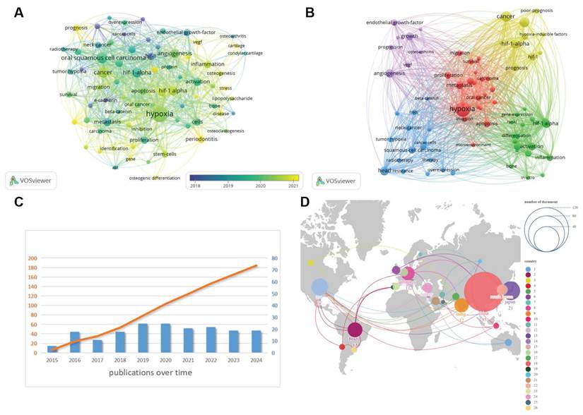
Bibliometric analysis is widely applied to evaluate research impact and identify emerging scientific trends. To characterize the development and research hotspots related to HIFs in maxillofacial bone disease and regeneration, publications indexed in the Web of Science (WoS) database between 2015 and 2024 were retrieved and analyzed (Fig. 1A).
Bibliometric analysis of the role of hypoxia-inducible factors in maxillofacial bone disease and regeneration. (A) Annual and cumulative trend of publication from 2015 to 2024; (B) keyword co-occurrence analysis map produced by VOSviewer and Pajek; (C) average publication year of each keyword produced by VOSviewer; (D) world map of global overview and international collaboration.
The search equation was constructed as follows: (“hypoxia-inducible factor” OR “HIF-1α” OR “HIF-1 alpha” OR “HIF-2α”) AND (“oral” OR “maxillofacial” OR “craniofacial” OR “periodontal” OR “alveolar bone” OR “jaw” OR “mandible” OR “bone regeneration” OR “osteogenesis”). The selected time frame (2015-2024) was intended to capture the most recent decade of research development and reflect contemporary trends in HIF-related maxillofacial bone research.
The search was limited to English-language publications indexed in SCI-Expanded and restricted to document types “Article” and “Review.” Conference abstracts, editorials, letters, and non-research materials were excluded. After exporting full bibliographic records and cited references, duplicate entries were removed using database filtering and manual verification. Titles and abstracts were subsequently screened to exclude studies unrelated to oral or craniofacial tissues, such as investigations focusing exclusively on systemic hypoxia without relevance to maxillofacial bone biology.
A total of 189 records met the inclusion criteria and were included in the final analysis. Keyword co-occurrence mapping, overlay visualization, and international collaboration network analysis were performed using VOSviewer and Pajek software to delineate thematic evolution and global collaboration patterns (Fig. 1B).
2.2 Global Collaboration and Research Output
Geospatial analysis of 189 high-impact publications (2015-2024) identifies China as the leading contributor (95 publications, 54.3% of total), followed by Japan (21), the United States (17), and Brazil (13) (Fig. 1C, D).
In addition to publication trends, clinically registered studies relevant to oral and maxillofacial tissues further illustrate translational engagement in this field. As summarized in Table 1, current investigations encompass HIF-related biomarker assessments in periodontal and peri-implant inflammation, as well as oxygenation-modulating adjunct therapies in jaw osteonecrosis and osteoradionecrosis.
Hypoxia-involivng therapies under clinical trials
| Therapies | Application | NCT number | Study title |
|---|---|---|---|
| Periodontal therapy with biomarker evaluation | Generalized Aggressive Periodontitis | NCT04020315 | GCF HIF-1α, VEGF and TNF-α Levels in G-AgP Patients Before and After Periodontal Treatment |
| Peri-implant biomarker analysis | Peri-implant Disease | NCT06863116 | Analysis of HIF-1α, malondialdehyde, and glutathione peroxidase 4 in Peri-Implant Crevicular Fluid |
| Hypoxia-profile guided radiotherapy (Nimorazole) | Head and Neck Squamous Cell Carcinoma | NCT02661152 | DAHANCA 30: Hypoxia-profile Guided Hypoxic Modification of Radiotherapy of head and neck squamous cell carcinoma (HNSCC) |
| Hypoxia imaging (MRI + PET validation) | Head and Neck Cancer | NCT06108089 | Novel Hypoxia Imaging for Head and Neck Cancer: Imaging Phenotype for Personalized Treatment |
| Pimonidazole-based hypoxia detection | Head and Neck Cancer | NCT02414048 | Hypoxia Analysis in Head and/or Neck Cancer |
By integrating keyword visualization with an analysis of authors' national affiliations, it becomes clear that a sizeable international research network has taken shape in this field. Early studies on HIFs and the hypoxic microenvironment primarily focused on tumor biology. However, recent investigations have increasingly targeted chronic inflammation, bone regeneration, and stem-cell differentiation in the oral and maxillofacial region, signifying a growing multidisciplinary convergence. From the perspective of global collaboration, Asian countries—particularly China and Japan—contribute a substantial number of publications in this field, and their partnerships with researchers in Europe and the Americas are expanding, suggesting that future research will likely feature more cross-regional, multi-center initiatives. Through synthesizing and visualizing these relevant studies, this work provides a clearer disciplinary framework and research roadmap for subsequent inquiries into the role of hypoxia-inducible factors in oral and maxillofacial bone disease and regeneration.
3. Causative factors contributing to oral and maxillofacial hypoxia
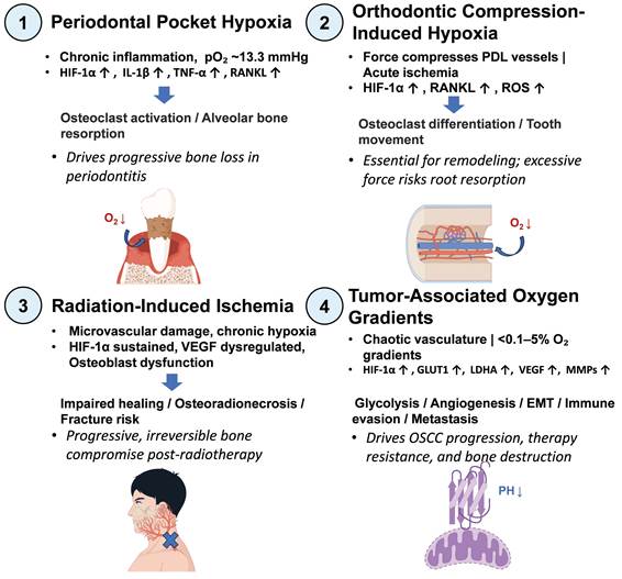
Hypoxia in oral and maxillofacial tissues can arise from systemic causes, but the most clinically relevant and tissue-specific drivers are those originating from local pathophysiological conditions. This review therefore focuses on four major categories of oral and maxillofacial hypoxia that directly impact bone metabolism and regeneration: periodontal pocket hypoxia, orthodontic compression-induced hypoxia, radiation-induced ischemia, and tumor-associated oxygen gradients (Fig. 2). Each of these microenvironments creates a distinct hypoxic signature that shapes cellular responses and disease outcomes.
Causative factors resulting in oral and maxillofacial hypoxia.
3.1 Periodontal pocket hypoxia
In periodontitis, the deepening of periodontal pockets restricts oxygen diffusion from the oral cavity, leading to a sharp drop in oxygen tension. Measurements have shown that untreated periodontal pockets can reach oxygen levels as low as 13.3 mmHg (1.8% O2) [18,19]. This chronic hypoxic niche fuels a self-reinforcing cycle of inflammation and tissue destruction: hypoxia stabilizes HIF-1α in periodontal ligament cells and immune cells, upregulating pro-inflammatory cytokines such as IL-1β, TNF-α, and osteoclastogenic factors like RANKL, while simultaneously impairing the antimicrobial functions of neutrophils[4,20]. The resulting environment promotes progressive alveolar bone resorption and impedes periodontal regeneration.
3.2 Orthodontic compression-induced hypoxia
Orthodontic tooth movement relies on the application of controlled mechanical forces that create areas of compression and tension within the periodontal ligament (PDL). On the compression side, blood vessels are occluded, causing acute local ischemia and a rapid decrease in oxygen availability[21,22]. This compression-induced hypoxia activates HIF-1α and its downstream target RANKL in PDL cells and osteocytes, triggering osteoclast differentiation and initiating bone resorption that allows tooth movement[23,24]. The duration and magnitude of force determine the severity of hypoxia; excessive or prolonged force can lead to pathological root resorption and delayed tissue repair [22].
3.3 Radiation-induced ischemia
Radiotherapy for head and neck cancer frequently causes irreversible damage to the microvasculature of the jawbones, leading to a state of chronic, progressive hypoxia known as osteoradionecrosis (ORN). Ionizing radiation induces endothelial cell apoptosis, capillary rarefaction, and fibrosis, which together reduce blood perfusion and oxygen delivery to the bone[25]. The resulting hypoxic environment is characterized by sustained HIF-1α stabilization, impaired osteoblast function, and a non-healing state that predisposes to infection and pathological fracture. ORN remains a challenging clinical complication with limited treatment options, and understanding the hypoxic pathophysiology is key to developing preventive strategies.
3.4 Tumor-associated oxygen gradients
Solid tumors of the oral cavity, particularly oral squamous cell carcinoma (OSCC), exhibit severe and heterogeneous hypoxia due to chaotic and dysfunctional tumor vasculature. Oxygen tensions within OSCC lesions can range from <0.1% to 5%, creating steep gradients that influence cancer cell behavior, metabolic adaptation, and therapeutic resistance[26,27]. Hypoxic areas stabilize HIF-1α, which orchestrates a transcriptional program that promotes glycolysis (glucose transporter 1 (GLUT1), LDHA), angiogenesis (VEGF), epithelial-mesenchymal transition, and immune evasion[28-30]. These adaptations enhance tumor aggressiveness, metastasis, and resistance to chemo- and radiotherapy. Moreover, the hypoxic microenvironment also affects surrounding stromal cells, including osteoclasts and osteoblasts, contributing to cancer-induced bone destruction [31].
4. Role of Hypoxia-Inducible Factors in Maxillofacial Bone Disorder
4.1 Epigenetic regulation of hypoxia in oral and maxillofacial bone formation
Hypoxia serves as a critical regulator of skeletal physiology, with embryonic osteogenesis predominantly occurring in hypoxic microenvironments that actively drive skeletal morphogenesis [32]. Under hypoxic conditions, the long non-coding RNA HOTAIRM1 is markedly upregulated, initiating a cascade through HIF-1α activation. This hypoxic signaling axis exerts dual epigenetic control by modulating histone-modifying enzymes: it enhances the expression of demethylases 6A/6B (KDM6A/B) while suppressing methyltransferase enhancer of zeste homolog 2 (EZH2) activity, collectively reducing global H3K27me3 levels. This epigenetic remodeling facilitates chromatin accessibility at key osteogenic loci (including alkaline phosphatase (ALP), macrophage colony-stimulating factor, Wnt-3a, Wnt-5a, Wnt-7a, and β-catenin), ultimately enhancing the osteogenic differentiation capacity of dental follicle stem cells. These mechanistic insights were further validated in a rat calvarial defect model, demonstrating hypoxia-driven bone regeneration [33].
Complementing these findings, pharmacological modulation using tetrazole-derived urea (TPPU), a soluble epoxide hydrolase inhibitor, was shown to potentiate osteogenesis through prostaglandin metabolism regulation. In vitro co-culture experiments revealed that TPPU treatment significantly enhanced both migratory capacity and tube-forming potential of human umbilical vein endothelial cells. Crucially, this pro-osteogenic effect was HIF-1α-dependent, as HUVEC-specific HIF-1α knockout abolished TPPU-mediated enhancement of osteogenic differentiation in co-cultured dental pulp stem cells. Molecular analyses confirmed coordinated downregulation of osteogenic markers (ALP, runt-related transcription factor-2 (RUNX2)) and angiogenic regulators (VEGF, vascular endothelial growth factor Receptor 2) following HIF-1α ablation, establishing endothelial HIF-1α as a central mediator in this conduction pathway [34].
4.2. Oral and maxillofacial Bone Injury and Inflammation
4.2.1. Hypoxic Regulation in Maxilla Bone
Hypoxic microenvironments constitute an essential component of maxillofacial bone repair, where fracture-associated oxygen tension reduction (pO₂ ≤0 mmHg at injury core) actively coordinates regenerative processes [35-40]. Mechanistically, bone healing is orchestrated through osteoblast-osteoclast crosstalk [41], with craniofacial defects predominantly undergoing intramembranous ossification—a direct MSC-to-osteoblast differentiation pathway bypassing chondrogenic intermediates [42-44]. Emerging evidence positions HIF-1α as a master regulator in this context. Study reported that mandibular osteotomy induced an increase in active osteoclasts and HIF-1α, which increased the expression level of dendritic cell-specific transmembrane protein (DC-STAMP) in active RAW264.7 cells to enhance osteoclastogenesis and cell fusion, causing the regional accelerated phenomenon to have an effect on alveolar bone remodeling[45]; research confirmed that HIF-1α also affects osteogenesis through cardiotrophin-1 signaling, playing a key role in the physiology and pathogenesis of bone resorption [46].
4.2.2. Hypoxic Regulation in Periodontal Bone Metabolism
Periodontal bone homeostasis is maintained through coupled osteoclastic resorption and osteoblastic formation, a dynamic equilibrium essential for mechanical integrity and mineral homeostasis [47,48]. Pathologically, alveolar bone loss—a hallmark of periodontitis and peri-implantitis—is intrinsically linked to hypoxic microenvironments, with oxygen tension in untreated periodontal pockets dropping to 13.3 mmHg [18,19]. As an adaptive response, hypoxia activates HIF-1α, which plays a dual regulatory role by regulating multiple proteins and signaling pathways involved in osteogenesis, bone resorption, and intraosseous angiogenesis, thus affecting the process of bone tissue repair in the diseased area. On the one hand, HIF-1α enhances the bone resorption activity of mature osteoclasts [49]. The HIF-1α/ angiopoietin like 4 (ANGPTL4) axis promotes facilitated the differentiation and fusion of RAW264.7 cells to osteoclasts [50]. On the other hand, HIF-1α also promotes vascular-osteogenic coupling: in canine extraction socket models, Toll like receptors 2 activation in bone marrow mesenchymal stem cells (BMSCs) under hypoxic conditions upregulates HIF-1α/ bone morphogenetic protein type 2 (BMP-2) signaling and promotes angiogenesis and osteogenesis [49].
The hypoxia-inflammation axis can also regulate periodontal regeneration through inflammatory crosstalk and mechanotransduction pathways: transforming growth factor beta 1 (TGF-β1) stabilizes HIF-1α in periodontal ligament stem cells (PDLSCs) under normoxic conditions, thereby reciprocally inhibiting TGF-β1/Smad3 signaling and forming a regulatory loop that inhibits osteogenesis [51]. Substance P, induced by cytokines and lipopolysaccharide (LPS), characterized as a pro-inflammatory neurotransmitter, plays a key role in inflammation [52], upregulates RANKL-induced expression of HIF-1α in Bone marrow-derived macrophages, and raises the RANKL/ osteoclastogenesis inhibitory factor (OPG) ratio, thereby exacerbating inflammatory bone loss [20]. Orthodontic force-induced hypoxia triggers a reactive oxygen species (ROS)/HIF-1α/RANKL cascade in compressed periodontal ligament (PDL) [22,23]. By quenching ROS and downregulating HIF-1α, superoxide dismutase 3 treatment can attenuate osteoclast differentiation [24].
Clinical studies have identified the rs11549467 single nucleotide polymorphism (SNP) in HIF-1α as a genetic determinant of residual alveolar ridge resorption, and enhanced HIF-1α transcriptional activity disrupts bone remodeling homeostasis through excessive angiogenesis [53]. Multigene interactions involving fibroblast growth factor receptor 1 oncogene partner / wt3.0 and VEGF SNPs further modulate racial differences in alveolar preservation [54].
4.2.3. Hypoxic Regulation in temporomandibular joint (TMJ) Disorders
In TMJ pathologies, spatial-temporal HIF-1α regulation governs chondro-osseous homeostasis [55]. Mandibular condylar resection models have shown that reduced HIF-1α expression in chondrocytes impairs trabecular neogenesis and downregulates angiogenic-osteogenic markers, thereby delaying condylar neck repair [56]. The up-regulation of the HIF-1α signaling pathway also promotes the formation of H-vessels [57]. Conversely, HIF-1α-overexpressing BMSCs synergized with collagen scaffolds to enhance cartilage regeneration and suppress subchondral sclerosis in rabbit TMJ osteochondral defects [58]. Notably, injury-induced HIF-1α accumulation in TMJ proliferative zones drives traumatic heterotopic ossification through angiogenesis-osteogenesis coupling [59]. Hypoxia-driven bone remodeling plays a pivotal role in temporomandibular joint osteoarthritis (TMJ-OA) pathogenesis. Mechanistic studies reveal that HIF-1α orchestrates subchondral angiogenesis through VEGF/Notch signaling cross-talk, providing a novel therapeutic target for the prevention and treatment of TMJ-OA [60]. HIF-2α emerges as a pivotal mediator in TMJ osteoarthritis under biomechanical stress. Mechanical stress activates NF-κB signaling, subsequently upregulating HIF-2α. This mechanohypoxic cascade transcriptionally activates cartilage-degrading enzymes, establishing a feedforward loop that exacerbates extracellular matrix degradation in mandibular condylar cartilage[47].
Beyond its role as a transcriptional activator of matrix-degrading enzymes, HIF-2α functions as a central hub integrating mechanical, inflammatory, and hormonal signals in TMJ pathology. Li et al. demonstrated that mechanical stress activates the NF-κB pathway, which directly upregulates HIF-2α expression, creating a mechano-hypoxic cascade that drives cartilage degradation[47]. This regulatory axis is particularly significant in TMJ osteoarthritis (TMJ-OA), where HIF-2α induces the expression of catabolic factors including MMP13, ADAMTS-4, and VEGF, leading to extracellular matrix breakdown and angiogenesis within the mandibular condylar cartilage.
HIF-2α also mediates estrogen's detrimental effects: Ye et al. found that high estrogen levels exacerbate condylar degeneration via the estrogen receptor beta (Erβ) /HIF-2α pathway, upregulating MMP-13, VEGF, and Col X, while estrogen deficiency attenuates these changes—explaining female predominance in TMJ disorders[61]. Additionally, early mechanical overloading elevates HIF-2α in chondrocytes, correlating with hypoxia and driven by TNF; this upregulation is blunted by intra-articular etanercept, highlighting crosstalk between inflammation and hypoxia[62]. These findings position HIF-2α as an early mediator and therapeutic target in TMJ pathology.
4.3. Hypoxic Regulation in Osteostatic Imbalance Disorders
Zhu et al. verified that HIF-1α up-regulated RANKL expression through activation of the janus kinase 2 (JAK2)/ Signal transducer and activator of transcription 3 (STAT3) signaling pathway and promoted the differentiation of RAW264.7 cells into osteoblasts in vitro. It provides a therapeutic strategy for the treatment of osteoporosis, bone fracture, osteoarthritis and orthodontic orthopedics [63].
Hypoxia and mouth-open breathing are closely associated with maxillofacial bone metabolism and are characteristic of obstructive sleep apnea-hypopnea syndrome (OSAHS). Chronic intermittent hypoxia in OSAHS induces metabolic reprogramming of osteoblasts. Bilateral nasal obstruction rat models exhibit reduced alveolar bone mineral density along with increased HIF-1α and VEGF expression. Dimethylcyclohexylglycine-mediated stabilization of HIF-1α reverses bone loss through a dual osteogenic-angiogenic mechanism [64].
The role of HIFs in bone homeostasis imbalance diseases is complex, involving the interaction and regulation of osteoblasts, osteoclasts, and chondrocytes. Regulating the expression and activity of HIFs may provide new ways and methods for preventing and treating these diseases.
4.4. HIFs in Oral and Maxillofacial Bone Tumors
Hypoxia-inducible factor-1α (HIF-1α) demonstrates persistent stabilization and accumulation within malignant cells under hypoxic tumor microenvironment conditions, with aberrant activation observed even in normoxic environments [26]. This constitutive activation arises through multiple interconnected mechanisms including genetic mutations, metabolic dysregulation, and crosstalk with other signaling pathways. Clinically, elevated HIF-1α expression correlates with unfavorable prognosis in oral carcinoma patients, serving as an independent predictor of poor clinical outcomes [27].
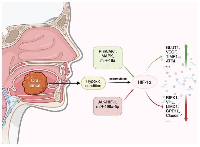
The oncogenic potency of HIF-1α overexpression manifests through enhanced hypoxic adaptation via transcriptional activation of downstream effectors. Notably, HIF-1α orchestrates metabolic reprogramming through upregulation of glucose transporter 1 (GLUT1), facilitating elevated glycolytic flux to sustain energy demands in rapidly proliferating HNSCC cells [28]. Concurrently, HIF-1α-driven vascular endothelial growth factor (VEGF) overexpression promotes pathological angiogenesis, establishing nutrient supply routes critical for tumor expansion and metastasis [29].
Mechanistically, HIF-1α stabilizes under hypoxic conditions through impaired von Hippel-Lindau (VHL)- LIM Domain Containing 1 complex function, a hallmark molecular defect observed in HNSCC that correlates with advanced pathological staging, therapeutic resistance, and reduced survival[65]. This stabilization enables HIF-1α-mediated transcriptional repression of glycerol-3-phosphate dehydrogenase 1-like protein, rewiring cellular oxygen utilization to favor tumor survival under metabolic stress [66].
The metastatic cascade in OSCC is profoundly influenced by HIF-1α through dual mechanisms: (1)induction of matrix metalloproteinases coupled with downregulation of intercellular junction proteins (e.g., Claudin-1), enhancing extracellular matrix degradation and epithelial-mesenchymal transition [67,68]; (2) paradoxical upregulation of tissue inhibitor of metalloproteinase-1 in periosteal cells, creating a reactive physical barrier that paradoxically facilitates localized tumor containment while promoting distant metastasis[31]. Spatial analysis reveals heightened HIF-1α expression at tumor-stroma interfaces, particularly in metastatic OSCC lesions, where it drives vasculogenic mimicry to supplement conventional angiogenesis in hypoxic peripheral regions [30].
HIF-1α confers chemoradioresistance through activation of Phosphoinositide 3-kinase-Akt (PI3K/AKT) and MAPK survival pathways while modulating immune cell recruitment to establish an immunosuppressive niche [27,30]. Emerging evidence identifies a reciprocal regulatory axis between HIF-1α and miR-199a-5p, where HIF-1α suppresses this tumor-suppressive miRNA through promoter targeting, thereby amplifying glycolytic metabolism and invasive potential in OSCC [69]. Conversely, adrenomedullin and RNA polymerase I and III subunit D gene silencing attenuates oncogenic phenotypes via JAK/HIF-1 signaling, highlighting cyclin D1 as a critical cell cycle mediator in HIF-driven proliferation[70].
The hypoxic paradigm extends to benign pathologies, with HIF-1α activation observed in odontogenic keratocysts (OKC). Activating transcription factor 4 (ATF4)-mediated HIF-1α upregulation correlates with M2 macrophage polarization and neovascularization, while NOTCH1 signaling cooperates with HIF-1α to sustain cystic epithelial proliferation [71,72]. Molecular profiling identifies ADAM Metallopeptidase Domain 1 as a hypoxia-responsive mediator of OKC invasiveness, suggesting conserved hypoxic adaptation mechanisms across neoplastic and developmental lesions [73-75].
Given the diversity and overlap of the mechanisms described above, the oncogenic actions of HIF-1α in oral and maxillofacial tumors can be viewed from several interconnected aspects. The tumor-promoting effects of HIF-1α involve several interconnected processes. These include metabolic reprogramming that allows tumor cells to adapt to oxygen deprivation and angiogenic activation that supports abnormal vascularization and nutrient supply. HIF-1α also promotes extracellular matrix remodeling and metastatic dissemination, facilitating tumor invasion and spread. In addition, it regulates immune and survival signaling pathways that contribute to an immunosuppressive microenvironment and therapy resistance. In general, these processes highlight the central role of HIF-1α in coordinating multiple pathways that collectively drive tumor progression (Fig. 3).
Cytokines associated with HIF-1α in oral and maxillofacial bone tumors.
5. Role of Hypoxia-Inducible Factor in Maxillofacial Bone Regeneration
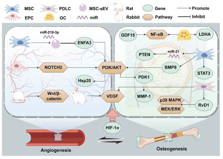
HIF-1α orchestrates bone regeneration through four interconnected functional axes: metabolic reprogramming, angiogenic signaling, osteoclast-osteoblast coupling, and immune modulation (Fig. 4). These axes provide a unified framework for understanding how hypoxia directs tissue repair versus pathology, and the subsequent sections examine each axis in detail within specific oral and maxillofacial contexts.
HIF-1α-centered signaling networks of the vascular-osteogenic coupled cytokines and pathways during hypoxia.
5.1 Metabolic Reprogramming
HIF-1α drives a fundamental shift from oxidative phosphorylation to aerobic glycolysis by upregulating glucose transporter 1 (GLUT1) and glycolytic enzymes such as lactate dehydrogenase A (LDHA) and pyruvate dehydrogenase kinase 1 (PDK1)[76,77]. This metabolic adaptation, known as the Warburg effect, maintains ATP production under low oxygen and provides biosynthetic precursors for proliferating cells. In osteoblasts, this glycolytic switch supports differentiation and matrix production; BMP9 enhances HIF-1α stability via PI3K-AKT, increasing PDK1 expression and glycolytic flux during osteogenesis[78]. In osteoclasts, hypoxia-induced glycolysis fuels the acid secretion required for bone resorption [79]. Notably, HIF-1α-mediated bone formation can occur independently of angiogenesis but requires intact metabolic reprogramming, underscoring the centrality of metabolic adaptation in hypoxic bone regeneration.
5.2 Angiogenic Signaling
HIF-1α is the master transcriptional regulator of angiogenesis, directly activating vascular endothelial growth factor (VEGF), angiopoietin-2, and placental growth factor to promote endothelial cell proliferation, migration, and tube formation [80,81]. In maxillofacial bone, this axis is critical for vascular-osteogenic coupling. Bone marrow-derived mesenchymal stem cells (MSCs) undergo HIF-1α-dependent transcriptional reprogramming under hypoxic conditions: while HIF-1α enhances MSC proliferation, migration, and pro-angiogenic paracrine activity, it concurrently suppresses osteogenic differentiation markers (BMP-2/Runx2), establishing a hypoxia-driven prioritization of vascular priming over matrix mineralization[16]. MSC-derived small extracellular vesicles (MSC-sEVs) under hypoxia are enriched with miR-210-3p via HIF-1α stabilization, activating the ephrin A3/PI3K/AKT pathway to promote vascularized bone regeneration[82]. Endothelial progenitor cells (EPCs) are mobilized from bone marrow through circulatory recruitment and execute targeted vascular reparative functions in hypoxic niches; HIF-1α activation in EPCs under hypoxia enhances vascularization via heat shock protein 20 upregulation through PI3K/Akt pathway activation [83], and EPC-derived NOTCH2 signaling is essential for angiogenesis-osteogenesis coordination during mandibular distraction osteogenesis [84]. In distraction osteogenesis, the hypoxic microenvironment drives HIF-1α-mediated mechanotransduction, with dual signaling cascades—HIF-1α/VEGF and HIF-1α/Wnt/β-catenin—synergistically promoting angiogenic-osteogenic coupling during regenerative callus maturation[85-87]. Remarkably, mechanical strain exacerbates hypoxia in regenerating tissues, and 10% uniaxial static strain enhances HIF-1α/VEGF expression in osteoblasts under hypoxia, suggesting mechanical stimulation may synergize with hypoxic signaling to optimize bone regeneration [88].
5.3 Osteoclast-Osteoblast Coupling
HIF-1α regulates both arms of bone remodeling, with context-dependent outcomes that determine whether net bone formation or resorption occurs. HIF-1α is indispensable for osteoclast activation: it enhances RANKL expression via JAK2/STAT3 and NF-κB signaling, upregulates osteoclast-specific enzymes including tartrate-resistant acid phosphatase and cathepsin K, and promotes osteoclast differentiation and function[12,63]. Hypoxia also increases DC-STAMP to promote osteoclast fusion, contributing to the regional acceleratory phenomenon following mandibular osteotomy [45], and induces growth differentiation factor 15 as a positive paracrine regulator of NF-κB-mediated osteoclast differentiation during bone remodeling [89]. In periodontitis, the HIF-1α/ANGPTL4 axis facilitates osteoclast differentiation and fusion [50], while orthodontic force-induced hypoxia triggers a ROS/HIF-1α/RANKL cascade in compressed periodontal ligament, activating osteoclastogenesis[22-24].
The conflicting evidence regarding hypoxia's effects on osteogenesis—sometimes promoting, sometimes inhibiting differentiation—can be reconciled by considering several contextual variables. First, oxygen concentration plays a critical role: mild hypoxia (2-5% O₂) generally promotes osteogenic differentiation of MSCs and PDLSCs by stabilizing HIF-1α at levels sufficient to activate VEGF and downstream Runx2 without triggering cellular stress responses[16,90,91]; in contrast, severe hypoxia (<0.5-1% O₂) suppresses osteogenesis by inducing excessive HIF-1α accumulation that activates Notch signaling, promotes apoptosis, and inhibits matrix mineralization [9,92]. Second, exposure duration fundamentally alters outcomes: transient hypoxia (24-72 hours) acts as a preconditioning stimulus that enhances MSC survival, proliferation, and subsequent osteogenic potential upon reoxygenation [10,16], whereas chronic hypoxia (>7 days) leads to sustained HIF-1α activation that downregulates Runx2 and osteocalcin expression, impairing terminal differentiation and bone formation [92,93]. Third, cell source and differentiation stage significantly influence hypoxic responses: undifferentiated MSCs and progenitor cells respond to hypoxia with enhanced proliferation and angiogenic paracrine activity, while committed osteoblasts and mature osteocytes may exhibit reduced matrix mineralization and increased RANKL expression under the same conditions [63,94]. Fourth, inflammatory context modulates HIF-1α function through cytokine crosstalk: in sterile inflammation or controlled repair settings, hypoxia promotes reparative osteogenesis via HIF-1α-mediated VEGF and BMP signaling [49,90]; however, in the presence of bacterial LPS or pro-inflammatory cytokines such as TNF-α and IL-1β, HIF-1α synergizes with NF-κB to drive osteoclast differentiation and bone resorption rather than formation [19,20]. Fifth, mechanical environment interacts with hypoxic signaling: mechanical loading during distraction osteogenesis enhances HIF-1α/VEGF expression and promotes osteogenesis under hypoxic conditions[88], while excessive orthodontic force can exacerbate hypoxia-induced root resorption [22]. Finally, experimental variables such as cell passage number, culture conditions, and oxygen measurement techniques contribute to inter-study variability and must be carefully controlled [95,96]. These contextual determinants, summarized in Table 2, explain the apparent contradictions in the literature and underscore the need for precise experimental design when evaluating HIF-targeted therapies for bone regeneration.
Contextual determinants of HIF-mediated osteogenic outcomes
| Determinant | Osteogenesis-promoting conditions | Osteogenesis-suppressing conditions | References |
|---|---|---|---|
| Oxygen concentration | Mild hypoxia (2-5% O₂) | Severe hypoxia (<0.5%O₂) | [9,16,90-92] |
| Exposure duration | Transient/intermittent (24-72 h) | Chronic/persistent (>7 days) | [10,16,92,93] |
| Cell type/stage | Undifferentiated MSCs, progenitor cells | Committed osteoblasts, mature osteocytes | [63,94] |
| Inflammatory context | Sterile inflammation, repair settings | Bacterial LPS, pro-inflammatory cytokines | [19,20,49,90] |
| Mechanical environment | Moderate mechanical loading | Excessive compression/abnormal load | [22,88] |
| Experimental conditions | 3D culture, low passage number | 2D culture, high passage number | [95,96] |
5.4 Immune Modulation
HIF-1α links hypoxia to inflammation by regulating innate and adaptive immune cells in the bone microenvironment. It promotes pro-inflammatory cytokine production (IL-1β, TNF-α) in macrophages and dendritic cells, enhances neutrophil survival under hypoxia, and influences T cell differentiation [13,15]. This immune-modulatory function is particularly evident in periodontitis, where Porphyromonas gingivalis-derived LPS synergizes with hypoxia to activate HIF-1/NF-κB in periodontal ligament cells, inducing MMP-1 and VEGF secretion and promoting immune cell infiltration and periodontal tissue destruction [19]. Substance P, induced by cytokines and LPS, upregulates HIF-1α and the RANKL/OPG ratio, exacerbating inflammatory bone loss [20]. HIF-1α also influences T helper 17 cells / regulatory T cells balance, contributing to inflammatory bone resorption [46]. In oral cancer, HIF-1α establishes an immunosuppressive niche by recruiting regulatory T cells and myeloid-derived suppressor cells and by upregulating immune checkpoint molecules, contributing to tumor immune evasion and progression [27,30]. Tumor-derived exosomal miR-21 from hypoxic environments activates cancer-associated fibroblasts, further promoting head and neck squamous cell carcinoma progression[97].
5.5 Mechanical Forces and Hypoxia Integration
Mechanical loading and trauma from fractures critically regulate bone homeostasis and regeneration through cellular differentiation and structural reorganization, and these mechanical signals converge with hypoxic signaling on HIF-1α[98]. In distraction osteogenesis, mechanical strain exacerbates local hypoxia in regenerating tissues, and 10% uniaxial static strain synergistically enhances HIF-1α/VEGF expression in osteoblasts under hypoxia, suggesting mechanical stimulation may optimize bone regeneration through HIF pathway activation[88]. Orthodontic force application compresses periodontal ligament vessels, inducing acute local hypoxia that activates HIF-1α/RANKL signaling and initiates bone resorption to facilitate tooth movement[22-24].The duration and magnitude of force determine the severity of hypoxia; excessive or prolonged force can lead to pathological root resorption and delayed tissue repair [22]. This mechano-hypoxic crosstalk highlights the therapeutic potential of modulating mechanical stimuli to optimize HIF-mediated regeneration in clinical settings.
5.6 Translational Applications: Harnessing HIF Pathways
With the increasing incidence of rejection and resorption associated with traditional bone grafting materials, tissue engineering strategies aiming to mimic or amplify HIF signaling have emerged as promising therapeutic approaches (Fig. 5). Hypoxia-mimetic agents (HMAs) that stabilize HIF-1α expression are increasingly utilized to enhance osteogenesis. Cobalt chloride (CoCl₂) activates the Akt/mTOR/4EBP-1 signaling pathway to upregulate HIF-1α, promoting osteogenic differentiation of human periodontal ligament stem cells and facilitating alveolar bone regeneration [99]. An injectable hydrogel-formulated inhibitor of prolyl-4-hydroxylase (PHD) has been shown to promote T regulatory cell recruitment and enhance alveolar bone regeneration during the resolution of experimental periodontitis, representing a promising localized delivery strategy for HIF stabilization (Fig. 5B) [100]. Semaphorin 3A (Sema3A) has been demonstrated to counteract hypoxia-impaired osteogenesis in periodontal ligament stem cells, suggesting synergistic utility with HIF-targeted therapies (Fig. 5C) [101].
HIFs in Tissue Engineering and Clinical Applications. (A) Hypoxia-mimetic agents (HMAs) CoCl₂ promotes osteogenesis in hPDLSCs [99]. (Reproduced with permission from Wiley). (B) Deferoxamine (DFO) promotes osteogenesis through HIF-1A stabilization [100]. (Reproduced with permission from PNAS). (C) Semaphorin 3A (Sema3A) counteracts hypoxia-impaired osteogenesis in PDLSCs[101]. (Reproduced with permission from Wiley). (D) Activation of HIF-1 attenuates periapical inflammation and bone loss[108]. (Reproduced with permission from IJOS). (E) Cobalt ions (Co²⁺), acting as HIF-1α stabilizers through PHD inhibition and VHL complex disruption [104]. (Reproduced with permission from Elsevier). (F) Interplay between non-coding RNAs and HIF signaling in maxillofacial bone biology[105]. (Reproduced with permission from Wiley). (G) MiR-21 upregulates HIF-1α expression and osteogenic differentiation[106]. (Reproduced with permission from Spandidos). (H) Autophagy regulates hypoxia-induced osteoclastogenesis through the HIF-1α/BNIP3 signaling pathway[109]. (Reproduced with permission from Wiley).
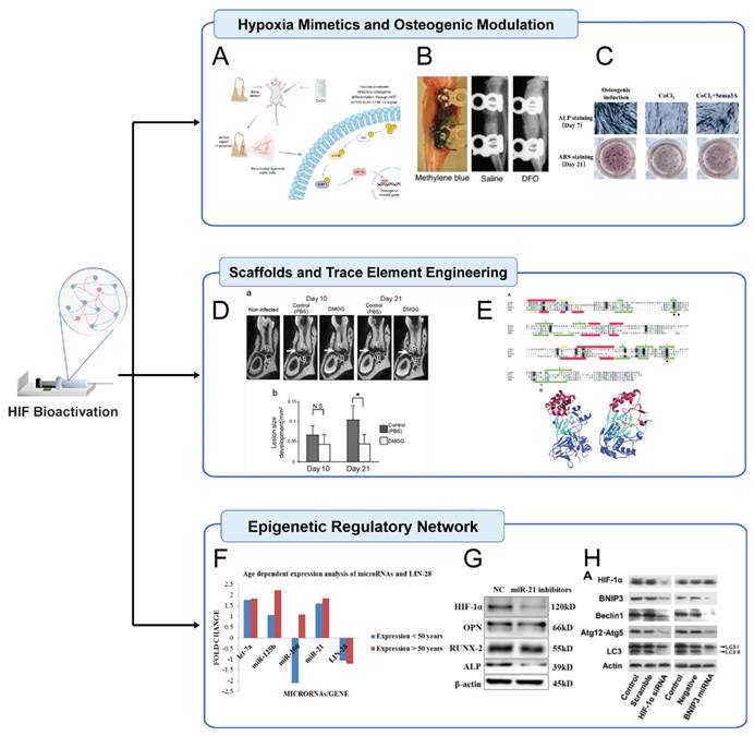
Beyond pharmacological agents, activation of HIF-1 signaling itself has been shown to attenuate periapical inflammation and bone loss, highlighting the therapeutic potential of endogenous HIF pathway activation in inflammatory bone diseases (Fig. 5D) [102]. The integration of trace elements into biomaterial scaffolds represents another frontier in bone tissue engineering. Titanium dioxide (TiO₂) coatings doped with copper not only confer antimicrobial properties but also upregulate HIF-1α/VEGF signaling and osteogenic markers in bone marrow-derived MSCs, achieving simultaneous antimicrobial, osteogenic, and angiogenic effects [103]. Cobalt ions incorporated into scaffolds act as HIF-1α stabilizers through prolyl hydroxylase domain inhibition and von Hippel-Lindau complex disruption, demonstrating dual benefits in promoting osteogenesis and angiogenesis (Fig. 5E) [104].
Emerging evidence also highlights the regulatory interplay between non-coding RNAs and HIF signaling. Dysregulated miRNAs, including let-7a, miR-125b, miR-100, and miR-21, are implicated in periodontal pathogenesis (Fig. 5F) [105]. Hypoxia-induced miR-21 upregulation in human periodontal ligament cells enhances HIF-1α expression and osteogenic differentiation, providing mechanistic insights into tissue remodeling during orthodontic treatment. In the context of oral cancer, microRNA-18a has been identified as a key regulator of metastatic properties in oral squamous cell carcinoma cells via modulation of HIF-1α expression (Fig. 5G) [106]. Furthermore, autophagy has been shown to regulate hypoxia-induced osteoclastogenesis through the HIF-1α/BNIP3 signaling pathway, providing mechanistic insights into bone remodeling under hypoxic conditions (Fig. 5H) [107]. However, the context-dependent roles of these pathways must be carefully considered, as tumor-derived exosomal miR-21 from hypoxic environments activates cancer-associated fibroblasts and promotes head and neck squamous cell carcinoma progression [97].
5.7 Differential Roles of HIF-α Isoforms in Oral and Maxillofacial Tissues
The HIF-α family includes HIF-1α, HIF-2α, and HIF-3α, each playing distinct roles in oral and maxillofacial biology. Understanding these isoform-specific functions is essential for precision therapeutics.
HIF-2α in cartilage and bone metabolism predominantly exerts catabolic effects. Unlike HIF-1α, which supports chondrocyte survival and matrix synthesis, HIF-2α drives cartilage degradation in osteoarthritis by transactivating genes for matrix-degrading enzymes, pro-inflammatory cytokines, and angiogenic factors[110]. Zhang et al. showed that HIF-2α promotes chondrocyte apoptosis and inflammation, while HIF-1α maintains matrix stability and viability[111]. The balance between these isoforms may thus determine tissue homeostasis versus degeneration.
HIF-3α is the least studied isoform and appears to function as a negative regulator of hypoxia signaling. It may be a target gene of HIF-1α and HIF-2α, forming a feedback loop that modulates their transcriptional activity. Under hypoxia, HIF-3α expression rises in a HIF-1α/2α-dependent manner and may compete for HIF-β binding or hypoxia response elements, attenuating target gene expression[111]. This mechanism could serve as an intrinsic brake on excessive angiogenesis, inflammation, or metabolic reprogramming. In OA, HIF-3α upregulation correlates with hypoxia duration, suggesting an adaptive role. However, its specific functions in oral and maxillofacial bone and cartilage remain largely unexplored, warranting further investigation.
5.8 Safety Considerations and Translational Challenges of HIF Modulation
Despite the therapeutic promise of HIF stabilization, several safety concerns must be addressed before clinical translation. Oncogenic risk is paramount: as detailed in Section 4.4, HIF-1α promotes metabolic reprogramming, angiogenesis, immune evasion, and metastasis in oral cancer[27,30]. Pharmacological HIF activators could theoretically accelerate occult tumor growth or create a permissive microenvironment for malignant transformation [112]. Uncontrolled angiogenesis driven by sustained HIF activation may lead to aberrant, leaky vessels rather than functional networks, potentially exacerbating inflammatory pathology rather than promoting repair [19,113]. Fibrotic responses have been associated with chronic HIF-1α activation through upregulation of pro-fibrotic factors including connective tissue growth factor and plasminogen activator inhibitor-1, raising concerns about pathological scarring and impaired tissue architecture [80]. Long-term safety of systemic HIF activators remains uncertain; clinical experience with PHD inhibitors for anemia management has revealed cardiovascular and thromboembolic risks warranting careful monitoring[112]. Tissue specificity presents a fundamental challenge: HIF-1α promotes bone formation in osteoblasts but drives resorption in osteoclast precursors, while HIF-2α exerts catabolic effects on cartilage[12,47]. These dichotomies demand precision targeting strategies.
Addressing these challenges requires spatiotemporally controlled delivery systems to restrict HIF modulation to target tissues and limit exposure duration[103,104]. Rigorous preclinical safety evaluation in large-animal models, patient stratification based on cancer risk, and biomarker development for early detection of adverse effects are essential prerequisites for clinical translation.
6. Conclusions and Perspective
Hypoxia-inducible factors (HIFs), particularly HIF-1α, have emerged as central regulators of both pathological progression and regenerative potential in oral and maxillofacial bone biology. This review has synthesized the dualistic roles of HIFs in orchestrating cellular responses to hypoxia, balancing destructive processes such as osteolysis and tumorigenesis with reparative mechanisms including angiogenesis-osteogenesis coupling. Mechanistically, HIF-1α drives adaptive transcriptional programs that modulate metabolic reprogramming, immune crosstalk, and vascular remodeling, with its activity finely tuned by oxygen tension, epigenetic modifications, and mechanical stimuli. In bone regeneration, HIF-1α enhances mesenchymal stem cell survival, osteogenic differentiation, and angiogenic paracrine signaling, whereas its dysregulation in pathologies such as periodontitis, osteoarthritis, and oral squamous cell carcinoma exacerbates inflammation, bone resorption, and metastatic potential. Despite significant advances, critical gaps remain that must be addressed to translate these mechanistic insights into clinical therapies.
Three key unresolved questions warrant urgent investigation. The first concerns the spatiotemporal determinants that govern whether HIF-1α activation leads to regeneration or pathology. Although contextual factors such as hypoxia severity, duration, and inflammatory milieu have been identified, the precise molecular thresholds that mark the transition from adaptive to maladaptive HIF signaling remain undefined. Second, the functional interplay between HIF-1α and HIF-2α in oral and maxillofacial tissues requires elucidation. Their opposing effects on cartilage—HIF-1α promoting chondrocyte survival and HIF-2α driving degradation—suggest that the balance between these isoforms may determine tissue fate, yet their co-regulation and potential crosstalk in bone are poorly characterized. Third, the feasibility of achieving cell-type specificity with HIF-targeted therapies without off-target effects remains an open challenge. The dual roles of HIF-1α in osteoblasts versus osteoclast precursors, and in normal versus malignant cells, demand precision targeting strategies that engage desired cell populations while sparing others.
Two translational priorities emerge from this analysis. The first is the development of localized, controlled delivery systems for HIF modulators to minimize systemic exposure and mitigate oncogenic risks. Biomaterial-based approaches—including hypoxia-responsive scaffolds, nanoparticle formulations, and injectable hydrogels—offer promising platforms for achieving spatiotemporal control over HIF activation [103]. The second priority is rigorous preclinical safety evaluation prior to clinical trials, with particular attention to tumor-promoting potential, vascular abnormalities, and fibrotic responses in relevant large-animal models.
Several specific experimental directions are needed to address these priorities. Future studies should establish dose-response curves for HIF stabilizers across different oxygen tensions and cell types using advanced 3D culture systems and microphysiological models [95,96]. Genetic tools such as cell-type-specific HIF knockout and knockin mice will be essential for dissecting isoform-specific functions in craniofacial development and disease. Biomaterial formulations for controlled HIF modulator release require systematic screening and optimization in preclinical bone defect models. The interplay between mechanical forces and HIF signaling in distraction osteogenesis and orthodontic tooth movement should be investigated using bioreactor systems that apply defined mechanical loads under controlled oxygen conditions[88]. Long-term safety studies in immunocompetent animal models are necessary to assess oncogenic risk and off-target effects. Finally, predictive biomarkers of HIF pathway activation and therapeutic response must be identified to enable patient stratification in future clinical trials.
HIFs occupy a pivotal position at the intersection of hypoxia, inflammation, and regeneration in oral and maxillofacial bone. Bridging mechanistic insights with bioengineering innovations will unlock their full therapeutic potential, transforming the management of craniofacial bone diseases from palliative care to functional restoration. The coming decade should witness the translation of HIF-targeted strategies from bench to bedside, guided by rigorous science and a commitment to precision medicine.
Acknowledgements
The graphics were created in BioRender. Huang, W. (2024) (https://biorender.com/j39r177) and Figdraw (http://www.figdraw.com) (UROPY79999, ASPAS020f4).
Diwen Shi: Conceptualization, Writing-review and editing, Visualization; Wenxuan Huang: Conceptualization, Writing-review and editing, Software; Yurong Kou: Supervision, Data curation; Lin Wu: conceptualization, Project administration, Funding acquisition.
Funding
This work was supported by National Key Research and Development Program of China, grant number 2022YFB3804304 and National Natural Science Foundation of China, grant number U24A20713.
AI usage statement
During the preparation of this manuscript, the authors used AI-assisted tools (including large language models) solely for the purpose of language polishing and improving the readability of the text. These tools were not used for data analysis, interpretation of results, generation of scientific content, or drawing of conclusions. All scientific content, data interpretation, and conclusions presented in this manuscript were generated, verified, and approved by the authors. The authors take full responsibility for the integrity and accuracy of the entire work.
No AI tools were used in the creation of the figures.
Competing Interests
The authors have declared that no competing interest exists.
References
1. Lee YK, Moon HJ. Reciprocal influence of masticatory apparatus, craniofacial structure and whole body homeostasis. Med Hypotheses. 2012;79:761-6
2. Mujcic H, Nagelkerke A, Rouschop KM. et al. Hypoxic activation of the PERK/eIF2α arm of the unfolded protein response promotes metastasis through induction of LAMP3. Clin Cancer Res. 2013;19:6126-37
3. Harris AL. Hypoxia-a key regulatory factor in tumour growth. Nat Rev Cancer. 2002;2:38-47
4. Perić Kačarević Ž, Rider P, Alkildani S. et al. An introduction to bone tissue engineering. Int J Artif Organs. 2020;43:69-86
5. Tian Y, Shao Q, Tang Y. et al. HIF-1α regulates osteoclast activation and mediates osteogenesis during mandibular bone repair via CT-1. Oral Dis. 2022;28:428-441
6. Semenza GL. HIF-1: mediator of physiological and pathophysiological responses to hypoxia. J Appl Physiol (1985). 2000;88:1474-80
7. Wang GL, Jiang BH, Rue EA. et al. Hypoxia-inducible factor 1 is a basic-helix-loop-helix-PAS heterodimer regulated by cellular O2 tension. Proc Natl Acad Sci U S A. 1995;92:5510-4
8. Semenza GL. HIF-1 and mechanisms of hypoxia sensing. Curr Opin Cell Biol. 2001;13:167-71
9. Wang P, Zhang XP, Liu F. et al. Progressive Deactivation of Hydroxylases Controls Hypoxia-Inducible Factor-1α-Coordinated Cellular Adaptation to Graded Hypoxia. Research (Wash D C). 2025;8:0651
10. Stegen S, Deprez S, Eelen G. et al. Adequate hypoxia inducible factor 1α signaling is indispensable for bone regeneration. Bone. 2016;87:176-86
11. Schipani E, Maes C, Carmeliet G. et al. Regulation of osteogenesis-angiogenesis coupling by HIFs and VEGF. J Bone Miner Res. 2009;24:1347-53
12. Miyauchi Y, Sato Y, Kobayashi T. et al. HIF1α is required for osteoclast activation by estrogen deficiency in postmenopausal osteoporosis. Proc Natl Acad Sci U S A. 2013;110:16568-73
13. Tannahill GM, Curtis AM, Adamik J. et al. Succinate is an inflammatory signal that induces IL-1β through HIF-1α. Nature. 2013;496:238-42
14. Koivunen P, Hirsilä M, Remes AM. et al. Inhibition of hypoxia-inducible factor (HIF) hydroxylases by citric acid cycle intermediates: possible links between cell metabolism and stabilization of HIF. J Biol Chem. 2007;282:4524-4532
15. Thompson AA, Binham J, Plant T. et al. Hypoxia, the HIF pathway and neutrophilic inflammatory responses. Biol Chem. 2013;394:471-7
16. Lampert FM, Kütscher C, Stark GB. et al. Overexpression of Hif-1α in Mesenchymal Stem Cells Affects Cell-Autonomous Angiogenic and Osteogenic Parameters. J Cell Biochem. 2016;117:760-8
17. He G, Nie JJ, Liu X. et al. Zinc oxide nanoparticles inhibit osteosarcoma metastasis by downregulating β-catenin via HIF-1α/BNIP3/LC3B-mediated mitophagy pathway. Bioact Mater. 2023;19:690-702
18. Cheng R, Liu W, Zhang R. et al. Porphyromonas gingivalis-Derived Lipopolysaccharide Combines Hypoxia to Induce Caspase-1 Activation in Periodontitis. Front Cell Infect Microbiol. 2017;7:474
19. Gölz L, Memmert S, Rath-Deschner B. et al. Hypoxia and P. gingivalis synergistically induce HIF-1 and NF-κB activation in PDL cells and periodontal diseases. Mediators Inflamm. 2015;2015:438085
20. Yan K, Lin Q, Tang K. et al. Substance P participates in periodontitis by upregulating HIF-1α and RANKL/OPG ratio. BMC Oral Health. 2020;20:27
21. Niklas A, Proff P, Gosau M. et al. The role of hypoxia in orthodontic tooth movement. Int J Dent. 2013;2013:841840
22. Krishnan V, Davidovitch Z. Cellular, molecular, and tissue-level reactions to orthodontic force. Am J Orthod Dentofacial Orthop. 2006;129:469.e1-32
23. Dandajena TC, Ihnat MA, Disch B. et al. Hypoxia triggers a HIF-mediated differentiation of peripheral blood mononuclear cells into osteoclasts. Orthod Craniofac Res. 2012;15:1-9
24. Jindarojanakul P, Kobayashi Y, Kamimoto H. et al. Changes in superoxide dismutase 3 (SOD3) expression in periodontal tissue during orthodontic tooth movement of rat molars and the effect of SOD3 on in vitro hypoxia-exposed rat periodontal ligament cells. Eur J Orthod. 2023;45:430-437
25. Lyons A, Ghazali N. Osteoradionecrosis of the jaws: current understanding of its pathophysiology and treatment. Br J Oral Maxillofac Surg. 2008;46:653-60
26. Kosim MY, Fukazawa T, Miyauchi M. et al. p53 status modifies cytotoxic activity of lactoferrin under hypoxic conditions. Front Pharmacol. 2022;13:988335
27. Singh P, Rajput M, Pandey M. Tumor hypoxia and role of hypoxia-inducible factor in oral cancer. World J Surg Oncol. 2024;22:18
28. Swartz JE, Wegner I, Noorlag R. et al. HIF-1a expression and differential effects on survival in patients with oral cavity, larynx, and oropharynx squamous cell carcinomas. Head Neck. 2021;43:745-756
29. Hwang SJ, Jung Y, Song YS. et al. Enhanced anti-angiogenic activity of novel melatonin-like agents. J Pineal Res. 2021;71:e12739
30. Murugesan A, Bharath C, Balakrishnan S. et al. Expression of HIF-1α and Nestin in oral squamous cell carcinoma and its association with vasculogenic mimicry. J Cancer Res Ther. 2024;20:176-180
31. Nakamura K, Tsukasaki M, Tsunematsu T. et al. The periosteum provides a stromal defence against cancer invasion into the bone. Nature. 2024;634:474-481
32. Endo Y, Zhu C, Giunta E. et al. The Role of Hypoxia and Hypoxia Signaling in Skeletal Muscle Physiology. Adv Biol (Weinh). 2024;8:e2200300
33. Leppäniemi A. Trauma surgery. Scand J Surg. 2002;91:5-6
34. Chen Z, Gan L, Chen X. et al. LncRNA HOTAIRM1 promotes dental follicle stem cell-mediated bone regeneration by regulating HIF-1α/KDM6/EZH2/H3K27me3 axis. J Cell Physiol. 2023;238:1542-1557
35. Kong L, Li J, Bai Y. et al. Inhibition of soluble epoxide hydrolase enhances the dentin-pulp complex regeneration mediated by crosstalk between vascular endothelial cells and dental pulp stem cells. J Transl Med. 2024;22:61
36. Yellowley CE, Genetos DC. Hypoxia Signaling in the Skeleton: Implications for Bone Health. Curr Osteoporos Rep. 2019;17:26-35
37. Dehne N, Brüne B. HIF-1 in the inflammatory microenvironment. Exp Cell Res. 2009;315:1791-7
38. Loi F, Córdova LA, Pajarinen J. et al. Inflammation, fracture and bone repair. Bone. 2016;86:119-30
39. Maes C, Carmeliet G, Schipani E. Hypoxia-driven pathways in bone development, regeneration and disease. Nat Rev Rheumatol. 2012;8:358-66
40. Sadiku P, Walmsley SR. Hypoxia and the regulation of myeloid cell metabolic imprinting: consequences for the inflammatory response. EMBO Rep. 2019;20:e47388
41. Miclau KR, Brazina SA, Bahney CS. et al. Stimulating Fracture Healing in Ischemic Environments: Does Oxygen Direct Stem Cell Fate during Fracture Healing? Front Cell Dev Biol. 2017;5:45
42. Epari DR, Lienau J, Schell H. et al. Pressure, oxygen tension and temperature in the periosteal callus during bone healing-an in vivo study in sheep. Bone. 2008;43:734-9
43. Fang J, Hall BK. Chondrogenic cell differentiation from membrane bone periostea. Anat Embryol (Berl). 1997;196:349-62
44. Marsell R, Einhorn TA. The biology of fracture healing. Injury. 2011;42:551-5
45. Tang Y, Li X, Cai Y. et al. Hypoxia-induced factor 1α promotes osteotomy-induced regional acceleratory phenomenon via DC-STAMP mediated membrane fusion. Oral Dis. 2023;29:2139-2153
46. Lin J, Huang J, Zhang Z. et al. Periodontal ligament cells under mechanical force regulate local immune homeostasis by modulating Th17/Treg cell differentiation. Clin Oral Investig. 2022;26:3747-3764
47. Li W, Wu N, Wang J. et al. Role of HIF-2α/NF-κB pathway in mechanical stress-induced temporomandibular joint osteoarthritis. Oral Dis. 2022;28:2239-2247
48. Eltzschig HK, Carmeliet P. Hypoxia and inflammation. N Engl J Med. 2011;364:656-65
49. Zhou Q, Gu X, Dong J. et al. The use of TLR2 modified BMSCs for enhanced bone regeneration in the inflammatory micro-environment. Artif Cells Nanomed Biotechnol. 2019;47:3329-3337
50. Qi X, Bie M, Jiang R. et al. HIF-1α regulates osteoclastogenesis and alveolar bone resorption in periodontitis via ANGPTL4. Arch Oral Biol. 2023;153:105736
51. Liu Z, Guo L, Li R. et al. Transforming growth factor-β1 and hypoxia inducible factor-1α synergistically inhibit the osteogenesis of periodontal ligament stem cells. Int Immunopharmacol. 2019;75:105834
52. Györfi A, Fazekas A, Rosivall L. Neurogenic inflammation and the oral mucosa. J Clin Periodontol. 1992;19:731-6
53. Paek J, Oh Y, Kim J. et al. Single nucleotide polymorphisms in HIF-1α gene and residual ridge resorption (RRR) of mandible in Korean population. Gene Expr. 2015;16:137-44
54. Suwanwela J, Lee J, Lin A. et al. A genetic association study of single nucleotide polymorphisms in FGFR1OP2/wit3.0 and long-term atrophy of edentulous mandible. PLoS One. 2011;6:e16204
55. Tang Y, Hong C, Cai Y. et al. HIF-1α Mediates Osteoclast-Induced Mandibular Condyle Growth via AMPK Signaling. J Dent Res. 2020;99:1377-1386
56. Hong C, Tang Y, Hu X. et al. Partial deficiency of HIF-1α in chondrocytes effected bone repair of mandibular condylar neck. Arch Oral Biol. 2021;122:105023
57. Leng S, Cong R, Xia Y. et al. Deferoxamine Accelerates Mandibular Condylar Neck Fracture Early Bone Healing by Promoting Type H Vessel Proliferation. J Oral Rehabil. 2024
58. Cheng MS, Yi X, Zhou Q. Overexpression of HIF-1alpha in Bone Marrow Mesenchymal Stem Cells Promote the Repair of Mandibular Condylar Osteochondral Defect in a Rabbit Model. J Oral Maxillofac Surg. 2021;79:345.e1-345.e15
59. Zhao Y, Liu P, Chen Q. et al. Development process of traumatic heterotopic ossification of the temporomandibular joint in mice. J Craniomaxillofac Surg. 2019;47:1155-1161
60. Chen Y, Zhao B, Zhu Y. et al. HIF-1-VEGF-Notch mediates angiogenesis in temporomandibular joint osteoarthritis. Am J Transl Res. 2019;11:2969-2982
61. Ye T, He F, Lu L. et al. The effect of oestrogen on mandibular condylar cartilage via hypoxia-inducible factor-2α during osteoarthritis development. Bone. 2020;130:115123
62. Sperry MM, Yu YH, Kartha S. et al. Intra-articular etanercept attenuates pain and hypoxia from TMJ loading in the rat. J Orthop Res. 2020;38:1316-1326
63. Zhu J, Tang Y, Wu Q. et al. HIF-1α facilitates osteocyte-mediated osteoclastogenesis by activating JAK2/STAT3 pathway in vitro. J Cell Physiol. 2019;234:21182-21192
64. Liu Z, Li Y. Expression of the HIF-1α/VEGF pathway is upregulated to protect alveolar bone density reduction in nasal-obstructed rats. Histol Histopathol. 2024;39:1053-1063
65. Mukhopadhyay D, Chakraborty B, Sarkar S. et al. Clinical implications of activation of the LIMD1-VHL-HIF1α pathway during head-&-neck squamous cell carcinoma development. Indian J Med Res. 2024;159:479-493
66. Liu H, Wang S, Cheng A. et al. GPD1L is negatively associated with HIF1α expression and predicts lymph node metastasis in oral and HPV- Oropharyngeal cancer. Oral Dis. 2021;27:1654-1666
67. Kumar A, Asiedu E, Hefni E. et al. Angiopoietin-like 4 is upregulated by amphiregulin and activates cell proliferation and migration through p38 kinase in head and neck squamous cell carcinoma. J Oral Pathol Med. 2024;53:366-375
68. Kim S, Park S, Moon EH. et al. Hypoxia disrupt tight junctions and promote metastasis of oral squamous cell carcinoma via loss of par3. Cancer Cell Int. 2023;23:79
69. Chen X, Yu J, Tian H. et al. The miR-199a-5p/HIF1α dual-regulatory axis participates in hypoxia-induced aggressive phenotypes of oral squamous cell carcinoma (OSCC) cells. Int J Clin Oncol. 2024;29:1244-1254
70. Shi M, Huang K, Wei J. et al. Identification and Validation of a Prognostic Signature Derived from the Cancer Stem Cells for Oral Squamous Cell Carcinoma. Int J Mol Sci. 2024;25:1301
71. Zhong WQ, Li ZZ, Jiang H. et al. Elevated ATF4 Expression in Odontogenic Keratocysts Epithelia: Potential Involvement in Tissue Hypoxia and Stromal M2 Macrophage Infiltration. J Histochem Cytochem. 2019;67:801-812
72. Miranda Da Costa NM, Saab Abe CT, Mitre GP. et al. HIF-1α is Overexpressed in Odontogenic Keratocyst Suggesting Activation of HIF-1α and NOTCH1 Signaling Pathways. Cells. 2019;8:687
73. Da Costa NM, Fialho AD, Proietti CC. et al. Role of hypoxia-related proteins in invasion of ameloblastoma cells: crosstalk between NOTCH1, hypoxia-inducible factor 1α, a disintegrin and metalloproteinase 12, and heparin-binding epidermal growth factor. Histopathology. 2016;69:99-106
74. Da Costa NMM, De Siqueira AS, Ribeiro ALR. et al. Role of HIF-1α and CASPASE-3 in cystogenesis of odontogenic cysts and tumors. Clin Oral Investig. 2018;22:141-149
75. Ribeiro AL, Nobre RM, Alves-Junior SM. et al. Matrix metalloproteinases, tissue inhibitors of metalloproteinases, and growth factors regulate the aggressiveness and proliferative activity of keratocystic odontogenic tumors. Oral Surg Oral Med Oral Pathol Oral Radiol. 2012;114:487-96
76. Amir MS, Chiba N, Seong CH. et al. HIF-1α plays an essential role in BMP9-mediated osteoblast differentiation through the induction of a glycolytic enzyme, PDK1. J Cell Physiol. 2022;237:2183-2197
77. Semenza GL. Hypoxia-inducible factors: roles in cardiovascular disease progression, prevention, and treatment. Cardiovasc Res. 2023;119:371-380
78. Cheng H, Jiang W, Phillips FM. et al. Osteogenic activity of the fourteen types of human bone morphogenetic proteins (BMPs). J Bone Joint Surg Am. 2003;85:1544-52
79. Tang Y, Zhu J, Huang D. et al. Mandibular osteotomy-induced hypoxia enhances osteoclast activation and acid secretion by increasing glycolysis. J Cell Physiol. 2019;234:11165-11175
80. Chen W, Wu P, Yu F. et al. HIF-1α Regulates Bone Homeostasis and Angiogenesis, Participating in the Occurrence of Bone Metabolic Diseases. Cells. 2022;11:3489
81. Qin Q, Liu Y, Yang Z. et al. Hypoxia-Inducible Factors Signaling in Osteogenesis and Skeletal Repair. Int J Mol Sci. 2022;23:11201
82. Zhuang Y, Cheng M, Li M. et al. Small extracellular vesicles derived from hypoxic mesenchymal stem cells promote vascularized bone regeneration through the miR-210-3p/EFNA3/PI3K pathway. Acta Biomater. 2022;150:413-426
83. Han Z, He X, Feng Y. et al. Correction to: Hsp20 Promotes Endothelial Progenitor Cell Angiogenesis via Activation of PI3K/Akt Signaling Pathway Under Hypoxia. Tissue Eng Regen Med. 2022;19:1389-1390
84. Jiang W, Zhu P, Zhang T. et al. MicroRNA-205 mediates endothelial progenitor functions in distraction osteogenesis by targeting the transcription regulator NOTCH2. Stem Cell Res Ther. 2021;12:101
85. Mori S, Akagi M, Kikuyama A. et al. Axial shortening during distraction osteogenesis leads to enhanced bone formation in a rabbit model through the HIF-1alpha/vascular endothelial growth factor system. J Orthop Res. 2006;24:653-63
86. Song J, Ye B, Liu H. et al. Fak-Mapk, Hippo and Wnt signalling pathway expression and regulation in distraction osteogenesis. Cell Prolif. 2018;51:e12453
87. Jiang X, Zhang Y, Fan X. et al. The effects of hypoxia-inducible factor (HIF)-1α protein on bone regeneration during distraction osteogenesis: an animal study. Int J Oral Maxillofac Surg. 2016;45:267-72
88. Zhang L, Peng Y, Guo T. et al. Uniaxial static strain enhances osteogenic and angiogenic potential under hypoxic conditions in distraction osteogenesis. J Orthop Surg Res. 2024;19:711
89. Hinoi E, Ochi H, Takarada T. et al. Positive regulation of osteoclastic differentiation by growth differentiation factor 15 upregulated in osteocytic cells under hypoxia. J Bone Miner Res. 2012;27:938-49
90. Zhou Y, Fan W, Xiao Y. The effect of hypoxia on the stemness and differentiation capacity of PDLC and DPC. Biomed Res Int. 2014;2014:890675
91. Xu Q, Liu Z, Guo L. et al. Hypoxia Mediates Runt-Related Transcription Factor 2 Expression via Induction of Vascular Endothelial Growth Factor in Periodontal Ligament Stem Cells. Mol Cells. 2019;42:763-772
92. Xu N, Liu H, Qu F. et al. Hypoxia inhibits the differentiation of mesenchymal stem cells into osteoblasts by activation of Notch signaling. Exp Mol Pathol. 2013;94:33-9
93. Zhang M, Shi X, Wu J. et al. CoCl(2) induced hypoxia enhances osteogenesis of rat bone marrow mesenchymal stem cells through cannabinoid receptor 2. Arch Oral Biol. 2019;108:104525
94. Semenza GL. Oxygen homeostasis. Wiley Interdiscip Rev Syst Biol Med. 2010;2:336-361
95. Kimura W, Xiao F, Canseco DC. et al. Hypoxia fate mapping identifies cycling cardiomyocytes in the adult heart. Nature. 2015;523:226-30
96. Daly AC, Sathy BN, Kelly DJ. Engineering large cartilage tissues using dynamic bioreactor culture at defined oxygen conditions. J Tissue Eng. 2018;9:2041731417753718
97. Ye B, Duan Y, Zhou M. et al. Hypoxic tumor-derived exosomal miR-21 induces cancer-associated fibroblast activation to promote head and neck squamous cell carcinoma metastasis. Cell Signal. 2023;108:110725
98. He X, Han Z, Jiang W. et al. Hypoxia improved vasculogenesis in distraction osteogenesis through Mesenchymal-Epithelial transition (MET), Wnt/β-catenin signaling pathway, and autophagy. Acta Histochem. 2020;122:151593
99. Fan Z, Liu Y, Lan Y. et al. CoCl(2)-Induced hypoxia promotes hPDLSCs osteogenic differentiation through AKT/mTOR/4EBP-1/HIF-1α signaling and facilitates the repair of alveolar bone defects. Cell Biol Int. 2024;48:808-820
100. Wan C, Gilbert SR, Wang Y. et al. Activation of the hypoxia-inducible factor-1alpha pathway accelerates bone regeneration. Proc Natl Acad Sci U S A. 2008;105:686-91
101. Chang X, Zhou F, Bu L. et al. Semaphorin 3A attenuates the hypoxia suppression of osteogenesis in periodontal ligament stem cells. J Periodontal Res. 2022;57:425-433
102. Hirai K, Furusho H, Hirota K. et al. Activation of hypoxia-inducible factor 1 attenuates periapical inflammation and bone loss. Int J Oral Sci. 2018;10:12
103. Wu Q, Li J, Zhang W. et al. Antibacterial property, angiogenic and osteogenic activity of Cu-incorporated TiO(2) coating. J Mater Chem B. 2014;2:6738-6748
104. Hewitson KS, Mcneill LA, Riordan MV. et al. Hypoxia-inducible factor (HIF) asparagine hydroxylase is identical to factor inhibiting HIF (FIH) and is related to the cupin structural family. J Biol Chem. 2002;277:26351-5
105. Venugopal P, Koshy T, Lavu V. et al. Differential expression of microRNAs let-7a, miR-125b, miR-100, and miR-21 and interaction with NF-kB pathway genes in periodontitis pathogenesis. J Cell Physiol. 2018;233:5877-5884
106. Zhang X, Chen D, Zheng J. et al. Effect of microRNA-21 on hypoxia-inducible factor-1α in orthodontic tooth movement and human periodontal ligament cells under hypoxia. Exp Ther Med. 2019;17:2830-2836
107. Zhao Y, Chen G, Zhang W. et al. Autophagy regulates hypoxia-induced osteoclastogenesis through the HIF-1α/BNIP3 signaling pathway. J Cell Physiol. 2012;227:639-48
108. Yang S, Kim J, Ryu JH. et al. Hypoxia-inducible factor-2alpha is a catabolic regulator of osteoarthritic cartilage destruction. Nat Med. 2010;16:687-93
109. Lee SY, Park KH, Yu HG. et al. Controlling hypoxia-inducible factor-2α is critical for maintaining bone homeostasis in mice. Bone Res. 2019;7:14
110. Arai Y, Cha R, Nakagawa S. et al. Cartilage Homeostasis under Physioxia. Int J Mol Sci. 2024;25:9551
111. Zhang XA, Kong H. Mechanism of HIFs in osteoarthritis. Front Immunol. 2023;14:1168799
112. You J, Li Y, Wang C. et al. Mild Thermotherapy-Assisted GelMA/HA/MPDA@Roxadustat 3D-Printed Scaffolds with Combined Angiogenesis-Osteogenesis Functions for Bone Regeneration. Adv Healthc Mater. 2024;13:e2400545
113. Johnson RW, Schipani E, Giaccia AJ. HIF targets in bone remodeling and metastatic disease. Pharmacol Ther. 2015;150:169-77
Author contact
Corresponding author: E-mail address: Lin Wu (lwuedu.cn); Yurong Kou (yrkouedu.cn); Diwen Shi (18841169761com); Wenxuan Huang (wwwxuan_huangcom). Tel: Corresponding author. Lin Wu, (+86)18040229400. Unified Service Platform: (024)96615. Address: No. 36 Sanhao Street, Heping District Shenyang, Liaoning, Zip Code: 100004 (Nanhu Campus).