Impact Factor ISSN: 1449-1907




Int J Med Sci 2026; 23(11):3554-3564. doi:10.7150/ijms.130407 This issue Cite
Research Paper
Epigenome-Wide DNA Methylation Profiling of Matched Primary Breast Cancers and Brain Metastases Reveals Epigenetic Markers linked to Metastatic Progression: A Preliminary Study
Beate Alinger-Scharinger1, Georg Hutarew1, Christoph Schwartz2,9, Alexander Romagna2,3, Ognian Kalev4, Serge Weis4, Simon P. Gampenrieder5,6,7, Gabriel Rinnerthaler5,6,7,8, Vanessa Castagnaviz5,6, Franz Gassner5,6,7, Roland Geisberger5,6,7, Nadja Zaborsky5,6,7, Richard Greil5,6,7, Karl Sotlar1  , Theo F. J. Kraus1
1. Institute of Pathology, University Hospital Salzburg, Paracelsus Medical University, Müllner Hauptstr. 48, A-5020 Salzburg, Austria.
2. Department of Neurosurgery, University Hospital Salzburg, Paracelsus Medical University, Salzburg, Austria.
3. Department of Neurosurgery, München Klinik Bogenhausen, Munich, Germany.
4. Division of Neuropathology, Neuromed Campus, Institute of Pathology and Molecular Pathology, Kepler University Hospital, Johannes Kepler University, Linz, Austria and Clinical Research Institute for Neurosciences, Johannes Kepler University Linz, Austria.
5. Clinic of Internal Medicine 3, University Hospital Salzburg, Paracelsus Medical University, Salzburg, Austria.
6. Salzburg Cancer Research Institute - Laboratory for Immunological and Molecular Cancer Research (SCRI-LIMCR), Salzburg, Austria.
7. Cancer Cluster Salzburg, Salzburg, Austria.
8. Division of Oncology, Department for Internal Medicine, Medical University Graz, Graz, Austria.
9. Department of Neurosurgery, Medical University of Innsbruck, Innsbruck, Austria.
Received 2025-12-22; Accepted 2026-6-16; Published 2026-10-2
Abstract
Introduction: Breast cancer represents the most prevalent malignancy among women globally, with a rising incidence of metastases to the central nervous system. The presence of brain metastases is associated with a poor prognosis, largely due to the limited availability of effective treatment modalities.
Methods: In this preliminary study, we performed an epigenome-wide DNA-methylation analysis of matched primary breast cancer and brain metastasis of the same patients applying the Illumina Infinium EPIC Bead Chip interrogating more than 850,000 CpG sites at single base resolution.
Results: We found that primary breast cancers and corresponding metastases are very similar on the global DNA methylation level. Detailed analysis showed aberrant methylation patterns that reflect the process of brain metastasis. We identified 107 nominally significant methylated CpG sites (DMCGs) within promoter and gene regions (Δβ ≥ 0.1, p-value < 0.05), when comparing primary breast tumors to their brain metastatic counterparts. However, due to the limited sample size, no individual CpG site retained statistically significant after correction for multiple testing.
Conclusion: Our study highlights specific aberrant DNA methylation patterns that are associated with brain metastasis in breast cancer. Characterizing these epigenetic alterations provides a promising avenue for identifying novel therapeutic targets and clinically relevant biomarkers.
Keywords: breast cancer, brain metastases, metastasis, methylome, epigenetic profiling
Introduction
Metastatic breast cancer (MBC) represents the second most common malignancy associated with the development of brain metastases (BM), following lung cancer [1-5]. Breast cancer brain metastases (BCBM) are clinically correlated with poor prognosis, frequently accompanied by neurological impairments that adversely affect both life expectancy and quality of life. Although direct spread from breast cancer (BC) to BM is not uncommon, BM typically arises as a late-stage event in the course of disease progression, often following metastases to the lung, liver, and/or bone, with bone being the most prevalent site of BC dissemination.
The increasing incidence of BCBM is likely attributable to advancements in systemic therapies, which have extended the survival of patients with MBC [6]. Specific BC subtypes, most notably human epidermal growth factor receptor 2 (HER2)-positive and triple-negative breast cancer (TNBC; hormone receptor [HR]-negative/HER2-negative), exhibit an elevated risk of BCBM development [7]. Furthermore, younger age at BC diagnosis, negative estrogen receptor (ER) status, and high expression of the proliferation marker Ki-67 have been significantly associated with increased susceptibility to BCBM [3].
The formation of BCBM involves a complex, multistep biological cascade, including epithelial-mesenchymal transition (EMT) and translocation of tumor cells across the blood-brain barrier (BBB) into the central nervous system. The molecular and cellular mechanisms governing these processes have been extensively characterized [2]. Of particular interest is the interaction between disseminated tumor cells, whether originating from primary or metastatic brain lesions, and the brain tumor microenvironment, which facilitates colonization and proliferation [8, 9].
While therapeutic strategies for primary breast tumors have improved considerably, the onset of brain metastases remains associated with poor clinical outcomes. This limitation is largely due to the restrictive nature of the BBB, which impedes the effective penetration of most targeted therapies into the central nervous system [10, 11]. Encouraging results from recent investigations into anti-HER2 therapies for BCBM have been reported [12, 13]. However, such systemic treatments tailored to specific BC subtypes face constraints, as genomic profiling has revealed significant biological differences between primary breast cancers (PBC) and BCBMs, including the loss of hormone receptor expression, alterations in HER2 status, and dysregulation of PI3K signaling pathways [14-16]. Additionally, profound neurological symptoms often hinder the administration of anticancer agents and diminish patients' quality of life.
Epigenetic regulation plays a pivotal role in fundamental biological processes such as cellular differentiation, imprinting, and gene activation or silencing. Similar to genetic mutations, epigenetic dysfunction can lead to disease, even in the absence of changes to the underlying DNA sequence. Aberrant epigenetic modifications are increasingly recognized as key contributors to tumorigenesis and metastatic progression. Therefore, investigating epigenetic alterations may offer novel insights for improving screening approaches for both primary BC and BCBM.
In this preliminary study, we conducted integrated, epigenome-wide DNA methylation profiling on highly selective tissue samples derived from matched pairs of primary breast cancers and corresponding brain metastases. Using the Illumina EPIC BeadChip array, we identified distinct methylation signatures in genomic pathways, highlighting epigenetic modifications associated with metastatic transition.
Methods
Tissue collection
We analyzed 20 anonymized tissue samples obtained from ten patients diagnosed with metastatic breast cancer. For each patient, paired samples were collected, comprising both primary breast tumor tissue and the corresponding brain metastasis tissue. Formalin-fixed, paraffin-embedded (FFPE) specimens, which had undergone routine histopathological diagnostic procedures, were retrieved from the archives of the Institute of Pathology and Molecular Pathology, Kepler University Hospital, Johannes Kepler University Linz, and the University Institute of Pathology, University Hospital Salzburg, Paracelsus Medical University.
This study was conducted in compliance with the principles outlined in the Declaration of Helsinki and received approval from the Ethics Committee of Salzburg (reference number: 415-E/2262/4-2018). Informed consent was obtained from all participants and/or their legal guardians prior to inclusion.
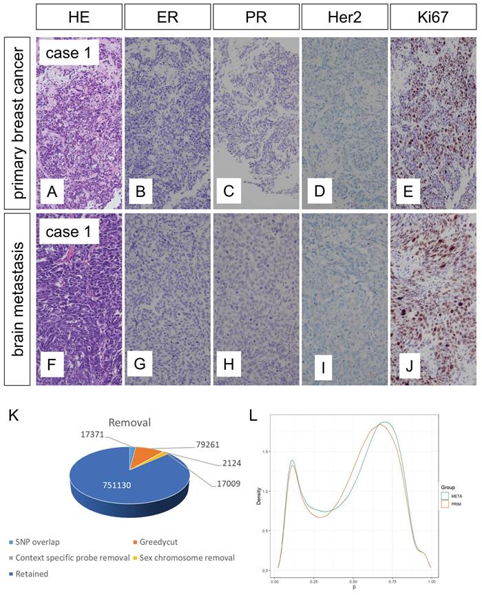
Inclusion criteria were the confirmed diagnosis of brain metastasis through standard diagnostic procedures and enough tumor material for isolation of at least 500ng of DNA both from primary BC and BCBM. No restrictions were applied regarding patient age, tumor classification, histological grading, tumor size, lymph node involvement, or the status of estrogen receptor, progesterone receptor, HER2, or Ki-67 expression in a representative primary breast cancer (Figure 1A-E) and the corresponding brain metastasis (Figure 1F-J). All tissue samples were pseudonymized before analysis to ensure non-identifiability. Detailed clinical and pathological characteristics of the patient cohort are presented in Supplementary Table S1.
Primary breast cancer and brain metastasis. Exemplified pictures of primary breast cancer (A-E) and brain metastasis (F-J) (Case 1). During preprocessing of the 866,895 probes 17,371 probes were removed that overlap with SNPs, 79,261 applying greedycut filtering, 2,124 context specific probes and 17,009 probes on sex-chromosomes resulting in a total of 751,130 retained probes (K). Global beta value distributions showed high densities with beta values <0.1 and >0.8 (L). HE: hemalaun-eosin stain; ER: estrogen receptor; PR: progesterone receptor; magnification: 20x.
Molecular genetic analysis
Molecular genetic analyses were performed as previously described [17-21]. Representative viable tumor tissue was microscopically identified and dissected. DNA extraction was performed applying the Maxwell system (Promega) according to the manufacturer's protocol. Methylation profiling was performed using the Infinium Methylation EPIC Bead Chip (Illumina) protocol according to the manufacturer's protocol [17-19]. The DNA input amount for bisulfite conversion was 500ng as standardized and accredited in our lab. Arrays were scanned on an Illumina NextSeq 550 DX device. Raw intensity data files (idat-files) were subjected to downstream analysis.
Computational data analysis
Methylation data were processed using the Illumina Genome Studio Methylation Module (2011.1) and RnBeads (version 2.4.0) [22] as previously described [23]. Methylation calls were transferred to beta values in the range from 0 to 1 (i.e. 0% of methylation to 100% of methylation) [22]. Probes were annotated to the four genomic regions tiling (genome tiling regions of length 5000), genes (version Ensemble genes 75), promoters (version Ensemble genes 75), and CpG islands (CpG island track of UCSC Genome Browser) [22]. We used “limma” for testing mean methylation differences [22]. Nominal p-values were applied as performed in other epigenome-wide association studies (EWAS) [24, 25]. To minimize technical bias, samples from different experimental groups were randomly distributed across processing batches and microarrays (Sentrix-ID/array barcode and the processing date can be found in Supplementary Table S1). Further analyses were performed using Morpheus and GraphPad Prism software suite (version 9).
Results
Patient characteristics
In the course of our study, we analyzed a well-defined but very heterogeneous collective of 20 anonymized tissue samples obtained from ten patients diagnosed with MBC (Supplementary Table S1). Patients' age ranged from 37-62 years, except one case that showed lobular histology all others were classified as no special type (NST) and immunohistochemical (IHC) subtypes varied between TNBC (4/10), HER2 positive (2/10) and HER2 negative (4/10) BC, respectively. The receptor status changed in 4/10 patients between primary BC and BCBM. Histological grade was G2 (4/10) and G3 (6/10) in primary BC samples and high histologic grade (G3) in all of the BCBM (10/10). The tumor cell content within the samples varied from a minimum of 50% to a maximum of 95% of tumor cells and the identified stromal TILs (tumor infiltrating lymphocytes) within the tissue samples showed a distribution from 1% to 40%.
Primary breast cancer and brain metastasis show high similarities on the global DNA methylation level
Methylation analysis of 20 samples from ten patients with each primary breast cancer and brain metastasis was performed. Applying the Illumina Infinium EPIC bead chip, we interrogated 866,895 methylation sensitive probes per sample. During preprocessing, we removed 17,371 probes that overlapped with SNPs, 79,261 probes applying greedycut filtering, 2,124 context specific probes and 17,009 probes on sex-chromosomes; this resulted in a total of 751,130 retained valid methylation probes (Figure 1K). An analysis of global beta value distributions showed high densities with beta values <0.1 and >0.8 (Figure 1L).
To assess the similarity between analyzed samples, we performed a principal component analysis (PCA). Focusing on methylation profiles at CpG islands, the PCA did not reveal two clearly distinct clusters separating primary breast cancer from corresponding brain metastases:
Unexpectedly, individual pairs of primary tumors and their matched brain metastases exhibited close spatial proximity within the PCA plot, indicating conserved epigenetic signatures between the two sample types (Figure 2A). This result suggests that, despite the biological transition associated with metastasis, the epigenetic landscape at promoter regions may retain subtype-specific patterns reflective of the primary tumor. Such observations could hold relevance for biomarker development and understanding the mechanisms underlying brain colonization.
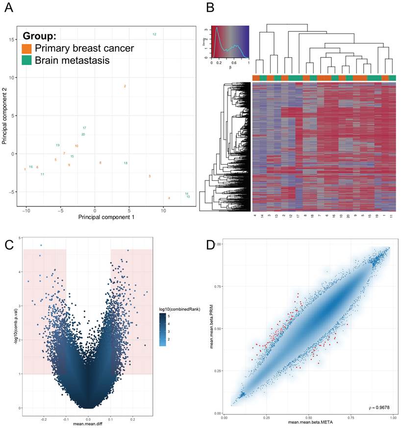
Methylation analysis. Principal component analysis (PCA) revealed that primary breast cancer and brain metastases do not form two distinct clusters. Instead, samples from individual patients, namely the primary tumor and its corresponding brain metastasis, were located in close proximity, suggesting a conserved epigenetic relationship between the two tissue types (pairs of primary cancer and metastasis: 1/11; 2/12; 3/13; 4/14; 5/15; 6/16; 7/17; 8/18; 9/19; 10/20.) (A). Clustering based on unsupervised hierarchical methods and random forest analysis further demonstrated the absence of two distinct groups. Instead, primary breast cancer and its corresponding brain metastases appeared in close proximity when considering the 1,000 most variable CpG sites, suggesting a shared epigenetic landscape between the matched tumor samples (B). Volcano plot shows nominally significantly methylated sites (Δβ ≥0.1, p <0.05) indicated in red (C). Scatter plot shows the combined rank highlighting the top 100 best ranking regions in red (D).
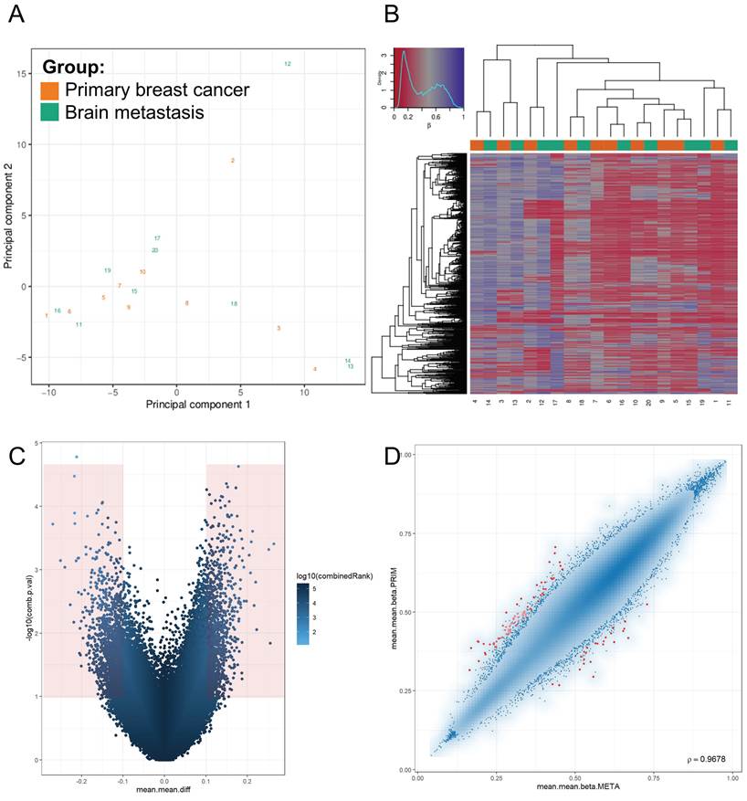
Subsequently, we carried out unsupervised hierarchical clustering leveraging a random forest algorithm applied to the top 1,000 most variable CpG sites. This analysis aimed to further interrogate methylation-based relationships between primary breast tumors and their matched brain metastases. Consistent with our principal component analysis findings, the clustering did not result in the emergence of two clearly distinct groups separating the primary tumors from the metastatic counterparts. Instead, samples from individual patients, i.e. those of the primary breast cancer and its corresponding brain metastasis, were frequently found in close proximity within the resulting dendrogram (CpG islands with the dissimilarity metric correlation based and the agglomeration strategy average) (Figure 2B). This spatial clustering suggests a preservation of epigenetic features during metastatic progression to the brain and reinforces the notion of patient-specific methylation patterns.
Together, these findings may reflect the evolutionary continuity between primary and metastatic disease within the same individual, and underscore the potential utility of DNA methylation signatures in tracking tumor origin and progression.
Primary breast cancer and the corresponding brain metastasis show distinct differences in methylation signatures
Differential DNA methylation analysis was conducted to compare primary breast cancer tissue with corresponding brain metastases. To identify significantly altered methylation sites, we performed pairwise comparisons using a threshold of Δβ ≥ 0.1 and p-value < 0.05. In the resulting volcano plot, probes meeting these criteria are highlighted in red (Figure 2C).
Subsequently, we generated a scatter plot to visualize methylation differences between primary breast cancer and brain metastasis tissue, emphasizing the top 100 regions ranked by combined statistical relevance (Figure 2D).
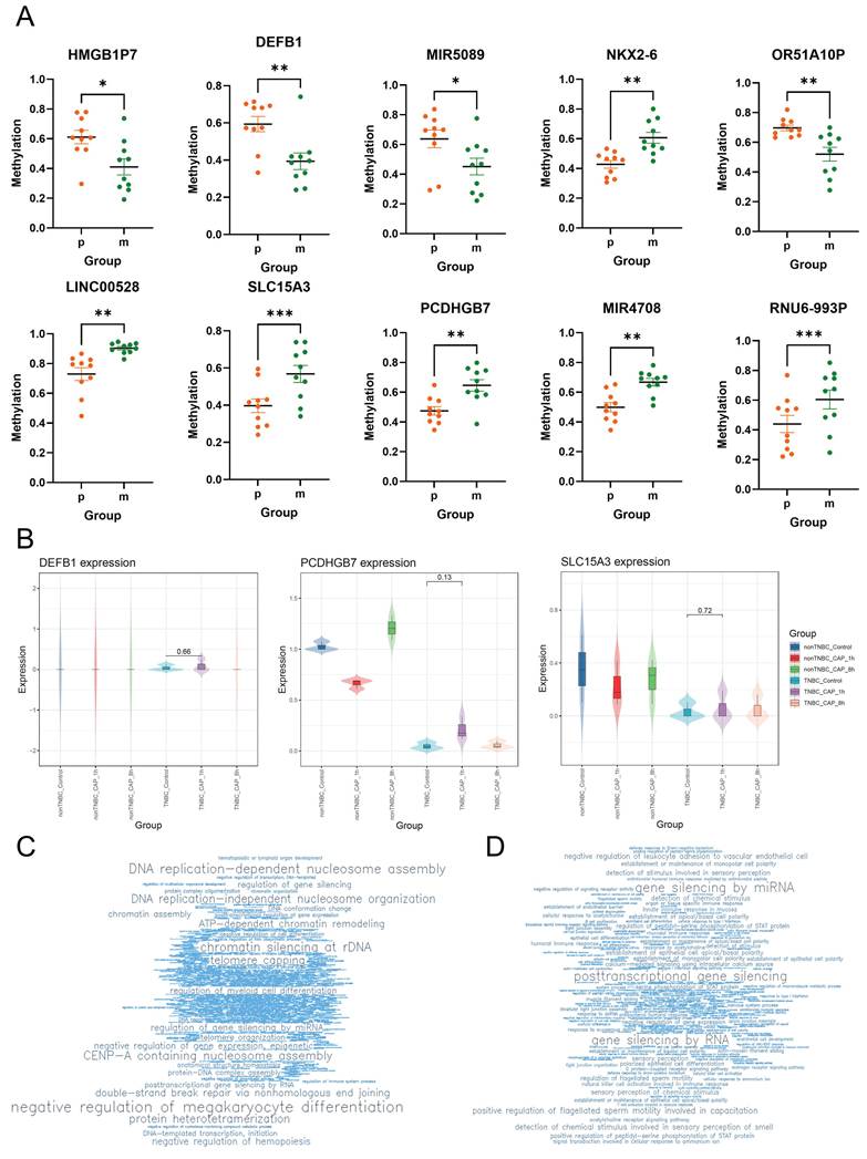
The analysis revealed a total of 107 nominally significantly methylated CpG sites (DMCGs) located within gene and promoter regions, comprising 97 within promoter regions and 10 within gene bodies, based on the defined statistical thresholds (Δβ ≥ 0.1; p-value < 0.05). Of note, the list of top ten nominal significantly methylated genes and promoters include HMGB1P7, DEFB1, MIR5089, NKX2-6, OR51A10P, LINC00528, SLC15A3, PCDHGB7, MIR4708, and RNU6-993P (Figure 3A, Supplementary Table S2). Methylation levels show significant differences in primary breast cancer compared with brain metastasis. The gene DEFB1 shows a hypomethylation in BCBM compared to primary BC whereas LINC00528 (long non-coding RNAs) shows significant hypermethylation in BCBM. The hypomethylated promoter regions of BCBM compared to BC comprise HMGB1P7, MIR5089 (miRNAs) and OR51A10P contrary to the promoters of NKX2-6, SLC15A3, PCDHGB7, MIR4708 and RNU6-993P which showed hypermethylation in BCBM.
Differential methylation analysis. Methylation levels of top 10 differentially methylated regions show significant differences in primary breast cancer compared with brain metastasis (A) (t-test; indicated are mean and SEM); p: primary breast cancer; m: brain metastasis. Transcriptomic screening of core differentially methylated loci under redox perturbation using CAPmed-BC (B). Violin plots display the expression levels of DEFB1, PCDHGB7, and SLC15A3 across non-TNBC and TNBC breast cancer models. Experimental groups include untreated controls (Control) and cells exposed to cold atmospheric plasma (CAP) after 1 hour (CAP_1h) and 8 hours (CAP_8h). Gene ontology (GO) enrichment analysis showed the top 1000 best ranking genes by word clouds of top terms being enriched in hypomethylated (C) and hypermethylated (D) pathways. * p < 0.05; ** p < 0.01; *** p < 0.001.
In silico screening of core loci for therapeutic potential under redox perturbation
To evaluate the translational and therapeutic potential of the identified differentially methylated regions, we cross-referenced our top loci with the specialized breast cancer multi-omics database CAPmed-BC. [68, 69] We focused on the three successfully mapped core genes: DEFB1, PCDHGB7, and SLC15A3 (Figure 3B).
The transcriptomic profiles under cold atmospheric plasma (CAP)-induced redox stress revealed distinct, subtype-specific regulation mechanisms. For DEFB1, expression levels were virtually zero across nearly all conditions in both non-TNBC and TNBC models, indicating robust baseline transcriptional silencing.
In contrast, PCDHGB7 displayed dynamic, time-dependent responsiveness. In non-TNBC cells, PCDHGB7 expression transiently dropped one hour post-treatment before undergoing a compensatory upregulation at eight hours. Conversely, the aggressive TNBC subtype showed a highly suppressed baseline expression of PCDHGB7, which was transiently alleviated by a multi-fold transcriptional induction one hour after redox perturbation.
Similarly, SLC15A3 exhibited a heavily repressed baseline state, particularly in TNBC cells where expression was mostly absent. However, redox stress triggered a subtle, stochastic activation of SLC15A3 in specific TNBC replicates after both one and eight hours.
Taking together, these in silico validation data demonstrate that the identified epigenetically altered loci are transcriptionally reactive to therapeutic oxidative stress challenges.
Enrichment analysis of differentially methylated CpGs (DMCGs) reveals distinct altered pathways in primary breast cancer and brain metastasis
Gene Ontology (GO) enrichment analysis was conducted to identify functionally enriched biological processes associated with differentially methylated genes (Figure 3C, D). In brain metastases compared with primary breast cancer tissue, hypermethylated genes were predominantly associated with the following GO terms: “DNA replication-dependent nucleosome assembly,” “chromatin silencing at rDNA,” “CENP-A-containing nucleosome assembly,” and “DNA replication-independent nucleosome organization.” Conversely, hypomethylated genes in brain metastases demonstrated enrichment in pathways related to “gene silencing by miRNA,” “posttranscriptional gene silencing,” and “gene silencing by RNA” (Supplementary Table S3).
Discussion
Breast cancer is a heterogeneous disease clinically classified by ER, PR, and HER2 status. TNBC and HER2-positive subtypes exhibit aggressive progression and a high incidence of brain metastases, posing substantial therapeutic challenges despite advances in systemic treatment. Epigenetic dysregulation, particularly via DNA methylation, can silence tumor suppressors or activate oncogenes without altering DNA sequences, contributing to carcinogenesis [26, 27]. These changes may also affect microRNA regulation, impacting apoptosis and proliferation.
In this study, we analyzed BCBM and primary BC samples using the Illumina Infinium Methylation EPIC Bead Chip. A total of 107 CpG sites showed nominal differential methylation (Δβ ≥ 0.1; p-value < 0.05), including 97 promoter and 10 gene regions. The top altered genes and promoters included HMGB1P7, DEFB1, MIR5089, NKX2-6, OR51A10P, LINC00528, SLC15A3, PCDHGB7, MIR4708, and RNU6-993P.
Having a closer look at the top ten differentially methylated genes, we found strong correlation being associated with tumorigenesis. DEFB1 supports antitumor immunity by activating adaptive responses, and shows tumor-suppressive function [28-31]. HMGB1P7H, although annotated as a pseudogene, may regulate gene expression similarly to microRNAs and lncRNAs [32, 33] and participate in maintaining genomic stability, activating oncogenic pathways and promoting tumor progression, therapy resistance, and immune evasion [34-44]. Olfactory-receptor pseudogene OR51A10P may also influence cancer biology by implicating in tumor signaling and biomarker development [45-48]. Two microRNAs showed altered methylation in BCBM: MIR5089 (hypomethylated) and MIR4708 (hypermethylated). MicroRNAs broadly regulate post-transcriptional gene expression and can function as oncogenes or suppressors [49, 50]. MIR5089-5p has been proposed as a circulating biomarker in breast cancer [51], while MIR4708 may contribute to TNBC risk stratification [52]. LINC00528, a hypermethylated lncRNA, likely acts as a tumor suppressor by modulating immune and adhesion pathways. Its reduced expression correlates with advanced HER2-positive BC and poor survival outcomes [53]. RNU6-993P, another pseudogene, lacks direct cancer evidence, though related U6 family members have diagnostic relevance [54, 55]. NKX2-6 encodes a transcription factor involved in neural developmental pathways [56], and related NKX2 genes can promote chemoresistance in breast cancer [57]. SLC15A3 (synonym PHT2), an immune-regulatory solute transporter, and other SLC family members have emerging roles as cancer biomarkers [59-62]. PCDHGB7 promotes apoptosis and cell-cycle arrest and increases TNBC sensitivity to carboplatin, supporting its utility as a serum biomarker [63, 64]. Though all of these genes have been reported to have a role in other cancer types, their relevance to brain metastasis requires further investigation.
A major limitation of this study is the absence of matched transcriptomic data; therefore, whether the identified differentially methylated sites affect gene expression is unknown. To support the biological relevance of our findings we performed validation of our dataset by searching in online databases. We found the three genes LINC00528, SLC15A3 and PCDHGB7 in an extended TCGA-BRCA (The Cancer Genome Atlas Breast Cancer) dataset with additional histologic type annotations in a total of 1,063 breast cancers. The genes are associated with a special gene expression signature containing significant upregulated genes associated with rare subtypes [65].
Prior studies highlight hypermethylated promoters as potential markers for BC progression and brain metastasis [66-67]. In our cohort, 107 CpG sites (Δβ ≥ 0.1; p < 0.05) were nominally significantly methylated between BCBM and primary BC tissues. Our epigenomic analysis revealed that, on a global scale, metastatic brain lesions exhibit greater similarity to their respective primary breast tumors than the primary tumors do to each other (Figure 2A, B). This observation underscores the clonal and lineage-dependent nature of metastatic progression, suggesting that key molecular features are preserved during dissemination. Matched primary and metastatic samples clustered together, reflecting patient-specific epigenetic patterns, but this does not preclude the existence of smaller, biologically relevant methylation changes associated with metastatic progression.
Despite this overarching resemblance, the data also highlight a subset of genes with distinct methylation and expression patterns that appear specific to the metastatic process itself. These loci may contribute to the acquisition of traits essential for successful colonization of distant organs, such as enhanced migratory capacity, immune evasion, or adaptation to the brain microenvironment. Notably, differential methylation was detected in several non-coding RNAs and pseudogenes, pointing to epigenetic reprogramming as a driver of metastatic competency. These findings suggest a dual landscape: while metastases largely recapitulate their tumor of origin, specific epigenetic alterations demarcate them as biologically distinct entities. This distinction may hold translational relevance for biomarker development and therapeutic targeting, particularly in the context of brain metastases, which remain a clinical challenge with limited treatment options.
A limitation of this study is on the one hand the small sample size of only ten matched cases and then that cohort includes tumors of all major breast cancer subtypes without stratification by ER, PR, or HER2 status. Because intrinsic subtypes display distinct baseline methylation landscapes, this heterogeneity may impede the detection of subtype-specific metastasis-associated alterations or diminish signals driven by particular biological contexts. Our findings nonetheless provide valuable preliminary insights into epigenetic alterations associated with brain metastasis in breast cancer. The restricted cohort size may constrain the generalizability of the results and preclude robust statistical power; however, the paired design and focus on methylation profiling across primary and metastatic sites offer a unique perspective that is rarely captured in larger datasets.
Future studies with larger, subtype-balanced cohorts will be essential to determine which components of the signature are universal versus subtype-restricted. These initial observations lay the groundwork for future investigations and underscore the importance of integrating epigenomic data into the study of metastatic progression. Furthermore, a major limitation of this study is the lack of direct in vitro functional assays. This must be clarified in further research. Despite its exploratory nature, the study contributes meaningfully to a growing body of literature and warrants dissemination to stimulate further research in this domain.
Supplementary Material
Supplementary tables.

Acknowledgements
AI disclosure
During the preparation of this work, the authors used “Copilot” and “Gemini” in order to improve readability and language. After using this tool, the authors reviewed and edited the content as needed and take full responsibility for the content of the publication.
Ethics statement and patient consent
This study was conducted in compliance with the principles outlined in the Declaration of Helsinki and received approval from the Ethics Committee of Salzburg (reference number: 415-E/2262/4-2018). Informed consent was obtained from all participants and/or their legal guardians prior to inclusion.
Author contributions
B.A.S., T.F.J.K., and K.S. analysed and interpreted the data. B.A.S. performed the histological and molecular analysis. K.S., R.G., C.S., A.R., O.K., S.W., S.P.G., and G.R. provided resources. B.A.S. and T.F.J.K. wrote the first draft of the manuscript, G.H., C.S., K.S., V.C., F.G., and N.Z. critically reviewed the manuscript.
Data availability
The datasets generated and analyzed during the current study are not publicly available in open repositories due to legal and ethical restrictions under the European General Data Protection Regulation (GDPR), as they contain sensitive human biometric data. However, the data are available for independent scientific verification from the corresponding author upon completion of a formal Data Transfer Agreement (DTA) and approval by our institutional data protection compliance office. Interested researchers may initiate this process by contacting the corresponding authors.
Competing Interests
The authors have declared that no competing interest exists.
References
1. Watase C, Shiino S, Shimoi T. et al. Breast Cancer Brain Metastasis-Overview of Disease State, Treatment Options and Future Perspectives. Cancers (Basel). 2021;13(5):1078 Published 2021 Mar 3. doi:10.3390/cancers13051078
2. Hosonaga M, Saya H, Arima Y. Molecular and cellular mechanisms underlying brain metastasis of breast cancer. Cancer Metastasis Rev. 2020;39(3):711-720 doi:10.1007/s10555-020-09881-y
3. Koniali L, Hadjisavvas A, Constantinidou A. et al. Risk factors for breast cancer brain metastases: a systematic review. Oncotarget. 2020;11(6):650-669 Published 2020 Feb 11. doi:10.18632/oncotarget.27453
4. Custódio-Santos T, Videira M, Brito MA. Brain metastasization of breast cancer. Biochim Biophys Acta Rev Cancer. 2017;1868(1):132-147 doi:10.1016/j.bbcan.2017.03.004
5. Cali Daylan AE, Leone JP. Targeted Therapies for Breast Cancer Brain Metastases. Clin Breast Cancer. 2021;21(4):263-270 doi:10.1016/j.clbc.2020.11.014
6. Rostami R, Mittal S, Rostami P, Tavassoli F, Jabbari B. Brain metastasis in breast cancer: a comprehensive literature review. J Neurooncol. 2016;127(3):407-414 doi:10.1007/s11060-016-2075-3
7. Pedrosa RMSM, Mustafa DA, Soffietti R, Kros JM. Breast cancer brain metastasis: molecular mechanisms and directions for treatment. Neuro Oncol. 2018;20(11):1439-1449 doi:10.1093/neuonc/noy044
8. Achrol AS, Rennert RC, Anders C. et al. Brain metastases. Nat Rev Dis Primers. 2019;5(1):5 Published 2019 Jan 17. doi:10.1038/s41572-018-0055-y
9. Quail DF, Joyce JA. The Microenvironmental Landscape of Brain Tumors. Cancer Cell. 2017;31(3):326-341 doi:10.1016/j.ccell.2017.02.009
10. Deeken JF, Löscher W. The blood-brain barrier and cancer: transporters, treatment, and Trojan horses. Clin Cancer Res. 2007;13(6):1663-1674 doi:10.1158/1078-0432.CCR-06-2854
11. Fong CW. Permeability of the Blood-Brain Barrier: Molecular Mechanism of Transport of Drugs and Physiologically Important Compounds. J Membr Biol. 2015;248(4):651-669 doi:10.1007/s00232-015-9778-9
12. Harbeck N, Ciruelos E, Jerusalem G. et al. Trastuzumab deruxtecan in HER2-positive advanced breast cancer with or without brain metastases: a phase 3b/4 trial. Nat Med. 2024;30(12):3717-3727 doi:10.1038/s41591-024-03261-7
13. Murthy RK, Loi S, Okines A. et al. Tucatinib, Trastuzumab, and Capecitabine for HER2-Positive Metastatic Breast Cancer. N Engl J Med. 2020;382(7):597-609 doi:10.1056/NEJMoa1914609
14. Morgan AJ, Giannoudis A, Palmieri C. The genomic landscape of breast cancer brain metastases: a systematic review. Lancet Oncol. 2021;22(1):e7-e17 doi:10.1016/S1470-2045(20)30556-8
15. Duchnowska R, Dziadziuszko R, Trojanowski T. et al. Conversion of epidermal growth factor receptor 2 and hormone receptor expression in breast cancer metastases to the brain. Breast Cancer Res. 2012;14(4):R119 Published 2012 Aug 16. doi:10.1186/bcr3244
16. Priedigkeit N, Hartmaier RJ, Chen Y. et al. Intrinsic Subtype Switching and Acquired ERBB2/HER2 Amplifications and Mutations in Breast Cancer Brain Metastases. JAMA Oncol. 2017;3(5):666-671 doi:10.1001/jamaoncol.2016.5630
17. Kraus TFJ, Machegger L, Pöppe J. et al. Diffuse midline glioma of the cervical spinal cord with H3 K27M genotype phenotypically mimicking anaplastic ganglioglioma: a case report and review of the literature. Brain Tumor Pathol. 2020;37(3):89-94 doi:10.1007/s10014-020-00365-z
18. Kraus TFJ, Schwartz C, Machegger L. et al. A patient with two gliomas with independent oligodendroglioma and glioblastoma biology proved by DNA-methylation profiling: a case report and review of the literature. Brain Tumor Pathol. 2022;39(2):111-119 doi:10.1007/s10014-021-00423-0
19. Hölzl D, Hutarew G, Zellinger B. et al. EGFR Amplification Is a Phenomenon of IDH Wildtype and TERT Mutated High-Grade Glioma: An Integrated Analysis Using Fluorescence In Situ Hybridization and DNA Methylome Profiling. Biomedicines. 2022;10(4):794 Published 2022 Mar 29. doi:10.3390/biomedicines10040794
20. Hölzl D, Hutarew G, Zellinger B. et al. Integrated analysis of programmed cell death ligand 1 expression reveals increased levels in high-grade glioma. J Cancer Res Clin Oncol. 2021;147(8):2271-2280 doi:10.1007/s00432-021-03656-w
21. Kraus TFJ, Langwieder CK, Hölzl D. et al. Dissecting the Methylomes of EGFR-Amplified Glioblastoma Reveals Altered DNA Replication and Packaging, and Chromatin and Gene Silencing Pathways. Cancers (Basel). 2023;15(13):3525 Published 2023 Jul 7. doi:10.3390/cancers15133525
22. Müller F, Scherer M, Assenov Y. et al. RnBeads 2.0: comprehensive analysis of DNA methylation data. Genome Biol. 2019;20(1):55 Published 2019 Mar 14. doi:10.1186/s13059-019-1664-9
23. Hutarew G, Hölzl D, Schiefer T. et al. Methylome Profiling of PD-L1-Expressing Glioblastomas Shows Enrichment of Post-Transcriptional and RNA-Associated Gene Regulation. Cancers (Basel). 2022;14(21):5375 Published 2022 Oct 31. doi:10.3390/cancers14215375
24. De Jager PL, Srivastava G, Lunnon K. et al. Alzheimer's disease: early alterations in brain DNA methylation at ANK1, BIN1, RHBDF2 and other loci. Nat Neurosci. 2014;17(9):1156-1163 doi:10.1038/nn.3786
25. Lunnon K, Smith R, Hannon E. et al. Methylomic profiling implicates cortical deregulation of ANK1 in Alzheimer's disease. Nat Neurosci. 2014;17(9):1164-1170 doi:10.1038/nn.3782
26. Jones PA, Baylin SB. The epigenomics of cancer. Cell. 2007;128(4):683-692 doi:10.1016/j.cell.2007.01.029
27. Lee CJ, Ahn H, Jeong D, Pak M, Moon JH, Kim S. Impact of mutations in DNA methylation modification genes on genome-wide methylation landscapes and downstream gene activations in pan-cancer. BMC Med Genomics. 2020;13(Suppl 3):27 Published 2020 Feb 24. doi:10.1186/s12920-020-0659-4
28. Adyns L, Proost P, Struyf S. Role of Defensins in Tumor Biology. Int J Mol Sci. 2023;24(6):5268 Published 2023 Mar 9. doi:10.3390/ijms24065268
29. Sun CQ, Arnold RS, Hsieh CL. et al. Discovery and mechanisms of host defense to oncogenesis: targeting the β-defensin-1 peptide as a natural tumor inhibitor. Cancer Biol Ther. 2019;20(6):774-786 doi:10.1080/15384047.2018.1564564
30. Yang D, Chertov O, Bykovskaia SN. et al. Beta-defensins: linking innate and adaptive immunity through dendritic and T cell CCR6. Science. 1999;286(5439):525-528 doi:10.1126/science.286.5439.525
31. Donald CD, Sun CQ, Lim SD. et al. Cancer-specific loss of beta-defensin 1 in renal and prostatic carcinomas. Lab Invest. 2003;83(4):501-505 doi:10.1097/01.lab.0000063929.61760.f6
32. Hu X, Yang L, Mo YY. Role of Pseudogenes in Tumorigenesis. Cancers (Basel). 2018;10(8):256 Published 2018 Aug 1. doi:10.3390/cancers10080256
33. Nakamura-García AK, Espinal-Enríquez J. Pseudogenes in Cancer: State of the Art. Cancers (Basel). 2023;15(16):4024 Published 2023 Aug 8. doi:10.3390/cancers15164024
34. Chen R, Kang R, Tang D. The mechanism of HMGB1 secretion and release. Exp Mol Med. 2022;54(2):91-102 doi:10.1038/s12276-022-00736-w
35. Zhang QY, Wu LQ, Zhang T, Han YF, Lin X. Autophagy-mediated HMGB1 release promotes gastric cancer cell survival via RAGE activation of extracellular signal-regulated kinases 1/2. Oncol Rep. 2015;33(4):1630-1638 doi:10.3892/or.2015.3782
36. Wu X, Wang W, Chen Y. et al. High Mobility Group Box Protein 1 Serves as a Potential Prognostic Marker of Lung Cancer and Promotes Its Invasion and Metastasis by Matrix Metalloproteinase-2 in a Nuclear Factor-κB-Dependent Manner. Biomed Res Int. 2018;2018:3453706 Published 2018 Apr 19. doi:10.1155/2018/3453706
37. Zhang J, Shao S, Han D. et al. High mobility group box 1 promotes the epithelial-to-mesenchymal transition in prostate cancer PC3 cells via the RAGE/NF-κB signaling pathway. Int J Oncol. 2018;53(2):659-671 doi:10.3892/ijo.2018.4420
38. Lv G, Wu M, Wang M. et al. miR-320a regulates high mobility group box 1 expression and inhibits invasion and metastasis in hepatocellular carcinoma. Liver Int. 2017;37(9):1354-1364 doi:10.1111/liv.13424
39. Huang M, Geng Y, Deng Q. et al. Translationally controlled tumor protein affects colorectal cancer metastasis through the high mobility group box 1-dependent pathway. Int J Oncol. 2018;53(4):1481-1492 doi:10.3892/ijo.2018.4502
40. Machado LR, Moseley PM, Moss R. et al. High mobility group protein B1 is a predictor of poor survival in ovarian cancer. Oncotarget. 2017;8(60):101215-101223 Published 2017 Aug 24. doi:10.18632/oncotarget.20538
41. Patra S, Roy PK, Dey A, Mandal M. Impact of HMGB1 on cancer development and therapeutic insights focused on CNS malignancy. Biochim Biophys Acta Rev Cancer. 2024;1879(3):189105 doi:10.1016/j.bbcan.2024.189105
42. Yuan S, Liu Z, Xu Z, Liu J, Zhang J. High mobility group box 1 (HMGB1): a pivotal regulator of hematopoietic malignancies. J Hematol Oncol. 2020;13(1):91 Published 2020 Jul 13. doi:10.1186/s13045-020-00920-3
43. Amornsupak K, Thongchot S, Thinyakul C. et al. HMGB1 mediates invasion and PD-L1 expression through RAGE-PI3K/AKT signaling pathway in MDA-MB-231 breast cancer cells. BMC Cancer. 2022;22(1):578 Published 2022 May 24. doi:10.1186/s12885-022-09675-1
44. Jiao D, Zhang J, Chen P. et al. HN1L promotes migration and invasion of breast cancer by up-regulating the expression of HMGB1. J Cell Mol Med. 2021;25(1):397-410 doi:10.1111/jcmm.16090
45. Malnic B, Godfrey PA, Buck LB. The human olfactory receptor gene family. Proc Natl Acad Sci U S A. 2004;101(8):2584-2589 doi:10.1073/pnas.0307882100
46. Maßberg D, Hatt H. Human Olfactory Receptors: Novel Cellular Functions Outside of the Nose. Physiol Rev. 2018;98(3):1739-1763 doi:10.1152/physrev.00013.2017
47. Sanz G, Leray I, Dewaele A. et al. Promotion of cancer cell invasiveness and metastasis emergence caused by olfactory receptor stimulation. PLoS One. 2014;9(1):e85110 Published 2014 Jan 8. doi:10.1371/journal.pone.0085110
48. Asadi M, Ahmadi N, Ahmadvand S. et al. Investigation of olfactory receptor family 51 subfamily j member 1 (OR51J1) gene susceptibility as a potential breast cancer-associated biomarker. PLoS One. 2021;16(2):e0246752 Published 2021 Feb 10. doi:10.1371/journal.pone.0246752
49. Saliminejad K, Khorram Khorshid HR, Soleymani Fard S, Ghaffari SH. An overview of microRNAs: Biology, functions, therapeutics, and analysis methods. J Cell Physiol. 2019;234(5):5451-5465 doi:10.1002/jcp.27486
50. Khordadmehr M, Shahbazi R, Ezzati H, Jigari-Asl F, Sadreddini S, Baradaran B. Key microRNAs in the biology of breast cancer; emerging evidence in the last decade. J Cell Physiol. 2019;234(6):8316-8326 doi:10.1002/jcp.27716
51. Ritter A, Hirschfeld M, Berner K. et al. Discovery of potential serum and urine-based microRNA as minimally-invasive biomarkers for breast and gynecological cancer. Cancer Biomark. 2020;27(2):225-242 doi:10.3233/CBM-190575
52. Andrade F, Nakata A, Gotoh N, Fujita A. Large miRNA survival analysis reveals a prognostic four-biomarker signature for triple negative breast cancer. Genet Mol Biol. 2020;43(1):e20180269 Published 2020 Mar 2. doi:10.1590/1678-4685-GMB-2018-0269
53. Zhang X, Zhang H, Li J. et al. 6-lncRNA Assessment Model for Monitoring and Prognosis of HER2-Positive Breast Cancer: Based on Transcriptome Data. Pathol Oncol Res. 2021;27:609083 Published 2021 Apr 13. doi:10.3389/pore.2021.609083
54. Lou G, Ma N, Xu Y. et al. Differential distribution of U6 (RNU6-1) expression in human carcinoma tissues demonstrates the requirement for caution in the internal control gene selection for microRNA quantification. Int J Mol Med. 2015;36(5):1400-1408 doi:10.3892/ijmm.2015.2338
55. Manterola L, Guruceaga E, Gállego Pérez-Larraya J. et al. A small noncoding RNA signature found in exosomes of GBM patient serum as a diagnostic tool. Neuro Oncol. 2014;16(4):520-527 doi:10.1093/neuonc/not218
56. McMahon AP. Neural patterning: the role of Nkx genes in the ventral spinal cord. Genes Dev. 2000;14(18):2261-2264 doi:10.1101/gad.840800
57. Lim JS, Jung GY, Park SY. Nkx-2.5 Regulates MDR1 Expression via Its Upstream Promoter in Breast Cancer Cells. J Korean Med Sci. 2019;34(12):e100 Published 2019 Mar 19. doi:10.3346/jkms.2019.34.e100
58. Song F, Yi Y, Li C. et al. Regulation and biological role of the peptide/histidine transporter SLC15A3 in Toll-like receptor-mediated inflammatory responses in macrophage. Cell Death Dis. 2018;9(7):770 Published 2018 Jul 10. doi:10.1038/s41419-018-0809-1
59. Nagata H, Kozaki KI, Muramatsu T. et al. Genome-wide screening of DNA methylation associated with lymph node metastasis in esophageal squamous cell carcinoma. Oncotarget. 2017;8(23):37740-37750 doi:10.18632/oncotarget.17147
60. Sutherland R, Meeson A, Lowes S. Solute transporters and malignancy: establishing the role of uptake transporters in breast cancer and breast cancer metastasis. Cancer Metastasis Rev. 2020;39(3):919-932 doi:10.1007/s10555-020-09879-6
61. Yan L, He J, Liao X. et al. A comprehensive analysis of the diagnostic and prognostic value associated with the SLC7A family members in breast cancer. Gland Surg. 2022;11(2):389-411 doi:10.21037/gs-21-909
62. Zhao X, Jin L, Liu Y, Liu Z, Liu Q. Bioinformatic analysis of the role of solute carrier-glutamine transporters in breast cancer. Ann Transl Med. 2022;10(14):777 doi:10.21037/atm-22-2620
63. Hou S, Shan M, Gao C. et al. PCDHGB7 Increases Chemosensitivity to Carboplatin by Inhibiting HSPA9 via Inducing Apoptosis in Breast Cancer. Dis Markers. 2019;2019:6131548 Published 2019 Jul 8. doi:10.1155/2019/6131548
64. Shan M, Yin H, Li J. et al. Detection of aberrant methylation of a six-gene panel in serum DNA for diagnosis of breast cancer. Oncotarget. 2016;7(14):18485-18494 doi:10.18632/oncotarget.7608
65. Thennavan A, Beca F, Xia Y, Recio SG, Allison K, Collins LC, Tse GM, Chen YY, Schnitt SJ, Hoadley KA, Beck A, Perou CM. Molecular analysis of TCGA breast cancer histologic types. Cell Genom. 2021Dec8;1(3):100067 doi: 10.1016/j.xgen.2021.100067. PMID: 35465400; PMCID: PMC9028992
66. Lehmann U, Länger F, Feist H, Glöckner S, Hasemeier B, Kreipe H. Quantitative assessment of promoter hypermethylation during breast cancer development. Am J Pathol. 2002;160(2):605-612 doi:10.1016/S0002-9440(10)64880-8
67. Van De Voorde L, Speeckaert R, Van Gestel D. et al. DNA methylation-based biomarkers in serum of patients with breast cancer. Mutat Res. 2012;751(2):304-325 doi:10.1016/j.mrrev.2012.06.001
68. Li J, Miao B, Wang S, Dong W, Xu H, Si C, Wang W, Duan S, Lou J, Bao Z, Zeng H, Yang Z, Cheng W, Zhao F, Zeng J, Liu XS, Wu R, Shen Y, Chen Z, Chen S, Wang M, Hiplot Consortium. Hiplot: a comprehensive and easy-to-use web service for boosting publication-ready biomedical data visualization. Brief Bioinform. 2022Jul18;23(4):bbac261 doi: 10.1093/bib/bbac261
69. Dai X, Wang M, Liu Y. A Hiplot-based web service for cold atmospheric plasma high-throughput data integration and analysis on breast cancer. Imeta. 2025;4(4):e70045 Published 2025 May 25. doi:10.1002/imt2.70045
Author contact
Corresponding authors: t.krausat (TFJK); k.sotlarat (KS).