Impact Factor ISSN: 1449-1907




Int J Med Sci 2026; 23(10):3101-3111. doi:10.7150/ijms.135847 This issue Cite
Review
Potential Role of Red Yeast Rice in the Prevention and Treatment of Alzheimer's Disease
Qining Quan1, Ronghua Pan2, Haolin Liu1, Lizhong Zheng2, Rui Zhang1 
1. Zhejiang Chinese Medical University, Hangzhou, Zhejiang 310053
2. Zhejiang Sanhe Bio-Tech Co., Ltd, Jiangshan, Zhejiang 324100
Received 2026-4-8; Accepted 2026-8-5; Published 2026-8-22
Abstract
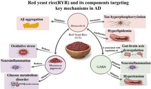
Red Yeast Rice (RYR) fermented from Monascus is a traditional fermented product with both dietary and medicinal uses and is widely used for lipid regulation. Clinically, it is utilized to lower blood lipid levels and manage hyperlipidemia. Contemporary research indicates that RYR is abundant in secondary metabolites, such as monacolin K, Monascus pigments, and γ-aminobutyric acid, which contribute to its lipid-lowering, blood sugar-reducing, anti-inflammatory, neuroprotective, and antihypertensive properties. These metabolites may play a significant role in the prevention and treatment of Alzheimer's disease (AD) through mechanisms that include cholesterol-dependent pathways, anti-neuroinflammation, antioxidant activity, neuroprotection, modulation of intestinal flora, inhibition of high-risk factors for AD, and suppression of AD-related β-amyloid peptide deposition and tau protein hyperphosphorylation. Consequently, RYR demonstrates potential for a multi-component synergistic approach to preventing and treating AD. However, current research on the use of RYR for AD prevention and treatment remains limited. This review focuses on the primary functional substances in RYR, elucidates the anti-AD effects and mechanisms of each metabolite, analyzes the advantages of its multi-component and multi-target properties, and provides a foundation for the further development of RYR.
Keywords: Red Yeast Rice, Monascus, Alzheimer's disease, Monascus pigments, monacolin K, Lovastatin
Introduction
With global population aging, the prevalence and mortality of Alzheimer's disease (AD) continue to increase. As of 2023, approximately 50 million people worldwide were affected by AD, and this number is projected to reach 152 million by 2050 [1]. AD not only seriously harms the personal health of patients, but also brings heavy care and economic burdens to families and society. It is currently estimated that the total social burden of AD patients worldwide exceeds 958 billion US dollars and is expected to increase significantly by 2050 [2]. Although some drugs for improving AD conditions, such as donepezil, have been developed, these drugs cannot fundamentally reverse the lesions of AD patients, and the toxic and side effects caused by long-term use are obvious. At present, there are still no drugs that can effectively prevent or treat Alzheimer's disease in clinical practice. Therefore, the search and research for multi-target and synergistic natural medicines for the prevention and treatment of AD is a future trend.
Red Yeast Rice (RYR), a traditional Chinese medicine with both medicinal and culinary origins, boasts a history of extensive applications spanning thousands of years. It is rich in various bioactive metabolites that influence lipid metabolism, neuroinflammation, oxidative stress, and the regulation of the gut-microbiome-brain axis. Consequently, this review investigates the potential of bioactive metabolites in RYR in the prevention and treatment of AD. By examining the pathological mechanisms and risk factors associated with AD, this review provides a comprehensive evaluation of the multiple functional components of RYR. Ultimately, we aim to elucidate the anti-AD effects and mechanisms of the secondary metabolites found in RYR, thereby providing a foundation for further research and the development of applications aimed at preventing and treating AD.
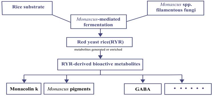
RYR is produced by fermenting rice with filamentous fungi of the genus Monascus. During fermentation, Monascus grows on the rice substrate and synthesizes a variety of bioactive metabolites, which are then accumulated in the fermented product. These RYR-derived metabolites include monacolin K, Monascus pigments, γ-aminobutyric acid (GABA), and other fermentation-related compounds. This process is presented in Figure 1. Therefore, in this review, the term “RYR-derived metabolites” refers to bioactive compounds generated during Monascus-mediated fermentation and retained in the final RYR product.
Schematic overview of the relationship among RYR, Monascus, and the major bioactive metabolites generated or enriched during Monascus-mediated fermentation.
Main functional substances and functions in RYR
RYR is a traditional fermented product that originated in China. It is produced through solid or liquid fermentation using specific Monascus species on steamed rice and other grain substrates. RYR has long been used as a food colorant, flavor enhancer, brewing material, and traditional Chinese medicinal product. Ongoing research into RYR components has revealed its potential effects in lowering blood lipids and blood sugar, as well as its anti-inflammatory and antioxidant properties. Concurrently, the optimization of Monascus strains and the application of pure fermentation technology, which incorporates rigorously screened and identified superior Monascus strain, have led to its increased use in the medical and health sectors. The anti-Alzheimer's disease (AD) potential and multi-target synergistic effects of RYR may be attributable to its abundant secondary metabolites, including monacolin K, Monascus pigments, and GABA. These functional compounds provide a foundational basis for the prevention and treatment of AD through the use of RYR.
Monacolin K
Japanese scientist Endo et al. (1979) first discovered and extracted monacolin K from RYR in 1979 during their search for hypolipidemic substances, subsequently confirming its hypolipidemic activity [3]. Alberts et al. (1980) later isolated mevinolin, now widely known as lovastatin, from cultures of Aspergillus terreus, and subsequently established that major monacolin K in RYR is the natural form of lovastatin (acid lovastatin) [4]. Since that time, monacolin K has been commercially purified and marketed under the trade name lovastatin. Monacolin K shares a similar chemical structure with 3-hydroxy-3-methylglutaryl coenzyme A (HMG-CoA) and competitively inhibits HMG-CoA reductase, the rate-limiting enzyme in the mevalonate pathway of cholesterol biosynthesis [5]. By inhibiting HMG-CoA reductase, monacolin K reduces mevalonate formation and downstream cholesterol synthesis, thereby exerting its lipid-lowering effect [5]. Commercial lactone lovastatin requires hydrolytic activation to its active acid form in plasma and liver cells. Long-term or high-dose lovastatin use may be associated with elevations in serum alanine aminotransferase (ALT) and aspartate aminotransferase (AST) levels and rare clinically significant liver injury or acute liver failure [6]. The acidic lovastatin produced by the natural fermentation process has lower crystallinity, higher oral bioavailability, and better safety in toxicological evaluation [7]. As a lipophilic statin and a secondary metabolite of Monascus-fermented RYR, monacolin K can cross the blood-brain barrier and has been reported to possess neuroprotective activity in neuronal cell models [8]. Cohort evidence suggests that lovastatin use may be associated with slower cognitive decline or potential clinical benefits in patients with AD [9]. Together, these experimental and clinical observations indicate that monacolin K/lovastatin may represent a promising RYR-derived candidate for AD-related intervention. However, further experimental and clinical studies are warranted to clarify the AD-related potential of monacolin K in RYR.
Monascus pigments
Monascus pigments are another major secondary metabolite in RYR [10]. More than 100 types of Monascus pigments have been identified so far, and they are divided into six major categories: monascorubramine, rubropunctamine, monascorubrin, rubropunctatin, ankaflavin and monascin [10]. Monascus pigments are polyketide compounds containing a variety of functional groups. These functional groups contribute to the antioxidant, anti-inflammatory, hypolipidemic, anti-diabetic, and gut microbiota-regulating effects of Monascus pigments [11]. Lin et al. (2023) demonstrated through in vivo experiments in mice and cell-based in vitro experiments that the anti-inflammatory and antioxidant effects of Monascus pigments can inhibit neuroinflammation induced by Aβ40 [12]. Their blood pressure and blood sugar-lowering effects may attenuate the induction and promotion of AD by metabolic syndrome [13]. Several studies have demonstrated that Monascus pigments can modulate intestinal microbiota [14], therefore potentially influencing AD-related pathology via the microbiota-gut-brain axis [15]. Nevertheless, direct research examining the anti-AD effects of Monascus pigments through this axis remains limited. Compared with monacolin K, Monascus pigments appear to have close relevance to anti-inflammation and antioxidant pathways, and complement the lipid-centered actions of monacolin K within the multi-component framework of RYR [10, 14].
γ-Aminobutyric acid
GABA is a non-protein amino acid, water-soluble, and an inhibitory neurotransmitter widely present in the mammalian central nervous system and peripheral tissues [16, 17]. When ingested exogenously, GABA can reduce blood pressure, influence neuroinflammation-related and gut microbiota-related pathways [18, 19]. Endogenously produced GABA in the brain is involved in the regulation of cerebral circulation, supports neuronal health and survival, and plays a crucial role in synaptic plasticity underlying learning and memory [17]. GABA in RYR may contribute to AD-related neuroprotection through several potential mechanisms, including suppression of neuroinflammation and attenuation of neuronal injury [19], modulation of impaired GABAergic signaling and excitatory/inhibitory imbalance [20], regulation of gut-brain axis communication [21], and improvement of AD-related metabolic risk factors such as hypertension and glucose dysregulation [22, 23].
Potential Preventive and Therapeutic Effects of the Main Active Substances in RYR on AD
Cholesterol-Dependent Pathway of Monacolin K in RYR
Studies have demonstrated a correlation between AD and hyperlipidemia [24-26]. Initial investigations by Sparks et al. (1994) and Kivipelto et al. (2001) suggested hyperlipidemia as a significant risk factor for AD [24, 25]. Subsequent research on apolipoprotein E (ApoE) has further substantiated the link between hypercholesterolemia and AD. Individuals carrying an ApoE-ε4 allele exhibit a threefold increased likelihood of developing AD compared to those without this allele [27, 28]. The underlying pathogenic mechanism may involve the APOE-ε4 variant's role in degrading the myelin sheath of oligodendrocytes, which is intricately associated with apolipoprotein's function in lipid transport and metabolism [26]. Consequently, maintaining normal blood lipid levels may represent a viable strategy for the prevention and treatment of AD.
The cholesterol-dependent pathway involves the reduction of Aβ peptide levels in the brain by monacolin K from RYR, attributed to its hypolipidemic effect, thereby lowering the risk of AD [29]. Buxbaum et al. (2001) conducted cell culture experiments using lipid-depleted serum with or without active lovastatin metabolites [30]. They found that lovastatin treatment significantly reduced intracellular Aβ peptide levels under cholesterol-depleted conditions [30]. Furthermore, in human subgroups with elevated low-density lipoprotein cholesterol, the application of lovastatin controlled-release formulation or a placebo revealed a decrease in serum Aβ peptide content among hypercholesterolemic patients [31]. A cohort study utilizing a longitudinal registry demonstrated that AD patients taking lovastatin exhibited higher Mini-Mental State Examination scores after 3 years compared to non-lovastatin users, with a more pronounced effect observed in patients with hyperlipidemia [32].
Evidence from in vivo and in vitro studies as well as clinical studies has suggested that the cholesterol-dependent mechanism of monacolin K has positive anti-AD effects in inhibiting Aβ peptide deposition and improving cognition [29-33]. This cholesterol-dependent mechanism likely involves lipid rafts, which serve as platforms for processing amyloid precursor protein (APP) into Aβ40 and Aβ42 and thus critically influence Aβ40 and Aβ42 formation [33]. Area-Gomez and Schon (2024) proposed the "C99-lipid raft-cholesterol" axis to explain how cholesterol dysregulation promotes Aβ40 and Aβ42 production [33]. C99 is a 99-aa membrane-bound C-terminal fragment, generated after APP is cleaved by BACE1 and it is an intracellular lipid regulatory substance that regulates lipid raft formation [33]. According to this model, increased cholesterol may facilitate APP endocytosis and intracellular trafficking, leading to C99 increase and the recruitment of cholesterol and other lipids to form lipid raft-like microdomains at mitochondria-associated endoplasmic reticulum membranes. These lipid rafts provide sites for γ-secretase-mediated processing of C99 into Aβ40 and Aβ42 [33]. Therefore, systemic lipid imbalances may increase Aβ40 and Aβ42 production and alter the Aβ42/Aβ40 ratio by disrupting the C99-lipid raft-cholesterol axis, thereby linking lipid dysregulation to AD pathogenesis [33]. These observations support the view that lipid dysregulation contributes importantly to AD pathogenesis and provide a rationale for further exploring lipid-targeted interventions, which aligns with the cholesterol-dependent mechanism of lovastatin in RYR.
Non-Cholesterol-Dependent Pathways of Monacolin K in RYR
Beyond its cholesterol-dependent mechanism related to AD pathology, monacolin K may also act through a cholesterol-independent pathway that mediates anti-inflammatory and neuroprotective effects [34-36]. In an animal study of cerebral malaria, researchers compared chloroquine alone to chloroquine plus lovastatin and found that the lovastatin group showed reduced pro-inflammatory cytokines and oxidative stress markers, along with reversal of cognitive deficits and blood-brain barrier disruption attributable to neuroinflammation [34]. These results indicate a pronounced anti-neuroinflammatory and neuroprotective effect. In an experimental rat model involving cognitive impairment, statins increased the anti-inflammatory cytokine IL-10 and reduced neuroinflammation probably via activation of the transforming growth factor-β (TGF-β)/Smad family member 3 (Smad3) pathway [35]. Statins also lowered hippocampal levels of chitinase-3-like protein 1 and curtailed excessive glial-cell activation, which may help suppress neuroinflammation in the brains of AD patients [35]. The principal mechanism by which lovastatin inhibits neuroinflammation is suppression of excessive glial activation, as evidenced by its ability to reduce glial release of proinflammatory factors such as TNF-α, IL-1β, and IL-6 [36].
Effects of Monacolin K on Aβ peptide and Tau Proteins
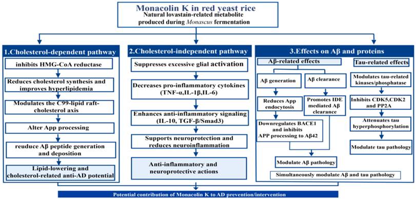
Excessive Aβ peptide deposition and phosphorylation of tau proteins are primary pathological hallmarks of AD and a key therapeutic target. Some studies indicate that Aβ peptide deposition and tau aggregation act synergistically in AD pathogenesis, implying that effective prevention and treatment should target both Aβ peptide and tau pathologies simultaneously [37]. Lovastatin may influence AD-related Aβ peptide metabolism through lipid-lowering and cholesterol-dependent mechanisms [29, 33], and it may also directly inhibit Aβ40 and Aβ42 formation and promote their clearance [38, 39]. The accumulation of Aβ peptides in the brain depends primarily on the balance between Aβ peptide clearance and APP processing [38]. Tamboli et al. (2010) reported that lovastatin promotes extracellular Aβ peptide degradation by stimulating exosome-associated insulin-degrading enzyme (IDE) secretion from microglia [40]. In a rat experiment, lovastatin significantly reduced brain Aβ42, but this clearance effect on Aβ42 disappeared after IDE gene knockout, indicating that lovastatin accelerates Aβ42 clearance via IDE [38]. Lovastatin also alters APP processing-related pathways to lower Aβ peptide levels [38, 39]. Endocytosis is a key step in converting APP to Aβ, and one study found that lovastatin inhibits APP endocytosis and downregulates endocytic factors, thereby reducing Aβ42 and Aβ40 formation [39]. Xu et al. (2021) experimentally showed that lovastatin down-regulates β-amyloid precursor protein-β-site APP cleaving enzyme 1 (BACE1), thereby inhibiting APP processing to Aβ1-42 [38]. Hyperphosphorylation of tau is another key therapeutic target in AD, and tau phosphorylation is governed by kinases and phosphatases; their overactivation induces tau hyperphosphorylation. An in vitro study found that lovastatin inhibits protein phosphatase 2A, cyclin-dependent kinase 5, and cyclin-dependent kinase 2, thereby reducing excessive tau phosphorylation [41]. Because Aβ peptide accumulation interacts with tau pathology during AD progression, lovastatin's concurrent modulation of both pathways may offer an advantage over agents that target only one pathological target. The three anti-AD pathways and mechanisms of the above monacolin K pathways are presented in the diagram (Figure 2).
Three pathways and main mechanisms by which monacolin K in RYR may modulate AD-related pathology.
Potential Preventive and Therapeutic Effects of Monascus pigments on AD
Direct evidence linking Monascus pigments to the modulation of Aβ peptide deposition or tau pathology remains limited. Nevertheless, Monascus pigments may influence AD-related risk pathways by regulating neuroinflammation, oxidative stress, and hyperglycemia-associated metabolic dysfunction [12, 42]. The chronic inflammatory response observed in AD patients can lead to the excessive activation of glial cells, resulting in an overproduction of inflammatory mediators [43]. This process exacerbates the accumulation of Aβ peptide and neurofibrillary tangles, ultimately causing neuronal damage and perpetuating a vicious cycle. These findings underscore the significance of neuroinflammation as a key mechanism in the pathogenesis of AD [43].
The anti-inflammatory effects of Monascus pigments have been substantiated by previous studies, suggesting their potential role in inhibiting the pathogenesis of chronic neuroinflammation involved in AD processes [44, 45]. Lin et al. (2023) reported that Monascus pigments can suppress inflammation induced by Aβ40 in mouse neuronal cells, exhibiting significant inhibitory effects on tumor necrosis factor α (TNF-α) and interleukin-6 (IL-6) [12]. Notably, the pigment's inhibitory effects on inflammatory factors in the brains of mice injected with Aβ40 in the hippocampus are pronounced [12]. Additional research has revealed the mechanisms through which Monascus pigments inhibit inflammation. A rat in vivo experiment demonstrated that Monascus pigments can activate pathways associated with peroxisome proliferator-activated receptor γ (PPARγ), leading to the inhibition of proinflammatory mediators such as tumor necrosis factor-α (TNF-α) and interleukin-6 (IL-6) produced by monocytes [46]. Additionally, they reduce inflammatory responses by inhibiting the activities of c-Jun N-terminal kinase (JNK), extracellular signal-regulated kinase (ERK), and Mitogen-Activated Protein Kinase p38 [13].
In AD-affected brains, redox imbalance leads to excessive reactive oxygen species (ROS) and reactive nitrogen species (RNS), which may damage neuronal cell membranes, mitochondrial membranes, and proteins, thereby contributing to neuronal dysfunction and degeneration [47]. The antioxidant properties of Monascus pigments may mitigate oxidative stress and neuroinflammation in the brains of AD patients. An in vitro study showed that a rubropunctatin derivative of Monascus pigments suppressed Aβ1-42-induced oxidative stress, cell death, and mitochondrial membrane damage in Neuro-2A cells [48], demonstrating a marked neuroprotective effect of Monascus pigments in vitro. The antioxidant activity of these pigments likely acts synergistically with their anti-inflammatory effects, thereby helping to attenuate late-stage AD-associated neuroinflammation and neuronal injury. Monascus pigments and monacolin K differ in their primary modes of action against AD-related pathologies: Monascus pigments may create a more favorable brain milieu for inhibiting Aβ peptide and tau pathology by reducing inflammation and oxidative stress [10]. In contrast, monacolin K may act more directly through the cholesterol-dependent pathway and Aβ peptide/tau-related mechanisms, as discussed above [29, 37-41].
Monascus pigments may provide adjunctive prevention and therapy for AD induced by diabetes or for AD patients with comorbid diabetes. Recent studies indicate that diabetes is a major risk factor for AD, increasing AD risk by approximately 1.52-1.92-fold [49, 50]. Evidence suggests that diabetes promotes AD through hyperglycemia-mediated dysregulation of PICALM and mTORC1, which disrupts endosomal function, enhances APP endocytosis, and impairs APP clearance in endosomes, leading to Aβ peptide accumulation in the brain [51]. Monascus pigments are currently considered one of the principal hypoglycemic components of RYR [52]. In particular, ankaflavin and monascin are the main anti-type 2 diabetes metabolites in RYR; they lower blood glucose by reversibly inhibiting α-glucosidase and, compared with acarbose, cause fewer adverse effects [42].
The hypolipidemic properties of Monascus pigments have been increasingly recognized. In a controlled study using a hyperlipidemic mouse model, the Monascus pigments-treated group exhibited superior lipid-lowering effects compared with the group treated with lovastatin alone [53]. This superiority is attributed to Monascus pigments' ability not only to reduce lipid accumulation via downregulation of phospholipase expression, but also to modulate lipid metabolic pathways, bile acid biosynthesis pathways, and bile secretion/metabolic pathways that are not influenced by lovastatin [53]. The lipid-lowering action of Monascus pigments can potentiate the effect of monacolin K; the two agents may engage in complex pharmacokinetic synergism. Consequently, in most experiments, composite RYR extracts demonstrate greater hypolipidemic efficacy than equivalent doses of purified lovastatin [54]. Some studies propose that the mechanism by which Monascus pigments lower lipids involves activation of PPAR-α and AMP-activated protein kinase (AMPK)-mediated responses, which promote fatty acid β-oxidation, prevent hepatic fatty acid accumulation, increase high-density lipoprotein levels, and decrease total cholesterol, triglycerides, and low-density lipoprotein content [55]. The presence of Monascus pigments enhances the cholesterol-dependent pathway through which monacolin K exerts its anti-AD effects, and this synergy increases the potential of RYR preparations for the prevention and treatment of AD.
Potential Preventive and Therapeutic Effects of GABA in AD
Regarding the neuroprotective effects of GABA in AD-related pathologies, studies demonstrate that oral or dietary GABA may suppress and alleviate the neuroinflammatory component of AD-related pathology [19, 56]. Although exogenous GABA is generally considered to have limited ability to cross the blood-brain barrier, some studies indicate that the GABA transporter GAT-3, which is highly expressed in astrocytes of the aged brain, can import peripherally derived GABA into the CNS. Once in the brain, GABA can downregulate histone deacetylases 2 and 3 (HDAC2/3) via the CP-CEBPα-miR-34a pathway, thereby reducing HDAC2/3-mediated promotion of proinflammatory cytokine expression and the resulting neuroinflammation and neuronal damage in AD patients [19]. Studies suggest that patients with AD exhibit impaired GABA synthesis and receptor function, alongside an excessive release of glutamate and glutamatergic overactivation, which may disrupt the excitatory/inhibitory balance in AD-related neural networks [57]. An in vivo study in rats demonstrated that chronic oral GABA supplementation increased hippocampal GABA levels and influenced certain memory-related behaviors [58]. In addition, indirect human neurophysiological evidence suggests a dietary intervention rich in GABA can modulate excitatory/inhibitory balance [20]. Therefore, dietary GABA in RYR may theoretically help modulate AD-related GABAergic dysfunction, but its ability to restore excitatory/inhibitory balance in patients with AD remains to be confirmed. Furthermore, orally administered GABA can influence the nervous system via the gut-brain axis [56]. AD-related gut microbiota dysbiosis may alter microbial GABA production by affecting GABA-producing taxa such as Bacteroides and Lactobacillus, thereby influencing GABAergic signaling along the gut-microbiome-brain axis [59]. Recent reviews suggest that oral or dietary GABA may influence cognition- and memory-related functions, with evidence from dementia mouse models and elderly populations [21, 56]. This potential cognitive effect may be partly linked to GABA-mediated modulation of the gut-microbiome-brain axis, although the precise mechanisms and direct clinical efficacy in patients with AD remain unclear [21, 56]. Therefore, the oral intake of GABA from RYR may reach the brain through specific transporters and act via the gut-brain axis, potentially contributing to anti-neuroinflammatory effects, modulation of GABAergic dysfunction, improvement of cognition- and memory-related neural function, and thereby increasing the plausibility of RYR's anti-AD activity.
From the perspective of GABA alleviating chronic metabolic disorders, studies have shown that hypertension is one of the risk factors for AD; long-term hypertension damages the cerebral blood supply system, particularly the circle of Willis, ultimately promoting Aβ peptide deposition and tau protein tangles that precipitate AD [60]. GABA exerts antihypertensive effects: in an experiment validating GABA's antihypertensive action, extracts from a high-GABA Monascus strain M9011 were administered orally to hypertensive mice. The results indicated that the GABA-rich extract not only completely prevented fructose-induced hypertension but also effectively reversed established fructose-induced hypertension [22]. Other studies have demonstrated that GABA promotes pancreatic β-cell proliferation and insulin secretion, thereby exerting anti-diabetic effects; this action can act synergistically with Monascus pigments from RYR to lower blood glucose [23], thereby potentiating RYR-associated glucose-lowering pathways relevant to AD prevention.
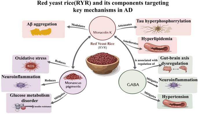
Monacolin K, Monascus pigments, and GABA in RYR exert a multitarget, synergistic effect against Alzheimer's disease (Figure 3), providing a theoretical basis for the potential role of RYR in the prevention and treatment of AD.
Mechanisms by which the active constituents of RYR exert therapeutic effects on AD.
Research on the Prevention and Treatment of AD with RYR Decoction Pieces
Potential Preventive and Therapeutic Effects of RYR Decoction Pieces on AD via Modulation of the Gut Microbiota
Studies have demonstrated that gut microbiota dysbiosis has been increasingly associated with AD pathogenesis [61]. The gut microbiome contributes to neurodevelopment, neurotransmission, and the maintenance of brain homeostasis through microbiota-gut-brain axis pathways [62]. Furthermore, it appears to modulate the bidirectional communication between the gastrointestinal tract and the central nervous system in both health and disease. The gut microbiota communicates bidirectionally with the central nervous system through neural pathways including both intrinsic and extrinsic components, endocrine, and immune pathways; this complex signaling network is referred to as the “microbiota-gut-brain” axis [62]. Dysbiosis of the gut microbiota, acting through this axis, has been associated with Aβ peptide deposition, tau protein phosphorylation, neuroinflammation, metabolic dysfunction, and oxidative stress [61]. Monascus-fermented RYR exerts modulatory effects on the gut microbiota. A study indicated that fermented RYR modulates gut microbial composition: lovastatin, Monascus pigments, red yeast polysaccharides, and GABA present in SWM008-fermented RYR can increase the abundance of beneficial taxa such as Duncaniella and Akkermansia while suppressing pathogenic bacteria [63]. Concurrently, these compounds enhance production of beneficial microbial metabolites, including vitamin K, nucleotide metabolites, and amino acid-derived biomolecules, which may help mitigate dysbiosis-driven induction of AD [63]. Exopolysaccharides secreted by Monascus during growth serve as significant components with potential prebiotic effects, especially those induced by genistein stimulation (exopolysaccharides, G-EMP). Research indicated that G-EMP can reach the colon, where it is selectively utilized by the gut microbiota [64]. This utilization helps preserve the intestinal barrier and inhibit inflammatory pathways, promoting the proliferation of beneficial bacteria, suppressing potentially harmful microbes, and increasing advantageous metabolites such as short-chain fatty acids [64]. The lipid-lowering effects of RYR can modulate lipid metabolic dysregulation in the intestines of high-fat diet-fed rats. Analysis of fecal microbiota from these rats revealed that RYR altered gut microbial composition and ameliorated gut dysbiosis induced by hyperlipidemia [65]. By modulating the gut microbiota, RYR may effectively reduce neuroinflammation and rectify brain dysfunction via the gut-microbiota-brain axis, thereby exerting anti-Alzheimer's disease effects.
Experimental Study on the Anti-AD and Neuroprotective Effects of Red Yeast Rice Decoction Pieces
Current research on RYR concerning its anti-Alzheimer's disease and neuroprotective properties remains limited, primarily focusing on in vitro and in vivo studies. One in vivo investigation revealed that RYR offers neuroprotective effects in a mouse model of Parkinson's disease through antioxidant and anti-neuroinflammatory mechanisms, suggesting its potential for the treatment or prevention of nervous system disorders [66]. Conditions of zinc deficiency in the brain, which have been associated with AD, contribute to age-related memory decline and hippocampal pathology [67]. In an in vivo study involving zinc-deficient rats, the administration of RYR significantly enhanced antioxidant enzyme activity and neuronal function, while also preserving cortical and hippocampal integrity, irrespective of zinc supplementation status [68]. Although these findings do not directly demonstrate anti-AD effects of RYR, they suggest that the antioxidative and anti-neuroinflammatory activities of RYR-related metabolites may warrant further investigation in AD-related contexts, particularly in relation to oxidative stress, neuroinflammation, memory impairment, and hippocampal injury [66, 68]. Lee et al. (2008) demonstrated in vitro that an extract of RYR fermented by Monascus purpureus NTU568 can alleviate Aβ40-induced toxicity in PC12 cells through its antioxidant and anti-inflammatory properties [69]. The study included a comparator group treated with an equivalent dose of lovastatin alone and found that the RYR extract resulted in a more pronounced reduction of Aβ40 toxicity, along with enhanced antioxidant and anti-inflammatory effects, which the authors attributed to synergistic interactions among multiple components [69]. In an in vivo study involving rats, an ethanol extract of RYR was administered to a hyperlipidemic AD rat model that received an intracerebroventricular injection of Aβ40; the RYR extract significantly reversed memory deficits. Mechanistic analyses indicated that these effects may be mediated by the inhibition of β-secretase activity, leading to decreased Aβ40 accumulation, and by the promotion of amyloid precursor protein processing in the hippocampus toward the production of the neuroprotective sAPPα [70, 71]. A recent experimental study in the amyloid precursor protein (APP) transgenic J20 mouse model of AD showed that RYR treatment reduced hippocampal Aβ peptide levels, enhanced brain cholinergic activity, attenuated neuroinflammation, and reduced release of proinflammatory cytokines [45]. The study proposed that the anti-AD mechanism of RYR may involve activation of PPAR-γ; among the potential downstream effects of PPAR-γ activation are inhibition of BACE1-mediated Aβ40 and Aβ42 production, reduction of tau hyperphosphorylation, and modulation of neuroinflammation [45]. These findings indicate the neuroprotective potential of RYR in vivo and in animal models, highlighting its multi-target anti-AD properties. However, clinical research remains limited.
Discussion
Potential advantages of RYR in AD-related modulation
The pathogenesis of AD is intricate and multifactorial, often occurring alongside metabolic disorders such as hyperlipidemia, hypertension, diabetes, and metabolic syndrome. These chronic metabolic conditions and AD interact synergistically, mutually reinforcing each other. Therefore, prevention and therapeutic strategies for AD should adopt a multifaceted approach targeting various angles and mechanisms. Current synthetic targeted pharmaceuticals encounter significant limitations, including restricted therapeutic efficacy and challenges in managing adverse reactions. A comprehensive therapeutic strategy that simultaneously modulates Aβ peptide production and clearance, inhibits excessive tau phosphorylation, alleviates neuroinflammation, improves metabolic disorders, and regulates gut microbiota homeostasis may hold greater promise than single-target drugs. Metabolites found in RYR—such as monacolin K, Monascus pigments, and GABA, may exhibit multi-effect, multi-target synergistic actions [45,69], providing antihyperlipidemic, antidiabetic, antihypertensive, antioxidant, anti-neuroinflammatory, anti-Aβ peptide deposition, tau phosphorylation-related, and gut microbiota-modulating properties [22, 42, 45, 65, 69]. However, current research on the synergistic effects of various RYR metabolites in AD-related pathologies remains limited, and further research is needed. These attributes indicate significant potential for RYR in the prevention and treatment of complex disorders such as AD. From the perspective of long-term disease progression, the onset of AD is protracted and insidious during its early stages. Once established, AD proves challenging to treat. Consequently, RYR may warrant further evaluation as a preventive measure and an early intervention for AD, potentially mitigating its progression associated with various metabolic disorders and facilitating timely intervention in the initial stages.
Safety standards and citrinin control
Although RYR is a natural fermented product with multi-component and multi-target characteristics, it should not be regarded as inherently free of safety concerns. Because monacolin K is chemically identical or closely related to lovastatin, monacolin K-containing RYR preparations may produce statin-like adverse effects, including hepatotoxicity, myopathy, and, rarely, rhabdomyolysis [72]. In addition, citrinin, a nephrotoxic mycotoxin that can be produced during Monascus fermentation, represents an important quality and safety concern for RYR preparations [73]. Citrinin contamination is particularly relevant when considering long-term preventive or adjunctive use of RYR in AD-related populations, who are often older and may have comorbid renal, hepatic, or metabolic disorders. Therefore, any future development of RYR-based interventions for AD should prioritize standardized production, quantitative monitoring of monacolin K and citrinin, and the use of well-characterized citrinin-free or low-citrinin-producing Monascus strains [74].
Limitations and perspectives
Currently, medicines derived from RYR have been utilized for lipid reduction with favorable outcomes. However, their application in the prevention and treatment of AD remains largely theoretical. While research has demonstrated that RYR and several of its constituents possess anti-AD properties, clinical studies are limited. Aside from lovastatin, which has been subjected to clinical trials for AD treatment [75], RYR preparations and other constituents have not undergone clinical evaluation. Consequently, advancing RYR toward clinical application for AD prevention and therapy presents several challenges. Future initiatives may need to initiate long-term epidemiological studies in high-risk populations or conduct trials involving combination drug regimens. Investigating the anti-AD activities of the various secondary metabolites in RYR, along with their potential synergistic interactions, could clarify the multi-target cooperative advantages of RYR for AD prevention and treatment, suggesting a promising avenue in the anti-AD domain.
Acknowledgements
ChatGPT5 was also used to enrich the colors of figures and enhance their clarity but was not used to produce any content or figure. The authors reviewed and edited the content as needed and take full responsibility for the content of the publication.
All figures were created with Adobe Illustrator. All authors promise that none of the figures were adapted or reproduced from other existing publications.
Funding statement
This work was supported by the General Research Project of Zhejiang Provincial Department of Education (Y202454941) and Zhejiang Chinese Medical University Research Project Talent Special Project (2023RCZXZK23).
Author contributions
Qining Quan and Ronghua Pan: Writing—Original draft preparation and Editing. Haolin Liu and Lizhong Zheng: Writing—Figures and Reviewing. Rui Zhang: Conceptualization, Supervision, Writing—Reviewing and Editing.
Competing Interests
The authors have declared that no competing interest exists.
References
1. Twarowski B, Herbet M. Inflammatory processes in alzheimer's disease-pathomechanism, diagnosis and treatment: a review. International Journal of Molecular Sciences. 2023;24:6518
2. Kwon KJ, Kim HY, Han S. et al. Future therapeutic strategies for alzheimer's disease: focus on behavioral and psychological symptoms. International Journal of Molecular Sciences. 2024;25:11338
3. Endo A. Monacolin K, a new hypocholesterolemic agent produced by Monascus species. The journal of antibiotics. 1979;32:852-4
4. Alberts AW, Chen J, Kuron G. et al. Mevinolin: a highly potent competitive inhibitor of hydroxymethylglutaryl-coenzyme A reductase and a cholesterol-lowering agent. Proc Natl Acad Sci U S A. 1980;77:3957-61
5. Huang Z, Xiao L, Mo W. et al. Molecular mechanism of Mok I gene overexpression in enhancing monacolin K production in Monascus pilosus. J Fungi (Basel). 2024;10:721
6. Tolman KG. The liver and lovastatin. Am J Cardiol. 2002;89:1374-80
7. Zheng Y, Zheng Y, Huang Z. et al. Investigating the biosynthesis pathways and hypolipidemic mechanisms of monacolin K in Monascus species. Phytomedicine. 2025;147:157170
8. Lin C, Lin Y, Lin R. et al. Neurocytoprotective effects of aliphatic hydroxamates from lovastatin, a secondary metabolite from Monascus-fermented red mold rice, in 6-hydroxydopamine (6-ohda)-treated nerve growth factor (ngf)-differentiated pc12 cells. Acs Chem Neurosci. 2015;20:716-24
9. Petek B, Häbel H, Xu H. et al. Statins and cognitive decline in patients with Alzheimer's and mixed dementia: a longitudinal registry-based cohort study. Alzheimer's Res Ther. 2023;15:220
10. Husakova M, Patakova P. Purified Monascus pigments biological activities and mechanisms of action. J Nat Prod. 2025;88:607-15
11. Majhi S, Dhale MA, Honganoor Puttananjaiah M. Inhibitory effect of Monascus purpureus pigment extracts against fungi and mechanism of action. Front Sustain Food Syst. 2023;7:1100961
12. Lin C, Lin P, Hsu Y. Monascus-fermented metabolites repressed amyloid β-peptide-induced neurotoxicity and inflammatory response in in vitro and in vivo studies. J Funct Foods. 2023;104:105509
13. Lin C, Lin T, Pan T. Alleviation of metabolic syndrome by monascin and ankaflavin: the perspective of Monascus functional foods. Food Funct. 2017;8:2102-9
14. Wu L, Zhou K, Yang Z. Monascuspiloin from Monascus-fermented red mold rice alleviates alcoholic liver injury and modulates intestinal microbiota. Foods. 2022;11:3048
15. Kowalski K, Mulak A. Brain-gut-microbiota axis in alzheimer's disease. J Neurogastroenterol Motil. 2019;25:48-60
16. Marina Diana A B, Joan Quílez B CD, Rafecas M. Gamma-aminobutyric acid as a bioactive compound in foods: a review. J Funct Foods. 2014;10:407-20
17. Watanabe M, Maemura K, Kanbara K. et al. Gaba and gaba receptors in the central nervous system and other organs. Int Rev Cytol. 2002;213:1-43
18. Ngo D, Vo TS. An updated review on pharmaceutical properties of gamma-aminobutyric acid. Molecules. 2019;24:2678
19. Zhang J, Wu S, Meng N. et al. The mechanism of gaba in attenuating neuroinflammation in alzheimer's disease: cp/cebpα/mir-34a-mediated suppression of hdac2/3 in astrocytes. Foods. 2026;15:837
20. Smith AK, Wade AR, Penkman KEH. et al. Dietary modulation of cortical excitation and inhibition. J Psychopharmacol. 2017;31:632-7
21. Liwinski T, Lang UE, Brühl AB. et al. Exploring the therapeutic potential of gamma-aminobutyric acid in stress and depressive disorders through the gut-brain axis. Biomedicines. 2023;11:3128
22. Hsieh P, Tai Y. Aqueous extract of Monascus purpureus m9011 prevents and reverses fructose-induced hypertension in rats. J Agric Food Chem. 2003;51:3945-50
23. Mick GJ, McCormick KL. The role of gaba in type 1 diabetes. Frontiers in Endocrinology (Lausanne). 2024;15:1453396
24. Sparks DL, Scheff SW, Hunsaker JC. et al. Induction of alzheimer-like beta-amyloid immunoreactivity in the brains of rabbits with dietary cholesterol. Exp Neurol. 1994;126:88-94
25. Kivipelto M, Helkala EL, Hanninen T. et al. Midlife vascular risk factors and late-life mild cognitive impairment: a population-based study. Neurology. 2001;56:1683-9
26. Cheng GW, Mok KK, Yeung SH. et al. Apolipoprotein e ε4 mediates myelin breakdown by targeting oligodendrocytes in sporadic alzheimer disease. Journal of Neuropathology & Experimental Neurology. 2022;81:717-30
27. Strittmatter WJ, Saunders AM, Schmechel D. et al. Apolipoprotein e: high-avidity binding to β-amyloid and increased frequency of type 4 allele in late-onset familial alzheimer disease. Proc Natl Acad Sci U S A. 1993;90:1977-81
28. Poirier J. Apolipoprotein e and alzheimer's disease. A role in amyloid catabolism. Ann N Y Acad Sci. 2000;924:81-90
29. Höglund K Blennow K. Effect of hmg-coa reductase inhibitors on beta-amyloid peptide levels: implications for alzheimer's disease. CNS Drugs. 2007;21:449-62
30. Buxbaum JD, Geoghagen NSM, Friedhoff LT. Cholesterol depletion with physiological concentrations of a statin decreases the formation of the alzheimer amyloid aβ peptide. Journal of Alzheimer's Disease. 2001;3:221-9
31. Buxbaum JD, Cullen E, Friedhoff LT. Pharmacological concentrations of the hmg-coa reductase inhibitor lovastatin decrease the formation of the alzheimer β-amyloid peptide in vitro and in patients. Frontiers in Bioscience. 2002;7:50-9
32. Petek B, Häbel H, Xu H. et al. Statins and cognitive decline in patients with alzheimer's and mixed dementia: a longitudinal registry-based cohort study. Alzheimer's Research & Therapy. 2023;15:220
33. Area-Gomez E, Schon EA. Towards a unitary hypothesis of alzheimer's disease pathogenesis. Journal of Alzheimer's Disease. 2024;98:1243-75
34. Reis PA, Estato V, Da Silva TI. et al. Statins decrease neuroinflammation and prevent cognitive impairment after cerebral malaria. PloS Pathog. 2012;8:e1003099
35. Catalão CHR, Da Costa LHA, Dos Santos JR. et al. Mitigating neuroinflammation in cognitive areas: exploring the impact of hmg-coa reductase inhibitor. Biochem J. 2024;481:1585-602
36. Pahan K, Sheikh FG, Namboodiri AM. et al. Lovastatin and phenylacetate inhibit the induction of nitric oxide synthase and cytokines in rat primary astrocytes, microglia, and macrophages. J Clin Invest. 1997;100:2671-9
37. Busche MA, Hyman BT. Synergy between amyloid-β and tau in alzheimer's disease. Nat Neurosci. 2020;23:1183-93
38. Xu Y, Dong Y, Wang C. et al. Lovastatin attenuates sevoflurane-induced cognitive disorder in aged rats via reducing Aβ accumulation. Neurochem Int. 2021;148:105078
39. Won JS, Im YB, Khan M. et al. Lovastatin inhibits amyloid precursor protein (app) β-cleavage through reduction of app distribution in lubrol wx extractable low density lipid rafts. J Neurochem. 2008;105:1536-49
40. Tamboli IY, Barth E, Christian L. et al. Statins promote the degradation of extracellular amyloid β-peptide by microglia via stimulation of exosome-associated insulin-degrading enzyme (IDE) secretion. Journal of Biological Chemistry. 2010;285:37405-14
41. Li R, Xu DE, Ma T. Lovastatin suppresses the aberrant tau phosphorylation from FTDP-17 mutation and okadaic acid-induction in rat primary neurons. Neuroscience. 2015;294:14-20
42. Wu S, Dong C, Zhang M. et al. Revealing the hypoglycemic effect of red yeast rice: perspectives from the inhibition of α-glucosidase and the anti-glycation capability by ankaflavin and monascin. Foods. 2024;13:1573
43. Chen Y, Yu Y. Tau and neuroinflammation in alzheimer's disease: interplay mechanisms and clinical translation. J Neuroinflammation. 2023;20:165
44. Husakova M, Patakova P. Purified Monascus pigments: biological activities and mechanisms of action. J Nat Prod. 2025;88:607-15
45. Fang M, Cheng IH, Chen C. Monascus purpureus fermented product ameliorates learning and memory impairment in the amyloid precursor protein transgenic j20 mouse model of alzheimer's disease. Fermentation (Basel). 2022;8:193
46. Lee B, Hsu W, Huang T. et al. Effects of monascin on anti-inflammation mediated by Nrf2 activation in advanced glycation end product-treated thp-1 monocytes and methylglyoxal-treated wistar rats. J Agric Food Chem. 2013;61:1288-98
47. Butterfield DA, Halliwell B. Oxidative stress, dysfunctional glucose metabolism, and alzheimer disease. Nat Rev Neurosci. 2019;20:148-60
48. Yunquan Zheng, Qisheng Pan, Liuda M. Monascus pigment rubropunctatin derivative fzu- h reduces ab(1-42)-induced neurotoxicity in neuro-2a cells. RSC Adv. 2018;8:17389
49. Kciuk M, Kruczkowska W, Gałęziewska J. et al. Alzheimer's disease as type 3 diabetes: understanding the link and implications. International Journal of Molecular Sciences. 2024;25:11955
50. Nguyen TT, Ta QTH, Nguyen TKO. et al. Type 3 diabetes and its role implications in alzheimer's disease. International Journal of Molecular Sciences. 2020;21:3165
51. Chae CW, Lee HJ, Choi GE. et al. High glucose-mediated PICALM and mTORC1 modulate processing of amyloid precursor protein via endosomal abnormalities. Br J Pharmacol. 2020;177:3828-47
52. Wu S, Sun Y, Chen D. et al. The noncovalent conjugations of human serum albumin (HSA) with ms/ak and the effect on anti-oxidant capacity as well as anti-glycation activity of Monascus yellow pigments. Food Funct. 2021;12:3692-704
53. Yang H, Pan R, Wang J. et al. Modulation of the gut microbiota and liver transcriptome by red yeast rice and Monascus pigment fermented by purple Monascus shm1105 in rats fed with a high-fat diet. Front Pharmacol. 2020;11:599760
54. Zheng Wenwei, Lin Jun, Lin Feng. et al. Comparison of lipid-lowering efficacy of functional Monascus and lovastatin[In Chinese]. China Brewing. 2022;41:116-9
55. Ablimit Arzugul, Wang Haijiao, Wang Congcong. et al. Research Progress in the Blood Lipid-Lowering Activity and Application Status of Monascus[In Chinese]. Journal of Food Science and Biotechnology. 2024;43:11-22
56. Almutairi S, Sivadas A, Kwakowsky A. The effect of oral gaba on the nervous system: potential for therapeutic intervention. Nutraceuticals. 2024;4:241-59
57. Czapski GA, Strosznajder JB. Glutamate and gaba in microglia-neuron cross-talk in alzheimer's disease. International Journal of Molecular Sciences. 2021;22:11677
58. Tabassum S, Ahmad S, Madiha S. et al. Impact of oral supplementation of glutamate and gaba on memory performance and neurochemical profile in hippocampus of rats. Pak J Pharm Sci. 2017;30:1013-21
59. Conn KA, Borsom EM, Cope EK. Implications of microbe-derived ɣ-aminobutyric acid (GABA) in gut and brain barrier integrity and gabaergic signaling in alzheimer's disease. Gut Microbes. 2024;16:2371950
60. Pacholko A, Iadecola C. Hypertension, neurodegeneration, and cognitive decline. Hypertension. 2024;81:991-1007
61. Nguyen NM, Cho J, Lee C. Gut microbiota and alzheimer's disease: how to study and apply their relationship. International Journal of Molecular Sciences. 2023;24:4047
62. Morais LH, Schreiber HL, Mazmanian SK. The gut microbiota - brain axis in behaviour and brain disorders. Nat Rev Microbiol. 2021;19:241-55
63. Yang P, You C, Lin Y. et al. Effects of Monascus pilosus SWM008-fermented red mold rice and its functional components on gut microbiota and metabolic health in rats. Foods. 2025;14:651
64. Xie Liuming, Cheng xianxiang, Qi xing. et al. Monascus purpureus ameliorates dextran sulfate sodium-induced colitis via protecting the intestinal barrier aganist oxidative stress, and regulating intestinal flora [In Chinese] CIFST. 2023: 590-1.
65. Zhou W, Guo R, Guo W. et al. Monascus yellow, red and orange pigments from red yeast rice ameliorate lipid metabolic disorders and gut microbiota dysbiosis in wistar rats fed on a high-fat diet. Food Funct. 2019;10:1073-84
66. Lin Q, Hou S, Dai Y. et al. Monascin exhibits neuroprotective effects in rotenone model of parkinson's disease via antioxidation and anti-neuroinflammation. Cellular, Molecular and Developmental Neuroscience. 2020;31:637-43
67. Rivers-Auty J, Tapia VS, White CS. et al. Zinc status alters Alzheimer's disease progression through NLRP3-dependent inflammation. The Journal of Neuroscience. 2021;41:3025-38
68. Lee B, Ho B, Wang C. et al. Red mold rice promoted antioxidase activity against oxidative injury and improved the memory ability of zinc-deficient rats. J Agric Food Chem. 2009;57:10600-7
69. Lee C, Wang J, Pan T. Red mold rice extract represses amyloid beta peptide-induced neurotoxicity via potent synergism of anti-inflammatory and antioxidative effect. Appl Microbiol Biotechnol. 2008;79:829-41
70. Lee CL, Kuo TF, Wang JJ. et al. Red mold rice promotes neuroprotective sappalpha secretion instead of alzheimer's risk factors and amyloid beta expression in hyperlipidemic abeta40-infused rats. J Agric Food Chem. 2010;58:2230-8
71. Lee CL, Kuo TF, Wang JJ. et al. Red mold rice ameliorates impairment of memory and learning ability in intracerebroventricular amyloid β-infused rat by repressing amyloid β accumulation. J Neurosci Res. 2007;85:3171-82
72. Filipi M, Aggett P, Aguilar F. Scientific opinion on the safety of monacolins in red yeast rice. EFSA J. 2018;16:e5368
73. Zek MT, Ałtyn I, Kosicki R. Dietary supplements based on red yeast rice-a source of citrinin? Toxins. 2021;13:497
74. Farawahida AH, Palmer J, Flint S. Monascus spp. and citrinin: identification, selection of Monascus spp. isolates, occurrence, detection and reduction of citrinin during the fermentation of red fermented rice. Int J Food Microbiol. 2022;379:109829
75. Sadowska A, Osiński P, Roztocka A. et al. Statins-from fungi to pharmacy. International Journal of Molecular Sciences. 2024;25:466
Author contact
Corresponding author: Rui Zhang, zhangruiedu.cn