Impact Factor ISSN: 1449-1907




Int J Med Sci 2026; 23(9):2921-2938. doi:10.7150/ijms.136917 This issue Cite
Review
Sjögren's Disease-Associated Renal Tubular Acidosis: An Integrated Immuno-Tubular Model of Tubular Dysfunction
Chien-Lin Lu1,2, Chia-Chao Wu3, Te-Chao Fang4,5,6, Kuo-Cheng Lu1,7 
1. Division of Nephrology, Department of Internal Medicine, Fu Jen Catholic University Hospital, Fu Jen Catholic University, New Taipei City 24352, Taiwan.
2. School of Medicine, College of Medicine, Fu Jen Catholic University, New Taipei City 24205, Taiwan.
3. Division of Nephrology, Department of Internal Medicine, Tri-Service General Hospital, National Defense Medical Center, Taipei 11490, Taiwan.
4. Division of Nephrology, Department of Internal Medicine, School of Medicine, College of Medicine, Taipei Medical University, Taipei 11031, Taiwan.
5. Division of Nephrology, Department of Internal Medicine, Taipei Medical University Hospital, Taipei Medical University, Taipei 11031, Taiwan.
6. Taipei Medical University-Research Center of Urology and Kidney, Taipei Medical University, Taipei 11031, Taiwan.
7. Division of Nephrology, Department of Medicine, Taipei Tzu Chi Hospital, Buddhist Tzu Chi Medical Foundation, New Taipei City 23142, Taiwan.
Received 2026-4-28; Accepted 2026-7-27; Published 2026-7-30
Abstract
Sjögren's disease (SjD) is traditionally viewed as an autoimmune exocrinopathy; however, renal involvement—particularly renal tubular acidosis (RTA)—is an underrecognized extraglandular manifestation that often precedes classical sicca symptoms and contributes to diagnostic delay. Distal RTA (dRTA) represents the dominant phenotype and arises from immune-mediated injury to α-intercalated cells, leading to disruption of key acidification components, including vacuolar H+-ATPase (V-ATPase), carbonic anhydrase II (CAII), and anion exchanger 1 (AE1). Beyond impaired distal proton secretion, systemic acidosis is proposed to induce adaptive responses in the proximal tubule, based largely on general renal physiology rather than SjD-specific data. Intracellular acidification may enhance sodium-dependent dicarboxylate cotransporter 1 (NaDC-1)-mediated citrate reabsorption and mitochondrial citrate metabolism. These processes may reduce urinary citrate excretion (hypocitraturia) and promote a lithogenic milieu characterized by calcium phosphate supersaturation. This integrated distal-proximal axis provides a plausible mechanistic link between tubular dysfunction and clinical manifestations such as nephrolithiasis, nephrocalcinosis, and hypokalemic paralysis. Incomplete dRTA further expands the disease spectrum and is frequently overlooked despite its clinical relevance. Current management is centered on alkali and potassium replacement, which correct metabolic abnormalities but do not address underlying immune-mediated injury. Treatment response is stage-dependent and often limited once fibrosis is established. This review proposes an integrated immuno-tubular framework that links clinical phenotype, pathogenesis, and therapeutic strategy, and highlights the need for early detection, biomarker-driven risk stratification—particularly using hypocitraturia—and incorporation of renal-specific endpoints to enable mechanism-based and precision-guided care.
Keywords: acidosis, citrate, hypocitraturia, renal tubular acidosis, Sjögren's disease, tubulointerstitial nephritis
1. Introduction
Sjögren's disease (SjD) is traditionally conceptualized as an autoimmune exocrinopathy defined by sicca symptoms; however, this gland-centric view does not adequately capture a subset of patients in whom extraglandular manifestations predominate [1, 2]. In this review, the term "Sjögren's disease" is used in place of the older "Sjögren's syndrome" to emphasize its systemic autoimmune nature rather than a symptom-defined syndrome. In clinical practice, renal tubular acidosis (RTA)—particularly distal RTA (dRTA)—often presents with hypokalemic paralysis, nephrolithiasis, or nephrocalcinosis, frequently in the absence of overt sicca features [3, 4]. In a compilation of 440 published case reports and small case series of SjD-associated RTA (rather than a systematically ascertained cohort), only 7.7% had a prior diagnosis of SjD, whereas hypokalemic paralysis was the initial presentation in 63.6% [3]. Because this dataset aggregates individually published cases rather than a prospectively ascertained population, these figures likely overstate the true frequency of clinically dramatic presentations such as hypokalemic paralysis due to reporting/publication bias, and are best interpreted as characterizing the published case literature rather than the general SjD-RTA population. As a result, patients are commonly evaluated within nephrological or neurological pathways without recognition of an underlying systemic autoimmune disorder, leading to diagnostic delay [5, 6]. Ongoing tubulointerstitial nephritis (TIN) may therefore progress to irreversible fibrosis before disease-modifying therapy is initiated [1, 2].
Despite its clinical importance, SjD-associated RTA remains conceptually fragmented. Clinical studies largely emphasize electrolyte disturbances and their downstream complications. In contrast, mechanistic work focuses on immune-mediated disruption of the distal acidification apparatus, including carbonic anhydrase II (CAII), vacuolar H+-ATPase (V-ATPase), and anion exchanger 1 (AE1) [2, 3]. These perspectives are rarely integrated into a framework that connects immune injury to impaired urinary acidification and clinical phenotypes. Subclinical forms such as incomplete dRTA—rare frequently overlooked despite their association with hypocitraturia and increased stone risk [7-9]. In a single-center Brazilian cohort of 42 patients, hypocitraturia predicted dRTA with 100% sensitivity and 91% specificity [7]; while this finding highlights that reliance on overt acidosis may underestimate disease burden, it derives from a small cohort and awaits external validation before its diagnostic performance can be generalized.
Therapeutic strategies remain largely empirical. Alkali replacement corrects metabolic abnormalities but does not address the underlying immune-mediated injury, and the role of immunosuppression in restoring tubular function is not well defined [2, 4]. In addition, renal-specific endpoints—such as urinary acidification capacity, tubular biomarkers, transporter expression, and nephrocalcinosis progression—are seldom incorporated into clinical studies, limiting mechanistic assessment of treatment response [10, 11].
This review proposes an integrated immuno-tubular framework for SjD-associated RTA that links clinical presentation, immunopathogenesis, and therapeutic strategy. RTA is conceptualized as a continuum from subclinical tubular dysfunction (incomplete dRTA) to overt metabolic acidosis, with emphasis on emerging biomarkers for earlier detection and risk stratification. This approach aims to support a shift from empiric management toward mechanism-based, precision-guided care.
2. Classification of RTA in Sjögren's Disease: Clinical Relevance and Phenotypic Spectrum
RTA in SjD encompasses multiple physiological defects, but their clinical distribution is highly uneven, with dRTA accounting for the vast majority of cases. Although all classical subtypes can occur, most patients present with distal tubular dysfunction, whereas proximal or hyperkalemic forms are uncommon and typically reflect broader or secondary processes.
2.1 Distal RTA (Type 1): The Dominant Phenotype
dRTA represents the principal renal phenotype in SjD and reflects impaired acid secretion by α-intercalated cells in the collecting duct. Proton secretion depends on V-ATPase and basolateral bicarbonate transport via AE1 [12, 13]. In SjD, dysfunction of this system occurs in the setting of autoimmune TIN, with evidence of immune-mediated disruption of V-ATPase, CAII, and AE1 [2, 14].
The biochemical profile is characterized by hyperchloremic normal anion-gap metabolic acidosis with an inappropriately high urine pH (>5.5) despite systemic acidemia [3]. Impaired ammonium excretion is typically reflected by a positive urinary anion gap (UAG) [13]. Hypokalemia is common and may be severe, resulting from increased distal potassium loss and occasionally presenting as hypokalemic paralysis [3, 15].
Persistently alkaline urine, together with hypercalciuria and hypocitraturia, promotes calcium phosphate precipitation, leading to nephrolithiasis and nephrocalcinosis. Chronic acidosis further contributes to bone demineralization and osteomalacia [16]. Clinically, dRTA frequently precedes classical sicca symptoms and is a major driver of delayed diagnosis in SjD [15, 17].
2.2 Proximal RTA (Type 2): Rare and Syndromic Presentation
Proximal RTA (pRTA) is uncommon in SjD and usually occurs as part of generalized proximal tubular dysfunction rather than as an isolated defect. It reflects impaired bicarbonate reabsorption in the proximal tubule, leading to bicarbonate wasting and normal anion-gap metabolic acidosis [18, 19]. In SjD, proximal involvement is most often associated with autoimmune TIN affecting proximal tubular segments [20].
Additional mechanisms may include reduced expression of endocytic receptors such as megalin and cubilin, potentially driven by Th17-mediated inflammation and ectopic germinal center formation [21]. Clinically, pRTA typically presents as Fanconi syndrome, with urinary losses of bicarbonate, phosphate, glucose, uric acid, and amino acids [1, 22]. Associated findings—such as hypophosphatemia, hypouricemia, and normoglycemic glycosuria—serve as key diagnostic clues [1, 20]. Compared with dRTA, proximal involvement is considerably less frequent and usually reflects more diffuse tubular injury rather than a dominant disease phenotype.
2.3 Type 4 RTA and Mixed Forms: Uncommon but Relevant Variants
Type 4 RTA is rare in SjD and is characterized by hyperchloremic normal anion-gap metabolic acidosis with hyperkalemia, typically resulting from aldosterone deficiency or resistance [13, 23]. Reduced aldosterone activity impairs sodium reabsorption via the epithelial sodium channel (ENaC), diminishing the lumen-negative potential and reducing distal hydrogen and potassium secretion [13]. Consistent with this mechanism, urine pH in type 4 RTA is typically appropriately low (< 5.5), reflecting preserved distal acidification despite reduced net acid excretion.
When present, type 4 RTA in SjD often reflects additional contributing conditions, including TIN, diabetes, obstructive uropathy, or medication effects such as renin-angiotensin system inhibitors or potassium-sparing diuretics [24, 25]. Large case series confirm its low prevalence, with only a small proportion of SjD-associated RTA cases demonstrating a hyperkalemic phenotype [3].
Mixed forms, combining distal and proximal defects, are more frequently recognized and usually indicate more extensive tubulointerstitial involvement [13]. These mixed forms reflect combined defects in both distal acid secretion and proximal bicarbonate handling and are therefore best interpreted as markers of more extensive tubulointerstitial injury rather than a distinct disease category. Clinically, the coexistence of hypokalemia with features of proximal tubular dysfunction or variable acidification capacity should prompt consideration of mixed RTA.
2.4 Incomplete Distal RTA: A Subclinical and Early Disease Form
Incomplete dRTA represents a subclinical defect in urinary acidification in which serum bicarbonate remains normal at baseline but maximal urinary acidification is impaired [26, 27]. Diagnosis requires provocative testing, such as ammonium chloride loading or the furosemide-fludrocortisone test, demonstrating failure to lower urine pH below approximately 5.3-5.5 [27]. Ammonium chloride loading remains the diagnostic gold standard but is frequently poorly tolerated, commonly causing nausea, vomiting, and abdominal discomfort, and it induces a systemic acid load that warrants caution in patients with hepatic impairment or baseline acidosis [28, 29]. The furosemide-fludrocortisone test avoids this acid load and is associated with substantially fewer gastrointestinal side effects, making it more feasible for routine outpatient use [28]. However, its diagnostic performance is test- and population-dependent: in a prospective cohort of kidney stone formers, furosemide-fludrocortisone testing showed 77% sensitivity, 85% specificity, and a high negative predictive value (98%) but a low positive predictive value (30%) against ammonium chloride, supporting its role in screening or exclusion rather than confirmation [30]. Notably, in primary Sjögren's disease specifically, furosemide-fludrocortisone testing performed less well by every diagnostic metric against ammonium chloride: sensitivity and specificity were 65% and 68%, respectively (compared with 77% and 85% in the stone-former cohort above), with a correspondingly lower negative predictive value (82% vs. 98%). Positive predictive value was numerically higher (46% vs. 30%), a difference attributable at least in part to the substantially higher prevalence of incomplete dRTA in the SjD cohort (25%) than in the stone-former cohort (8%), which mathematically raises PPV and lowers NPV independent of test accuracy. The authors concluded that the furosemide-fludrocortisone test should not replace ammonium chloride loading for confirmatory diagnosis in this population [9]. In routine clinical practice, the furosemide-fludrocortisone test is therefore reasonably used as an initial, better-tolerated screening tool, with ammonium chloride loading reserved for confirmatory testing when the diagnosis remains uncertain. Rather than a discrete entity, incomplete dRTA is best understood as part of a continuum of distal acidification capacity reflecting partial α-intercalated cell dysfunction [31, 32].
Despite the absence of overt acidosis, incomplete dRTA is clinically relevant. Patients often present with nephrolithiasis or nephrocalcinosis driven by persistently alkaline urine and hypocitraturia [13, 33]. In autoimmune settings, including SjD, it may represent early tubular injury preceding overt dRTA [2]. Recognition is particularly important in patients with unexplained recurrent calcium stones, as early intervention may prevent progression. These features underscore incomplete dRTA as an underrecognized early disease phenotype and a potential target for early detection.
The clinical, biochemical, and phenotypic distinctions among RTA subtypes in SjD are summarized in Table 1.
Clinical and Biochemical Features of RTA Subtypes in Sjögren's Disease.
| Feature | Distal RTA (Type 1) | Proximal RTA (Type 2) | Mixed RTA | Type 4 RTA | Incomplete dRTA |
|---|---|---|---|---|---|
| Relative frequency in SjD | Predominant phenotype (~80-90%) [3] | Rare [1, 20] | Occasional [13] | Very rare [3] | 6-25% [7-9] (variable across cohorts) |
| Primary defect | Distal acid secretion failure (α-intercalated cells) | Impaired proximal bicarbonate reabsorption | Combined proximal and distal defects | Aldosterone deficiency or resistance | Partial distal acidification defect |
| Serum HCO3- | ↓ | ↓ | ↓ | ↓ | Normal |
| Serum K+ | ↓ (often marked) | ↓ (usually mild) | ↓ | ↑ | Normal or ↓ |
| Urine pH | > 5.5 (inappropriately elevated) | Variable (< 5.5 after steady state) | Variable | Typically <5.5 (may be variable if a secondary or mixed tubular process is present) | >5.5 (after acid load) |
| Urine anion gap (UAG) | Positive | Variable | Variable | Positive (reflecting reduced NH4+ excretion) | Positive |
| FEHCO3- | < 5% | > 10-15% | Variable (may be elevated if proximal component present) | < 5% | < 5% |
| Key clinical features | Hypokalemic paralysis, nephrolithiasis, nephrocalcinosis | Fanconi syndrome (phosphaturia, glycosuria) | Overlapping features | Hyperkalemia, mild acidosis | Recurrent stones, nephrocalcinosis |
| Pathophysiology in SjD | TIN with V-ATPase/AE1/CAII dysfunction | Diffuse tubular injury | Extensive tubulopathy | Usually secondary or comorbid | Early or subclinical tubular injury |
| Clinical implications | Dominant phenotype; may represent initial presentation of SjD | Suggests broader proximal tubular involvement | Suggests more extensive disease | Consider evaluation for comorbidities or drug exposure | Often underrecognized; potential target for early detection |
Abbreviations: AE1, anion exchanger 1; CAII, carbonic anhydrase II; FEHCO3-, fractional excretion of bicarbonate; NH4+, ammonium; RTA, renal tubular acidosis; SjD, Sjögren's disease; TIN, tubulointerstitial nephritis; UAG, urine anion gap; V-ATPase, vacuolar H+-ATPase.
3. Pathogenesis of RTA in Sjögren's Disease: An Integrated Immuno-Tubular Model
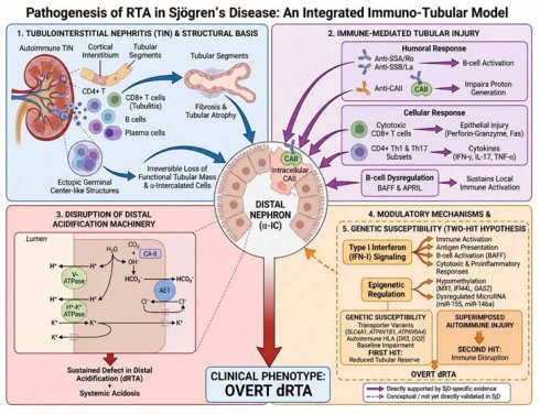
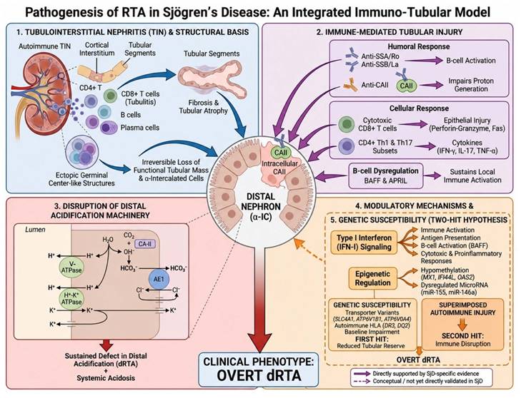
RTA in SjD arises from immune-mediated injury targeting distal tubular cells, with disruption of acidification machinery representing the functional endpoint. Rather than a single mechanism, dRTA reflects a coordinated network of immunological, molecular, and structural processes targeting α-intercalated cells in the distal nephron. These integrated immuno-tubular mechanisms are summarized in Figure 1.
Integrated immuno-tubular model of renal tubular acidosis in Sjögren's disease. Renal tubular acidosis (RTA) in Sjögren's disease (SjD) arises from converging structural, immune, molecular, and genetic mechanisms targeting the distal nephron. (1) Tubulointerstitial nephritis (TIN) with loss of α-intercalated cells; (2) Immune-mediated tubular injury; (3) Disruption of distal acidification machinery (V-ATPase, CAII, and AE1); (4) Type I interferon and epigenetic amplification; (5) Genetic susceptibility via a two-hit model. Solid borders/arrows denote components (1-4) directly supported by SjD-specific evidence; the dashed border/arrow denotes the two-hit genetic susceptibility model, which remains conceptual and has not been directly validated in SjD. Abbreviations: AE1, anion exchanger 1; α-IC, α-intercalated cell; APRIL, a proliferation-inducing ligand; BAFF, B-cell activating factor; CAII, carbonic anhydrase II; dRTA, distal renal tubular acidosis; H+-K+-ATPase, non-gastric H+,K+-ATPase; IFN-I, type I interferon; RTA, renal tubular acidosis; SjD, Sjögren's disease; TIN, tubulointerstitial nephritis; V-ATPase, vacuolar H+-ATPase.
3.1 Tubulointerstitial Nephritis as the Structural Substrate
Autoimmune TIN forms the structural basis of renal involvement in SjD and directly impairs tubular architecture and function [1, 34]. Inflammatory infiltrates are composed predominantly of CD4+ T lymphocytes, with contributions from CD8⁺ T cells, B cells, and plasma cells [20]. CD8+ T cells frequently infiltrate tubular epithelium (tubulitis), supporting direct cytotoxic injury, while B-cell aggregates may form ectopic germinal center-like structures, indicating localized immune activation [35].
Inflammation is typically distributed in the cortical interstitium surrounding tubular segments involved in urinary acidification [20]. Persistent injury promotes interstitial fibrosis and tubular atrophy, resulting in irreversible loss of functional tubular mass [1, 34]. Loss of viable α-intercalated cells progressively limits acidification capacity and underlies the development of persistent dRTA in advanced disease [3, 36].
3.2 Immune-Mediated Tubular Injury
Tubular injury in SjD is driven by coordinated humoral and cellular immune responses targeting renal epithelial cells [1, 20]. Autoantibodies such as anti-SSA/Ro and anti-SSB/La reflect B-cell activation and are associated with renal involvement [37]. Among these, anti-CAII antibodies are mechanistically relevant, as they impair intracellular proton generation and are associated with reduced urinary acidification and increased urine pH [38].
Cellular immunity amplifies this process. Cytotoxic CD8+ T cells induce epithelial injury through perforin-granzyme and Fas-mediated pathways, while CD4+ T-cell subsets, including Th1 and Th17 cells, promote inflammation via cytokines such as interferon-γ, interleukin-17, and tumor necrosis factor-α [39, 40]. In parallel, B-cell dysregulation through B-cell activating factor (BAFF) and a proliferation-inducing ligand (APRIL) sustains local immune activation and supports ectopic lymphoid structures [41, 42]. These combined mechanisms result in persistent epithelial injury and progressive tubular dysfunction.
3.3 Disruption of Distal Acidification Machinery
dRTA reflects failure of coordinated proton secretion by α-intercalated cells in the collecting duct. This process depends on apical V-ATPase, intracellular CAII, and basolateral AE1, which together maintain proton secretion and bicarbonate transport [43, 44]. Disruption of any component reduces net acid excretion and impairs urinary acidification in the setting of systemic acidosis [32, 45]. These processes converge on α-intercalated cells in the distal nephron, which represent the functional locus of acidification failure in SjD-associated dRTA.
In SjD, immune-mediated injury affects multiple elements of this system. Reduced or mislocalized V-ATPase impairs apical proton secretion, while inhibition of CAII limits intracellular proton generation [43]. AE1 dysfunction disrupts basolateral bicarbonate extrusion, leading to intracellular alkalinization and secondary V-ATPase mislocalization [46]. In some cases, selective loss of AE1 or combined transporter defects further reduce the population of functional α-intercalated cells [47]. The net effect is a sustained defect in distal acidification that defines the clinical phenotype of dRTA.
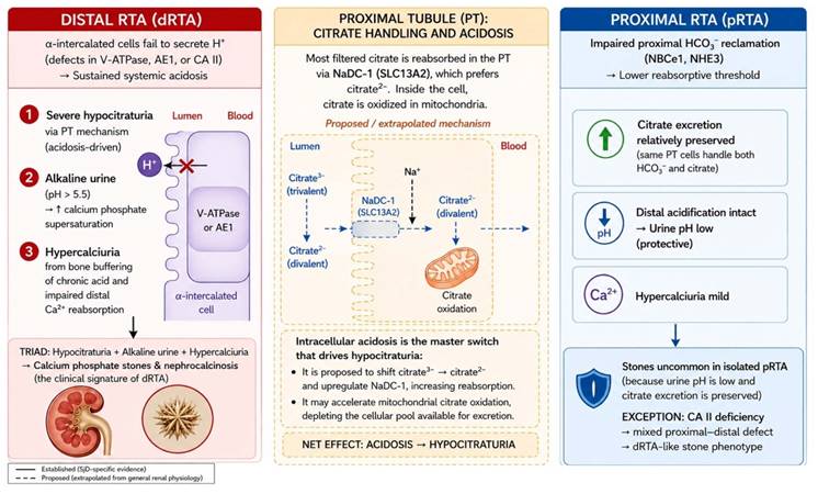
However, impaired distal acidification alone does not fully account for the characteristic lithogenic phenotype observed in SjD-associated dRTA. The proximal contribution to this phenotype should be regarded as a proposed, rather than an established, mechanism: direct SjD-specific evidence for NaDC-1 regulation and mitochondrial citrate flux is currently lacking, although pH-dependent proximal citrate handling is well supported by general renal physiology studies. On this basis, systemic acidosis is proposed to induce adaptive proximal tubular responses that may increase citrate reabsorption and reduce urinary citrate excretion, resulting in hypocitraturia (Figure 2) [7, 45, 48]. Beyond this proposed NaDC-1-mediated pathway, hypocitraturia in dRTA is also compounded by concurrent hypokalemia, which independently promotes proximal citrate reabsorption through intracellular acidification and increased NHE3-mediated hydrogen secretion, irrespective of systemic pH, and by dietary factors such as high animal-protein or low-alkali intake [49]. Hypercalciuria, the other key contributor to the lithogenic phenotype, arises predominantly from chronic metabolic acidosis itself: sustained buffering of the systemic acid load mobilizes calcium and phosphate from bone, while acidosis independently reduces distal tubular calcium reabsorption, together increasing urinary calcium excretion largely independent of dietary calcium intake [50]. Together with alkaline urine and hypercalciuria, hypocitraturia favors calcium phosphate supersaturation and provides a plausible, though not yet directly confirmed, link between distal tubular dysfunction and nephrolithiasis or nephrocalcinosis [7, 48].
Proposed model of distal acidification failure and acidosis-driven proximal citrate handling in Sjögren's disease-associated renal tubular acidosis. Schematic of the distal acidification defect (V-ATPase, CAII, AE1) and its proposed downstream link to proximal citrate handling and hypocitraturia. Solid lines/borders denote the distal pathway, directly supported by SjD-specific evidence; dashed lines/borders denote the proximal citrate-handling pathway, extrapolated from general renal physiology and not yet directly confirmed in SjD. Abbreviations: AE1, anion exchanger 1; CAII, carbonic anhydrase II; dRTA, distal renal tubular acidosis; NaDC-1, sodium-dependent dicarboxylate cotransporter 1; NBCe1, electrogenic sodium bicarbonate cotransporter 1; NHE3, sodium/hydrogen exchanger 3; PT, proximal tubule; pRTA, proximal renal tubular acidosis; V-ATPase, vacuolar H+-ATPase.
3.4 Modulatory Mechanisms: Interferon and Epigenetic Regulation
Type I interferon (IFN-I) signaling is a central amplifier of immune activation in SjD, with widespread upregulation of interferon-stimulated genes in both immune and epithelial compartments [51, 52]. IFN-I enhances antigen presentation, promotes B-cell activation via BAFF, and increases cytotoxic and proinflammatory responses [53]. In tubular epithelial cells, interferon signaling induces cytokine production and cell death pathways, contributing to sustained tubulointerstitial injury [54]. These pathways act as upstream modulators that amplify and sustain immune-mediated tubular injury.
Epigenetic mechanisms reinforce this inflammatory state. Hypomethylation of interferon-responsive genes—including MX1, IFI44L, and OAS2—enhances transcriptional responsiveness to IFN signaling [51, 55, 56]. Dysregulated microRNA expression, particularly increased miR-155 and altered miR-146a, modulates pathways such as nuclear factor κB (NF-κB) and Janus kinase/signal transducer and activator of transcription (JAK/STAT), influencing cytokine production and cell survival [57, 58]. These processes sustain chronic inflammation and perpetuate tubular injury.
3.5 Genetic Susceptibility and the Two-Hit Hypothesis
Genetic factors may plausibly contribute to susceptibility to RTA in SjD by affecting both tubular transport and immune regulation; however, this remains a conceptual framework rather than an established disease mechanism, and direct validation in SjD is currently lacking. Variants in genes encoding acidification transporters—such as solute carrier family 4 member 1 (SLC4A1; AE1), ATP6V1B1, and ATP6V0A4—impair proton secretion and can produce subclinical defects in tubular acidification [45, 59-61]. In parallel, autoimmune susceptibility is associated with HLA haplotypes, particularly HLA-DR3 and DQ2, which predispose to dysregulated immune responses [62, 63]. This baseline impairment may be conceptualized as reduced tubular reserve, which predisposes to overt dysfunction upon immune-mediated injury, although this link has not been directly demonstrated in SjD-associated RTA.
On this basis, a two-hit model has been proposed, in which reduced tubular reserve is unmasked by superimposed autoimmune injury: baseline impairment of acidification capacity would represent the first hit, while immune-mediated disruption of transport proteins would constitute the second, leading to overt dRTA once functional compensation is exceeded. This model is illustrated with a dashed border in Figure 1 to distinguish it from the directly evidenced mechanisms (1-4), as no study has yet demonstrated a direct interaction between transporter gene variants and autoimmune tubular injury in SjD-associated dRTA. Direct validation of this model in SjD remains limited, and integration of genetic and immunological data will be required to define its clinical relevance.
4. Clinical Presentation and Diagnostic Approach to RTA in Sjögren's Disease
RTA in SjD spans a wide clinical spectrum, from incidental biochemical abnormalities to life-threatening complications. Diagnosis requires integration of clinical presentation, biochemical findings, and autoimmune evaluation to facilitate timely recognition.
4.1 Clinical Presentation: From Subclinical Disease to Acute Manifestations
RTA in SjD most commonly presents through hypokalemia rather than acid-base abnormalities. As noted above, in the 440-case published compilation, hypokalemic paralysis was recorded as the presenting feature in 63.6% of published cases, with respiratory paralysis in 8.4% [3]; given the case-report-based nature of this dataset, these frequencies should not be equated with true population prevalence. Clinical manifestations include fatigue, muscle weakness, acute flaccid quadriparesis, and, in severe cases, respiratory failure requiring ventilatory support [64, 65]. These manifestations typically occur in distal RTA with marked hypokalemia and normal anion-gap metabolic acidosis. Hypokalemic paralysis may represent the first manifestation of previously unrecognized SjD, particularly in the absence of sicca symptoms [6, 17].
Chronic distal RTA produces renal and skeletal complications through persistently alkaline urine, hypercalciuria, and hypocitraturia. This environment favors calcium phosphate stone formation and nephrocalcinosis, presenting as renal colic, hematuria, recurrent stones, or incidental imaging findings [45]. Nephrolithiasis and nephrocalcinosis are associated with renal dysfunction in SjD [66]. Long-standing acidosis contributes to bone demineralization, osteomalacia, and chronic bone pain [45, 67]. The presence of unexplained distal RTA, hypokalemic paralysis, nephrocalcinosis, or recurrent calcium stones should prompt evaluation for SjD.
4.2 Biochemical Confirmation of Renal Tubular Acidosis
Diagnosis is typically established by demonstrating hyperchloremic normal anion-gap metabolic acidosis with impaired renal acid handling. Key laboratory findings include reduced serum bicarbonate in the setting of preserved glomerular filtration, while potassium levels help distinguish subtypes—hypokalemia in distal and proximal RTA, and hyperkalemia in type 4 RTA [68]. Alternative causes, including extrarenal bicarbonate loss and advanced chronic kidney disease, should be excluded before confirming a tubular origin [69, 70].
Urinary acidification assessment is central to evaluation. In distal RTA, urine pH remains inappropriately elevated (> 5.3-5.5) despite systemic acidemia [45]. Reduced ammonium excretion is inferred from a positive UAG, whereas a negative UAG suggests extrarenal bicarbonate loss [68]. When results are inconclusive, the urine osmolal gap can provide an estimate of ammonium excretion [71].
The fractional excretion of bicarbonate (FEHCO3) helps differentiate subtypes. Values < 5% support distal RTA, whereas values > 10-15% indicate proximal RTA [68, 72]. Additional urinary findings aid in identifying underlying tubular disorders: low-molecular-weight proteinuria, glycosuria, phosphaturia, and aminoaciduria suggest Fanconi syndrome, whereas isolated tubular abnormalities without significant glomerular proteinuria are more typical of SjD-associated TIN [45].
4.3 Etiological Evaluation: Autoimmunity, Imaging, and Differential Diagnosis
After biochemical confirmation, evaluation focuses on identifying SjD and excluding alternative causes. Serological testing is central. Anti-SSA/Ro and anti-SSB/La antibodies are frequently detected and correlate with renal involvement and disease severity [73, 74]. Additional findings—including antinuclear antibodies (ANA), rheumatoid factor, and polyclonal hypergammaglobulinemia—support systemic autoimmune activity [74, 75]. Complement consumption (low C3/C4) and cryoglobulinemia are associated with renal involvement and increased lymphoma risk [66, 74]. Anti-CAII antibodies provide mechanistic support for tubular dysfunction [38].
Imaging and histology further refine diagnosis. Ultrasonography or computed tomography is typically used to detect nephrolithiasis and nephrocalcinosis, which are strongly linked to renal dysfunction in SjD [66, 73]. Renal biopsy is reserved for cases with diagnostic uncertainty, unexplained decline in kidney function, or when immunosuppressive therapy is considered. Histopathology typically demonstrates plasma cell-rich TIN and allows assessment of inflammatory activity and fibrosis [74, 76]. Biopsy findings directly inform therapeutic decisions: active, cellular TIN supports initiation of immunosuppressive therapy, whereas predominant interstitial fibrosis and tubular atrophy indicate a lower likelihood of treatment response and favor continued metabolic (alkali-based) management alone (see Section 5.1).
A structured differential diagnosis is essential. Drug-induced RTA should be excluded, particularly with exposure to amphotericin B, tenofovir, or ifosfamide [45, 77]. Other autoimmune diseases, including systemic lupus erythematosus and immunoglobulin G4-related disease (IgG4-related disease), may produce similar tubulointerstitial pathology and should be distinguished based on serology and histology [74]. In younger patients or those with a suggestive family history, hereditary distal RTA should be considered, with genetic testing for SLC4A1 and V-ATPase subunits when appropriate [59, 78]. A summary of key distinguishing features across these differential diagnoses is provided in Table 2.
Differential Diagnosis of Distal RTA: SjD-Associated versus Other Autoimmune, Hereditary, and Drug-Induced Causes.
| Category | Example Causes | Key Distinguishing Features vs. SjD-Associated dRTA | Diagnostic Clue |
|---|---|---|---|
| SjD-associated dRTA | Autoimmune TIN in primary Sjögren's disease | Sicca symptoms; anti-SSA/Ro, anti-SSB/La positivity; plasma cell-rich TIN on biopsy | Anti-SSA/SSB, biopsy, sicca assessment |
| Other autoimmune disease | SLE; IgG4-related disease | SLE: anti-dsDNA, hypocomplementemia, multi-organ involvement. IgG4-RD: elevated serum IgG4, storiform fibrosis with IgG4+ plasma cell-rich infiltrate on biopsy [74] | Serology (anti-dsDNA, complement, IgG4) and histology |
| Hereditary dRTA | SLC4A1 (AE1), ATP6V1B1, ATP6V0A4 mutations | Earlier onset (often childhood); family history; may be associated with sensorineural hearing loss (ATP6V1B1); absence of autoimmune serology [59, 78] | Genetic testing; family history; audiometry |
| Drug-induced RTA | Amphotericin B, tenofovir, ifosfamide | Clear temporal relationship to drug exposure; typically reversible after drug withdrawal; absence of autoimmune serology or TIN on biopsy [45, 77] | Medication history; resolution after discontinuation |
5. Therapeutic Strategies for RTA in Sjögren's Disease: A Clinical Framework
Management of RTA in SjD requires simultaneous control of metabolic abnormalities and assessment of underlying immune activity. While alkali therapy is generally required in patients with overt dRTA, immunomodulatory treatment is reserved for selected cases with active TIN or progressive renal involvement. Current practice is largely based on observational data, with limited randomized evidence.
5.1 Principles of Management
Management generally involves two parallel priorities: correction of metabolic derangements and identification of patients who may benefit from immunosuppression. Alkali and potassium replacement correct acidosis and hypokalemia, reduce recurrent weakness, and limit long-term complications including nephrolithiasis, nephrocalcinosis, and bone disease [1, 2]. Maintenance therapy is often required long term in most patients, typically at 1-2 mEq/kg/day in dRTA [79]. In acute presentations such as hypokalemic paralysis or respiratory failure, prompt potassium and alkali replacement leads to rapid clinical improvement [14, 80].
Immunosuppressive therapy is considered in patients with biopsy-proven active TIN, declining kidney function, or persistent tubular dysfunction despite adequate metabolic control [2, 76]. Corticosteroids are the most commonly used initial agent, with mycophenolate mofetil, azathioprine, cyclophosphamide, or rituximab used in selected cases with severe, relapsing, or mixed renal involvement [1, 42, 76]. Treatment response is strongly stage-dependent: active inflammatory TIN may improve, whereas established fibrosis is typically associated with persistent acidification defects and ongoing alkali requirement [10, 81].
5.2 Metabolic Correction: Alkali and Potassium Management
Alkali replacement corrects systemic acidosis and remains the cornerstone of therapy in all patients with overt dRTA. The treatment target is normalization of serum bicarbonate, generally ≥ 22 mmol/L [82]. Sodium bicarbonate and potassium citrate are both effective; however, potassium-containing alkali is generally preferred in patients with hypokalemia or stone disease because it corrects potassium deficit, increases urinary citrate, and reduces calcium stone risk [13, 83]. Sodium-based alkali may increase urinary calcium and should be used cautiously in patients with nephrolithiasis or volume-sensitive conditions [13].
Potassium management depends on severity. Chronic hypokalemia is typically managed with oral potassium salts, whereas severe hypokalemia or hypokalemic paralysis requires intravenous replacement with cardiac monitoring [82]. Correction of acidosis is necessary for sustained potassium control, as persistent acidosis promotes renal potassium wasting [13, 83].
Long-term therapy improves biochemical control, reduces stone risk, and stabilizes nephrocalcinosis [84, 85]. Monitoring should include serum bicarbonate and potassium, urinary calcium and citrate, kidney function, and treatment adherence, as incomplete correction is common in routine practice [82].
5.3 Immunomodulatory Therapy: Indications and Treatment Hierarchy
No formal treatment guidelines exist for SjD-associated RTA. The treatment hierarchy described below is therefore derived from observational cohorts, case series, and extrapolation from the management of TIN in other autoimmune contexts, rather than from formal, evidence-graded recommendations, and should be interpreted accordingly. Direct, controlled evidence linking specific immunosuppressive agents to renal tubular outcomes is limited: reported benefit is largely confined to case series and individual reports of improved urinary acidification, with corticosteroids showing the most consistent benefit when initiated during active inflammation, and no agent, including rituximab, has been evaluated in controlled trials with renal tubular function as a defined endpoint.
Immunosuppression is reserved for patients with evidence of active TIN, particularly when associated with declining kidney function or persistent tubular dysfunction. Treatment decisions are individualized based on disease activity and reversibility [86, 87]. Earlier treatment appears to be associated with better renal recovery, whereas delayed therapy in the presence of fibrosis is linked to incomplete response [87].
Corticosteroids are the most commonly used initial agent and are reported to be most effective during the inflammatory phase [86, 88]. In patients with relapse, steroid dependence, or incomplete response, steroid-sparing agents such as mycophenolate mofetil or azathioprine are commonly used, based on case-series experience [89]. Hydroxychloroquine is often used for systemic disease control but has not been shown to directly improve tubular acidification [90]. Calcineurin inhibitors have been reported in refractory cases but require caution due to nephrotoxicity, and supporting data are limited to small series [86]. Biologic therapies, including rituximab or tumor necrosis factor-α inhibitors, have shown benefit in individual cases or small series, although their role in SjD-associated RTA remains uncertain and is not supported by controlled trial data [91, 92]. The available evidence on renal outcomes and reversibility of tubular dysfunction for each agent is summarized in Table 3.
Evidence for Renal Outcomes and Reversibility of Tubular Dysfunction with Immunosuppressive Agents in SjD-Associated RTA.
| Agent | Role in Treatment Hierarchy | Reported Renal/Tubular Outcome | Evidence Level | Key Limitation |
|---|---|---|---|---|
| Corticosteroids | Most commonly used initial agent, especially during active inflammatory phase | Most consistent reported improvement in tubular function when initiated early | Case series [86-88] | No controlled comparison; benefit declines with established fibrosis |
| Mycophenolate mofetil / Azathioprine | Steroid-sparing, for relapse or incomplete response | Used to maintain control after steroid response; renal-specific outcome data limited | Case series [89] | No systematic renal outcome data; efficacy inferred from clinical experience |
| Hydroxychloroquine | Adjunct for systemic disease control | No direct evidence of improved tubular acidification | [90] | Not renal-outcome directed |
| Calcineurin inhibitors | Refractory cases | Reported benefit in small series | Small case series [86] | Nephrotoxicity risk; very limited data |
| Rituximab | Off-label, selected refractory cases | Individual reports of recovery of urinary acidification | Case reports/small series [93] | Inconsistent systemic benefit in RCTs; renal tubular outcomes not systematically evaluated [94, 95] |
| TNF-α inhibitors | Investigational | Benefit in individual cases | Case reports [91, 92] | Role in SjD-associated RTA remains uncertain |
5.4 Advanced and Emerging Therapies: Limited Evidence
Targeted therapies have been explored in refractory or severe disease, but renal-specific data remain limited. Rituximab has been reported in case-based and small series to improve renal involvement in individual cases, including recovery of urinary acidification [93]. However, randomized trials in SjD have shown inconsistent systemic benefit, and renal tubular outcomes have not been systematically evaluated [94, 95]. At present, rituximab is best considered an off-label option in selected refractory cases.
Other targeted approaches, including B-cell activating factor-directed therapy and Janus kinase inhibition, are supported by mechanistic rationale but lack renal outcome data. Agents targeting the BAFF axis (e.g., belimumab, ianalumab, telitacicept) have shown effects on systemic disease activity but their effects on tubular endpoints have not been evaluated. Similarly, Janus kinase inhibitors may suppress interferon-driven inflammation but remain investigational in SjD-associated RTA [96, 97]. Their clinical role will depend on future studies incorporating renal-specific endpoints.
5.5 Long-Term Management and Monitoring
Long-term management focuses on sustained metabolic control, prevention of complications, and monitoring of disease progression. Treatment targets include normalization of serum bicarbonate and potassium and control of urinary calcium excretion, although adequate control is achieved in only approximately half of patients in cohort studies [82, 84]. Lifelong alkali therapy is often required, with dose adjustment based on disease stability and stone activity [13, 98].
Nephrocalcinosis is common and may affect up to 88% of patients, often in association with nephrolithiasis and declining kidney function [84]. Chronic acidosis contributes to bone demineralization and fracture risk, which can be mitigated with sustained correction and appropriate vitamin D supplementation [99, 100]. Standard chronic kidney disease management should be applied in patients with progressive renal involvement [1].
Long-term follow-up may also include surveillance for lymphoma in selected patients with persistent systemic activity or B-cell-driven disease features [98, 101]. Treatment response is best assessed using combined biochemical and clinical parameters, as structural damage due to fibrosis often limits reversibility despite adequate therapy.
6. Clinical Integration: A Mechanism-Informed Diagnostic and Management Pathway
The mechanistic framework described above can be translated into a structured clinical pathway for SjD-RTA (Figure 3). Evaluation begins when clinical suspicion is raised by hypokalemic paralysis, recurrent calcium phosphate nephrolithiasis or nephrocalcinosis, unexplained normal anion-gap metabolic acidosis, persistently alkaline urine, or hypocitraturia. Because renal manifestations may precede sicca symptoms, early recognition requires a high index of suspicion across clinical settings. Biochemical confirmation relies on identifying hyperchloremic metabolic acidosis with preserved glomerular filtration, supported by urine pH, urinary anion gap, and, when appropriate, fractional excretion of bicarbonate and imaging findings.
Mechanism-informed clinical pathway for the diagnosis and management of Sjögren's disease-associated renal tubular acidosis. Stepwise pathway from clinical suspicion through biochemical confirmation, RTA subtype classification, serologic/histologic evaluation for SjD, risk stratification (hypocitraturia as a lithogenic risk biomarker), and management (metabolic correction with or without immunosuppression). Abbreviations: ANA, antinuclear antibody; eGFR, estimated glomerular filtration rate; FEHCO₃, fractional excretion of bicarbonate; HCO₃, bicarbonate; IFTA, interstitial fibrosis and tubular atrophy; IgG, immunoglobulin G; NAGMA, normal anion-gap metabolic acidosis; RTA, renal tubular acidosis; SjD, Sjögren's disease; UAG, urinary anion gap.
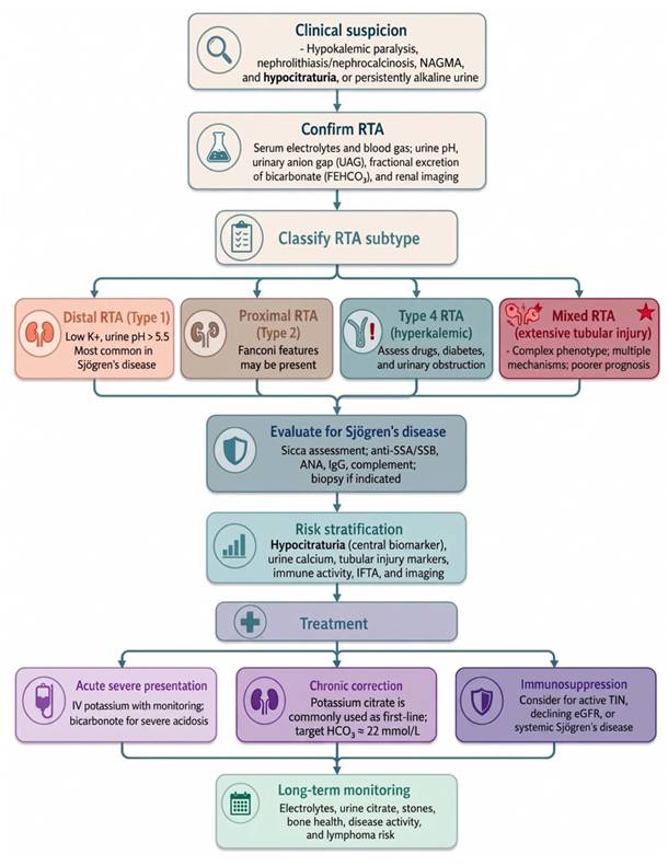
Following confirmation, classification of RTA subtype provides diagnostic direction. dRTA represents the dominant phenotype in SjD, whereas proximal, mixed, and type 4 forms suggest broader or alternative tubular involvement and should prompt evaluation for contributing conditions such as medications, diabetes, or urinary obstruction. Once RTA is established, focused evaluation for underlying SjD is warranted, including serologic testing (anti-SSA/Ro, anti-SSB/La, ANA), immunologic markers, and, when indicated, tissue biopsy. Recognition of the autoimmune context informs both prognosis and treatment selection.
Risk stratification links pathophysiology to clinical outcomes and guides management intensity. Hypocitraturia serves as a clinically useful biomarker, reflecting the combined effects of distal acidification failure and proximal tubular adaptation and associating with lithogenic risk. Management proceeds along two complementary axes: correction of metabolic derangements, typically with potassium-based alkali therapy, and consideration of immunosuppressive treatment in selected patients with active tubulointerstitial inflammation or progressive renal dysfunction.
Long-term management requires integrated monitoring of electrolytes, urinary parameters, stone burden, and systemic disease activity. This pathway provides a practical framework for translating mechanistic insights into individualized, mechanism-informed clinical care.
7. Special Clinical Contexts and Unresolved Challenges in Sjögren's Disease-Associated RTA
RTA in SjD is associated with persistent diagnostic and therapeutic challenges, particularly in early recognition, management of special populations, and limited evidence for treatment selection. These issues affect both clinical outcomes and the ability to implement individualized care.
7.1 Diagnostic Challenges and Interdisciplinary Gaps
Renal manifestations in SjD often precede systemic diagnosis and may remain unrecognized for years. dRTA frequently presents with hypokalemic paralysis, nephrolithiasis, or nephrocalcinosis, while SjD is only identified later following autoimmune evaluation [6, 48]. Renal involvement is well documented to precede, coincide with, or follow sicca symptom onset, though the literature does not converge on a fixed interval: in a large multicenter cohort, renal involvement preceded SjD diagnosis in 17% of affected patients, was identified concurrently with SjD diagnosis in 53%, and developed at a median of 8.7 years after SjD diagnosis in the remaining 30% [102]; conversely, an earlier biopsy series found that SjD diagnosis preceded renal manifestations by a median of 5.5 years [103]. Nephrolithiasis and nephrocalcinosis are reported in approximately 5-21% of patients across small, single-center cohort series [73], with wide variability attributable to differences in imaging modality and referral patterns; a similar pattern is also frequently described in the published case-report literature, where it often represents the presenting clinical clue [3].
Renal involvement is not the only extraglandular presentation that can precede or occur independently of overt sicca symptoms, and awareness of these additional clues may further support earlier interdisciplinary recognition of SjD. Musculoskeletal symptoms are common and may initially draw attention away from an underlying autoimmune diagnosis: non-erosive polyarthralgia occurs in up to 96% of patients and frank polyarthritis in approximately 16.6% [104]. Cutaneous vasculitis, most often manifesting as palpable purpura, is reported in 10-30% of patients and, together with urticarial vasculitis (0.8-21%), frequently co-occurs with anti-SSA/Ro and anti-SSB/La positivity [104]. Peripheral neuropathy is similarly informative: pure sensory neuropathy occurs in 40-49% and sensorimotor polyneuropathy in up to 56% of neurologically affected patients, and may itself be the presenting feature in the absence of sicca complaints [104]. Hypocomplementemia (low C3 in 10-15%, low C4 in 5-20%) and cryoglobulinemia frequently accompany these presentations and additionally flag increased lymphoma risk [104]. Beyond dRTA, other urological manifestations may serve as clues. Interstitial cystitis-like urinary symptoms are more common in SjD than in the general population: a Finnish population-based study found such symptoms up to 15-20 times more frequent among SS patients than controls [105], and a subsequent large Taiwanese nationwide cohort confirmed increased risk of both bladder pain syndrome/interstitial cystitis (HR 2.34) and overactive bladder (HR 1.68) in primary SjD [106]. Interstitial cystitis has occasionally been reported as the presenting clue to previously unrecognized SjD rather than a later complication [107], and in the largest biopsy-confirmed case series, bladder involvement was rarely severe enough to cause acute obstructive renal failure [108]. Acute presentations such as respiratory failure due to severe hypokalemic paralysis from distal RTA have similarly been reported as the initial manifestation of previously unrecognized SjD—in some cases with no prior sicca symptoms and initial misdiagnosis as a neurologic condition such as Guillain-Barré syndrome [109-111]. As with renal involvement, recognizing these atypical presentations requires a high index of suspicion, particularly because even overt sicca symptoms are associated with substantial diagnostic delay in their own right, with a median lag of 115 weeks (over two years) between symptom onset and diagnosis reported in a population-based cohort [112].
This presentation pattern leads to fragmented care, with patients evaluated in nephrology, rheumatology, dermatology, or neurology settings without consideration of an underlying autoimmune disorder. Delay in diagnosis allows progression from active inflammation to fibrosis, reducing the likelihood of treatment response [74, 113]. Early recognition remains challenging and requires consideration of SjD in patients with unexplained dRTA, hypokalemia, or recurrent calcium stones, even in the absence of sicca symptoms [2]. Integration of nephrology and rheumatology pathways remains inconsistent, and standardized referral strategies are lacking. Strengthening this interdisciplinary integration will likely require several concrete strategies. Standardized referral criteria could be established so that unexplained dRTA, persistent hypokalemia, or recurrent calcium nephrolithiasis automatically prompts serologic screening (anti-SSA/Ro, anti-SSB/La, ANA) and rheumatology referral, while conversely, rheumatologists managing confirmed SjD could incorporate routine screening for urinary acidification defects (urine pH, urinary anion gap) as part of standard extraglandular surveillance. Joint nephrology-rheumatology clinics or regular multidisciplinary case conferences may facilitate earlier recognition and shared management of biopsy-proven TIN. Embedding structured clinical decision-support prompts into electronic health records—triggered by combinations such as hypokalemia with alkaline urine, or nephrocalcinosis on imaging—could further reduce reliance on individual clinician awareness. Finally, multicenter registries linking nephrology and rheumatology cohorts would support standardized, prospective validation of candidate biomarkers such as hypocitraturia and enable systematic study of referral pathways and their impact on diagnostic delay.
7.2 Special Populations: Pregnancy and Pediatric Disease
Pregnancy may unmask or exacerbate dRTA due to physiological changes in renal function and electrolyte handling. Patients may present with hypokalemia, metabolic acidosis, or myopathy, and SjD-associated dRTA is occasionally first recognized during pregnancy [114-116]. Management often requires escalation of bicarbonate and potassium supplementation to maintain maternal stability [114, 117]. Maternal acidosis and hypokalemia may affect fetal outcomes, and anti-SSA/Ro positivity necessitates monitoring for neonatal lupus and congenital heart block [2, 114]. Immunosuppressive therapy should be selected with attention to fetal safety, with prednisone, hydroxychloroquine, or azathioprine preferred when indicated [114].
Pediatric SjD represents a distinct phenotype in which extraglandular manifestations, including RTA and TIN, may predominate. Presentations include hypokalemic paralysis, nephrocalcinosis, nephrolithiasis, nephrogenic diabetes insipidus, and metabolic bone disease [12, 118-121]. Chronic acidosis may impair growth and skeletal development. Evaluation should include autoimmune testing in children with unexplained RTA and exclusion of hereditary causes when indicated [82]. Management focuses on sustained metabolic correction, with immunosuppressive therapy considered in cases with active inflammatory disease [119, 122].
7.3 Incomplete RTA: Screening and Treatment Uncertainty
Incomplete dRTA represents a subclinical defect in urinary acidification without overt metabolic acidosis. Its reported prevalence varies considerably across dedicated SjD cohort studies [3, 7, 9]; by contrast, the 440-case published compilation does not report a comparable incomplete-dRTA prevalence figure and is therefore not included in this comparison. For example, prevalence was 25% in a Dutch cohort using the ammonium chloride gold standard [9], 11.3% for combined complete and incomplete dRTA in a Norwegian cohort [123], and under 6% in an Indian prospective cohort [8] and a Brazilian cohort applying different diagnostic criteria [7]. This heterogeneity most likely reflects differences in acidification test method, urine pH threshold, and case ascertainment rather than true differences in underlying disease burden, and the figures cited above should be interpreted as approximate rather than precise population prevalence estimates. Diagnosis requires provocative testing, although the furosemide-fludrocortisone test is often used in practice due to better tolerability [2, 9]. Persistent urine pH >5.5 and hypocitraturia may identify patients requiring further evaluation [7, 9].
Clinical implications remain uncertain. Patients may develop nephrolithiasis, nephrocalcinosis, and bone disease despite normal serum bicarbonate [9, 31]. In the single-center cohort noted above, hypocitraturia showed high sensitivity and specificity for identifying dRTA [7], though this finding has not yet been replicated in an independent SjD cohort. However, progression from incomplete to overt dRTA is variable, and predictors of progression are not well defined [8]. Evidence for pharmacologic intervention is limited, and the role of routine treatment remains unclear. On this basis, urinary citrate assessment cannot yet be recommended as a routine screening test in SjD patients without overt acidosis; its current value lies primarily in risk stratification among patients with an established RTA diagnosis or clinical suspicion (e.g., recurrent nephrolithiasis, persistently alkaline urine), rather than as a universal screening tool. Prospective, multi-center studies validating its sensitivity and specificity in unselected SjD populations are needed before broader screening use can be justified.
7.4 Therapeutic Uncertainty and Evidence Gaps
Reported prevalence estimates for both subclinical and overt RTA-related complications in SjD should be interpreted with caution. As summarized in Table 4, incomplete dRTA prevalence has ranged from under 6% to 25% across four cohort studies, despite all using some form of formal urinary acidification testing. Most available data are derived from small, single-center cohorts (typically fewer than 100 patients) or from pooled compilations of published case reports, both of which are subject to selection and publication bias toward more symptomatic presentations. These studies also apply variable diagnostic criteria—ranging from the ammonium chloride loading test considered the gold standard, to the better-tolerated but less sensitive furosemide-fludrocortisone test, to non-standardized criteria based on persistent urinary pH elevation alone—as well as differing case-ascertainment strategies, such as testing only patients with an elevated baseline urine pH versus systematically screening all enrolled patients.
Reported Prevalence of Incomplete Distal RTA Across Sjögren's Disease Cohorts, with Study-Level Quality Appraisal.
| Study | Design | n | Diagnostic Criterion Used | Reported Prevalence | Quality tier | Appraisal note |
|---|---|---|---|---|---|---|
| Both et al., 2015 [9](Netherlands) | Prospective, SjD-specific cohort | 57 | Furosemide-fludrocortisone testing with confirmatory ammonium chloride loading | 25% (incomplete dRTA) | High | Prospective design; standardized, validated provocative testing protocol; largest SjD-specific cohort among those cited |
| Aasarød et al., 2000 [123] (Norway) | Retrospective cohort | 62 | Combined complete + incomplete dRTA; urinary acidification testing | 11.3% (combined complete and incomplete dRTA) | Moderate | Older retrospective series; does not separate incomplete from complete dRTA, limiting direct comparability |
| Jain et al., 2018 [8](India) | Prospective cohort | 70 | Urinary acidification/citrate-based criteria (no confirmatory provocative test) | 5.7% (incomplete dRTA alone; 4/70) | Moderate | Prospective design but diagnostic criteria differ from the provocative-testing standard, likely underestimating true prevalence |
| Coradin et al., 2024 [7] (Brazil) | Single-center, retrospective | 42 | Hypocitraturia-based case definition | 4.9% (complete RTA; incomplete dRTA not separately reported) | Low-Moderate | Small single-center sample; biomarker-based rather than provocative-testing-based definition; findings not yet externally validated |
Quality tier reflects an informal appraisal (not a formal GRADE assessment) based on study design, sample size, and whether a standardized confirmatory provocative acidification test was used. Estimates from higher-tier cohorts should be weighted more heavily when interpreting the overall prevalence range. Abbreviations: dRTA, distal renal tubular acidosis; SjD, Sjögren's disease.
Imaging modality and referral patterns for detecting nephrolithiasis and nephrocalcinosis are similarly inconsistent across studies. As a result, the epidemiological ranges reported earlier in this review should be regarded as approximate, cohort-specific estimates rather than precise, generalizable population prevalence figures, and future studies would benefit from standardized diagnostic protocols to enable meaningful cross-study comparison [7-9, 123]
Reversibility of tubular dysfunction appears to be strongly influenced by disease stage. Active inflammatory TIN may respond to immunosuppression, whereas advanced fibrosis is associated with persistent functional impairment. Interstitial fibrosis and tubular atrophy (IFTA) exceeding 50-75% is associated with minimal recovery despite treatment in several series [124]. Across immune-mediated kidney diseases, chronic structural damage is a stronger determinant of outcome than residual inflammation [125, 126]. In SjD-associated RTA, normalization of urinary acidification is uncommon, and predictors of response remain poorly defined [2].
Biomarkers that distinguish reversible inflammation from irreversible fibrosis are not established. Candidate markers include soluble interleukin-2 receptor (sIL-2R) and urinary tubular injury markers such as kidney injury molecule-1 (KIM-1), neutrophil gelatinase-associated lipocalin (NGAL), and β2-microglobulin [127, 128]. These markers reflect injury but have limited specificity for predicting treatment response. Molecular approaches, including urinary microRNA profiling, remain investigational [129].
The absence of randomized trials limits evidence-based treatment selection. Most data are derived from observational studies or extrapolated from other autoimmune kidney diseases. Clinical trial design is complicated by disease rarity and heterogeneity. Strategies such as adaptive trial designs, enriched cohorts, and surrogate endpoints have been proposed, but standardized renal outcomes—including acidification capacity, tubular biomarkers, and kidney function trajectory—are not yet consistently applied [128, 130, 131].
7.5 Summary of Evidence Gaps and Future Research Priorities
The current evidence base for SjD-associated RTA is derived almost entirely from observational cohorts, case series, and case reports, and several evidence gaps warrant explicit summary. Epidemiologically, reported prevalence estimates for incomplete dRTA vary widely, ranging from under 6% to 25% across cohort studies (Table 4), a discrepancy that most likely reflects heterogeneous diagnostic criteria and small, single-center study populations rather than true differences in disease burden; standardized, multi-center prospective studies are therefore needed to establish reliable prevalence figures. Mechanistically, key components of the proposed pathophysiological model remain conceptual rather than directly validated: the proximal citrate-handling pathway is extrapolated from general renal physiology rather than confirmed in SjD-specific studies, and the genetic two-hit hypothesis linking transporter gene variants to autoimmune tubular injury has not been directly demonstrated in any clinical study. Diagnostically, candidate biomarkers such as urinary citrate and tubular injury markers, including β2-microglobulin, N-acetyl-β-D-glucosaminidase, kidney injury molecule-1, and neutrophil gelatinase-associated lipocalin, require prospective, multi-center validation before they can be incorporated into routine screening or risk-stratification protocols. Therapeutically, no randomized controlled trials have been conducted in this population, and available treatment data are derived from case series or extrapolated from the management of other autoimmune tubulointerstitial diseases, with no agent yet evaluated against a predefined renal or tubular functional endpoint (Table 3, Section 5.3). Addressing these gaps will require adaptive trial designs, enriched patient cohorts, and standardized renal outcome measures, such as acidification capacity, tubular injury biomarkers, and estimated glomerular filtration rate trajectory, to enable future evidence-based recommendations for this condition.
8. Future Directions in Sjögren's Disease-Associated RTA: From Mechanisms to Precision Medicine
Progress in SjD-associated RTA depends on improving early detection, linking mechanisms to measurable phenotypes, and incorporating renal-specific endpoints into clinical studies.
Hypocitraturia may serve as a practical marker for early disease detection. Earlier studies reported hypocitraturia in 74% of patients with primary SjD, and, as noted above, a single-center cohort of 42 patients reported high sensitivity and specificity for identifying dRTA [7, 132]; prospective, multi-center validation of this diagnostic performance is needed before urinary citrate can be considered for routine screening. Additional urinary markers, including β₂-microglobulin, α1-microglobulin, and N-acetyl-β-D-glucosaminidase (NAG), reflect tubular injury, while anti-CAII antibodies are associated with impaired urinary acidification [38, 74, 133]. Integration of urinary citrate, tubular injury markers, and serological features may facilitate identification of subclinical disease before overt metabolic acidosis develops.
Mechanistic studies need to define how immune-mediated injury translates into specific defects in tubular acidification. Current evidence implicates disruption of CAII, V-ATPase, and AE1, but is largely derived from small series and indirect models [2, 10, 134]. High-resolution approaches, including single-cell and spatial transcriptomics, may clarify immune-epithelial interactions and distinguish reversible inflammation from fibrosis, providing a basis for targeted intervention.
Therapeutic development is limited by the absence of renal-specific endpoints in SjD trials. Targeted approaches directed at B-cell activation, B-cell activating factor (BAFF) signaling, and Janus kinase-signal transducer and activator of transcription (JAK-STAT) pathways are supported by mechanistic rationale, but their effects on tubular acidification and long-term renal outcomes remain undefined. Evaluation of these therapies requires endpoints that capture tubular function, including urine acidification capacity, bicarbonate and potassium control, urinary citrate, estimated glomerular filtration rate trajectory, and imaging-based assessment of nephrocalcinosis.
Precision approaches will depend on prospective cohorts and multicenter registries that define disease trajectories and treatment response. Risk models incorporating clinical, biochemical, serological, and genetic variables have identified factors associated with RTA development, including disease duration, autoimmune features, and metabolic abnormalities [135, 136]. Integration of these datasets with molecular profiling may further improve patient stratification, although external validation is required before clinical implementation.
9. Conclusion
SjD-associated RTA should be understood as an immune-tubular disorder rather than a purely electrolyte abnormality. Disruption of distal acidification reflects immune-mediated injury to tubular epithelial cells and may precede classical sicca symptoms, contributing to frequent diagnostic delay and underrecognition, particularly in subclinical forms such as incomplete dRTA. While alkali therapy corrects metabolic disturbances, treatment response is largely determined by disease stage, with limited reversibility once fibrosis is established. This framework shifts the clinical perspective from descriptive electrolyte disturbance to a mechanism-oriented model of tubular injury, with implications for both diagnosis and therapeutic strategy.
Key Messages
1. SjD-associated RTA is an immune-tubular disorder rather than a purely electrolyte abnormality, arising from immune-mediated injury to tubular epithelial cells and manifesting predominantly as distal RTA.
2. Renal involvement, including subclinical incomplete dRTA, frequently precedes classical sicca symptoms and contributes to diagnostic delay and underrecognition.
3. An integrated distal-proximal tubular axis, with hypocitraturia as a candidate biomarker, provides a plausible mechanistic link between tubular dysfunction and nephrolithiasis, nephrocalcinosis, and hypokalemic paralysis.
4. Alkali therapy corrects metabolic abnormalities but does not reverse the underlying immune injury; treatment response is stage-dependent, with limited reversibility once fibrosis is established.
5. Early recognition, biomarker-driven risk stratification, and closer nephrology-rheumatology collaboration are needed to enable mechanism-based, precision-guided care.
Acknowledgements
Author Contributions
Conceptualization: Chien-Lin Lu, Kuo-Cheng Lu; Literature review and synthesis: Chien-Lin Lu, Chia-Chao Wu, Te-Chao Fang; Writing—original draft preparation: Chien-Lin Lu; Writing—critical revision and intellectual content: Chien-Lin Lu, Kuo-Cheng Lu; Supervision: Chien-Lin Lu, Kuo-Cheng Lu; All authors have read and agreed to the published version of the manuscript.
Declarations Disclosure of AI usage
During preparation, the authors used ChatGPT (OpenAI) only for language editing, organization, and presentation clarity. All scientific content, analyses, interpretations, and references were verified and approved by the authors.
Competing Interests
The authors have declared that no competing interest exists.
References
1. Aiyegbusi O, McGregor L, McGeoch L, Kipgen D, Geddes C, Stevens K. Renal Disease in Primary Sjögren's Syndrome. Rheumatology and Therapy. 2020;8:63-80
2. Ungureanu O, Ismail G. Distal Renal Tubular Acidosis in Patients with Autoimmune Diseases-An Update on Pathogenesis, Clinical Presentation and Therapeutic Strategies. Biomedicines. 2022 10
3. Sandhya P. Comprehensive analysis of clinical and laboratory features of 440 published cases of Sjögren's syndrome and renal tubular acidosis. International Journal of Rheumatic Diseases. 2022;26:278-85
4. Ram R, Swarnalatha G, Dakshinamurty K. Renal Tubular Acidosis in Sjögren's Syndrome: A Case Series. American Journal of Nephrology. 2014;40:123-30
5. Ravisankar A, Thiruvadi V, Sethuram S, Ravikumar NPG. Sjogren's Syndrome Presenting as Hypokalemic Paralysis Secondary to Distal Renal Tubular Acidosis: A Case Report. Cureus. 2024 16
6. Subhani B, Maathury S, Dissanayake U. Hypokalaemic paralysis due to underlying distal renal tubular acidosis as the first presentation of primary Sjogren's syndrome: a case report. Asian Journal of Internal Medicine. 2024
7. Coradin R, Lopes M, Goldani J, Ventura PE, Keitel E. Hypocitraturia as a biomarker of renal tubular acidosis in patients with Sjögren's disease. Advances in Rheumatology. 2024;64:1-7
8. Jain A, Srinivas BH, Emmanuel D, Jain VK, Parameshwaran S, Negi VS. Renal involvement in primary Sjogren's syndrome: a prospective cohort study. Rheumatol Int. 2018;38:2251-62
9. Both T, Hoorn E, Zietse R, Van Laar J, Dalm V, Brkić Z. et al. Prevalence of distal renal tubular acidosis in primary Sjögren's syndrome. Rheumatology. 2015 54 5: 933-9
10. Hamada S, Takata T, Yamada K, Yamamoto M, Mae Y, Iyama T. et al. Renal tubular acidosis without interstitial nephritis in Sjögren's syndrome: a case report and review of the literature. BMC Nephrology. 2023 24
11. Caretti R, Fiechter C, Babek N, Smith T, Sadiek H. A Case Report of Nearly Missed Renal Tubular Acidosis in the Setting of Sjögren's Syndrome. Cureus. 2023 15
12. Giglio S, Montini G, Trepiccione F, Gambaro G, Emma F. Distal renal tubular acidosis: a systematic approach from diagnosis to treatment. Journal of Nephrology. 2021;34:2073-83
13. Palmer B, Kelepouris E, Clegg D. Renal Tubular Acidosis and Management Strategies: A Narrative Review. Advances in Therapy. 2020;38:949-68
14. Rehman MU, Irfan M, Tabassum S, Nasir M. A Rare Renal Twist in Sjögren Syndrome: Type I RTA. Journal of Bahria University Medical and Dental College. 2025
15. Sharda K, Saxena P, Sharma A, Swami V. Type 1 Renal Tubular Acidosis with Hypokalemic Quadriparesis in Sjogren Syndrome. The Journal of the Association of Physicians of India. 2022 70 4: 11-2
16. Tsai P, Maalouf NM. Calcium Phosphate Nephrolithiasis: A Comprehensive Review. Kidney Med. 2026;8:101301
17. Ho K, Dokouhaki P, McIsaac M, Prasad B. Renal tubular acidosis as the initial presentation of Sjögren's syndrome. BMJ Case Reports. 2019 12
18. Kashoor I, Batlle D. Proximal renal tubular acidosis with and without Fanconi syndrome. Kidney Research and Clinical Practice. 2019;38:267-81
19. Philip P, Jacob C. Proximal renal tubular acidosis with primary Fanconi syndrome. International Journal of Contemporary Pediatrics. 2018;5:1131
20. Evans R, Laing C, Ciurtin C, Walsh S. Tubulointerstitial nephritis in primary Sjögren syndrome: clinical manifestations and response to treatment. BMC Musculoskeletal Disorders. 2016 17
21. Wang J, Wen Y, Zhou M, Shi X, Jiang L-P, Li M. et al. Ectopic germinal center and megalin defect in primary Sjogren syndrome with renal Fanconi syndrome. Arthritis Research & Therapy. 2017 19
22. Jung S, Park E, Kim J, Lee T, Ihm C, Lee SH. et al. Renal Tubular Acidosis in Patients with Primary Sjögren's Syndrome. Electrolytes & Blood Pressure: E & BP. 2017;15:17-22
23. Batlle D, Arruda J. Hyperkalemic Forms of Renal Tubular Acidosis: Clinical and Pathophysiological Aspects. Advances in chronic kidney disease. 2018 25 4: 321-33
24. Lagunas-Muñoz J, Muñoz R. Hyperkalemic Renal Tubular Acidosis (RTA Type IV). Renal Tubular Acidosis in Children. 2022
25. Cansu Ü, Cansu G, Güvenir S, Korkmaz C. Hyperkalemia in type 4 renal tubular acidosis associated with systemic lupus erythematosus. Rheumatology International. 2020;40:1895-901
26. D'Ambrosio V, Azzarà A, Sangiorgi E, Gurrieri F, Hess B, Gambaro G. et al. Results of a Gene Panel Approach in a Cohort of Patients with Incomplete Distal Renal Tubular Acidosis and Nephrolithiasis. Kidney and Blood Pressure Research. 2021;46:469-74
27. Dhayat N, Gradwell M, Pathare G, Anderegg M, Schneider L, Luethi D. et al. Furosemide/Fludrocortisone Test and Clinical Parameters to Diagnose Incomplete Distal Renal Tubular Acidosis in Kidney Stone Formers. Clinical journal of the American Society of Nephrology: CJASN. 2017 12 9: 1507-17
28. Walsh SB, Shirley DG, Wrong OM, Unwin RJ. Urinary acidification assessed by simultaneous furosemide and fludrocortisone treatment: an alternative to ammonium chloride. Kidney Int. 2007;71:1310-6
29. Györke ZS, Sulyok E, Guignard JP. Ammonium chloride metabolic acidosis and the activity of renin-angiotensin-aldosterone system in children. Eur J Pediatr. 1991;150:547-9
30. Dhayat NA, Gradwell MW, Pathare G, Anderegg M, Schneider L, Luethi D. et al. Furosemide/Fludrocortisone Test and Clinical Parameters to Diagnose Incomplete Distal Renal Tubular Acidosis in Kidney Stone Formers. Clin J Am Soc Nephrol. 2017;12:1507-17
31. Fuster D, Moe O. Incomplete Distal Renal Tubular Acidosis and Kidney Stones. Advances in chronic kidney disease. 2018 25 4: 366-74
32. Watanabe T. Improving outcomes for patients with distal renal tubular acidosis: recent advances and challenges ahead. Pediatric Health, Medicine and Therapeutics. 2018;9:181-90
33. Magni G, Unwin R, Moochhala S. Renal tubular acidosis (RTA) and kidney stones: Diagnosis and management. Archivos espanoles de urologia. 2021 74 1: 123-8
34. Fogo A, Lusco M, Najafian B, Alpers C. AJKD Atlas of Renal Pathology: Kidney Disease in Primary Sjögren Syndrome. American journal of kidney diseases: the official journal of the National Kidney Foundation. 2017 69 6
35. Yin L-C, Liao H. Lymphocytic interstitial pneumonitis, retroperitoneal B cell lymphoma, and type I renal tubular acidosis as initial presentations in Sjögren's syndrome. Egyptian Rheumatology and Rehabilitation. 2025 52
36. Skopouli F. Kidney injury in Sjögren's syndrome. Nephrology, dialysis, transplantation: official publication of the European Dialysis and Transplant Association - European Renal Association. 2001;16(Suppl 6):63-4
37. Duan N, Li Z, Fan Y, Jiang Y, Li H. Related factors of renal injury in primary Sjögren's syndrome. Immunity & Ageing: I & A. 2023 20
38. Jin Y, Dai Y-J, Chen J, Li J, Zhang X, Sun X. et al. Anti-carbonic anhydrase II antibody reflects urinary acidification defect especially in proximal renal tubules in patients with primary Sjögren syndrome. Medicine. 2023 102
39. Kaneko N, Chen H, Perugino C, Maehara T, Munemura R, Yokomizo S. et al. Cytotoxic CD8+ T cells may be drivers of tissue destruction in Sjögren's syndrome. Scientific Reports. 2022 12
40. Psianou K, Panagoulias I, Papanastasiou A, De Lastic A-L, Rodi M, Spantidea P. et al. Clinical and immunological parameters of Sjögren's syndrome. Autoimmunity reviews. 2018 17 10: 1053-64
41. Du W, Han M, Zhu X, Xiao F, Huang E, Che N. et al. The Multiple Roles of B Cells in the Pathogenesis of Sjögren's Syndrome. Frontiers in Immunology. 2021 12
42. Zhan Q, Zhang J, Lin Y, Chen W, Fan X, Zhang D. Pathogenesis and treatment of Sjogren's syndrome: Review and update. Frontiers in Immunology. 2023 14
43. Trepiccione F, Prosperi F, De La Motte L, Hübner C, Chambrey R, Eladari D. et al. New Findings on the Pathogenesis of Distal Renal Tubular Acidosis. Kidney Diseases. 2017;3:98-105
44. Wagner C, Silva PI, Bourgeois S. Molecular Pathophysiology of Acid-Base Disorders. Seminars in nephrology. 2019 39 4: 340-52
45. Wagner C, Unwin R, Lopez-Garcia S, Kleta R, Bockenhauer D, Walsh S. The pathophysiology of distal renal tubular acidosis. Nature Reviews Nephrology. 2023;19:384-400
46. Batlle D, Haque S. Genetic causes and mechanisms of distal renal tubular acidosis. Nephrology, dialysis, transplantation: official publication of the European Dialysis and Transplant Association - European Renal Association. 2012 27 10: 3691-704
47. Han J, Kim G, Kim J, Jeon U, Joo K, Na K. et al. Secretory-defect distal renal tubular acidosis is associated with transporter defect in H(+)-ATPase and anion exchanger-1. Journal of the American Society of Nephrology: JASN. 2002 13 6: 1425-32
48. Silveira M, Seguro A, Gomes S, Vaisbich M, Andrade L. Distal Renal Tubular Acidosis Associated with Autoimmune Diseases: Reports of 3 Cases and Review of Mechanisms. The American Journal of Case Reports. 2021 23
49. Kim GH. Renal Mechanisms for Hypercalciuria Induced by Metabolic Acidosis. Am J Nephrol. 2022;53:839-46
50. Zuckerman JM, Assimos DG. Hypocitraturia: pathophysiology and medical management. Rev Urol. 2009;11:134-44
51. Imgenberg-Kreuz J, Sandling J, Almlöf J, Nordlund J, Signér L, Norheim K. et al. Genome-wide DNA methylation analysis in multiple tissues in primary Sjögren's syndrome reveals regulatory effects at interferon-induced genes. Annals of the Rheumatic Diseases. 2016;75:2029-36
52. Mavragani C, Crow M. Activation of the type I interferon pathway in primary Sjogren's syndrome. Journal of autoimmunity. 2010 35 3: 225-31
53. Marketos N, Cinoku I, Rapti A, Mavragani C. Type I interferon signature in Sjögren's syndrome: pathophysiological and clinical implications. Clinical and experimental rheumatology. 2019
54. Córdoba-David G, García-Giménez J, Castelo-Branco RC, Carrasco S, Cannata P, Ortiz A. et al. Crosstalk between TBK1/IKKε and the type I interferon pathway contributes to tubulointerstitial inflammation and kidney tubular injury. Frontiers in Pharmacology. 2022 13
55. Teruel M, Barturen G, Martínez-Bueno M, Castellini-Pérez O, Barroso-Gil M, Povedano E. et al. Integrative epigenomics in Sjögren´s syndrome reveals novel pathways and a strong interaction between the HLA, autoantibodies and the interferon signature. Scientific Reports. 2021 11
56. Wang Y, Riaz F, Wang W, Pu J, Liang Y, Wu Z. et al. Functional significance of DNA methylation: epigenetic insights into Sjögren's syndrome. Frontiers in Immunology. 2024 15
57. Ibáñez-Cabellos J, Seco-Cervera M, Osca-Verdegal R, Pallardó F, García-Giménez J. Epigenetic Regulation in the Pathogenesis of Sjögren Syndrome and Rheumatoid Arthritis. Frontiers in Genetics. 2019 10
58. Li P, Han M, Zhao X, Ren G, Mei S, Zhong C. Abnormal Epigenetic Regulations in the Immunocytes of Sjögren's Syndrome Patients and Therapeutic Potentials. Cells. 2022 11
59. Palazzo V, Provenzano A, Becherucci F, Sansavini G, Mazzinghi B, Orlandini V. et al. The genetic and clinical spectrum of a large cohort of patients with distal renal tubular acidosis. Kidney international. 2017 91 5: 1243-55
60. Chen L, Wang H-L, Zhu Y-B, Jin Z, Huang J, Lin X-F. et al. Screening and function discussion of a hereditary renal tubular acidosis family pathogenic gene. Cell Death & Disease. 2020 11
61. Yang M, Sheng Q, Ge S, Song X, Dong J, Guo C. et al. Mutations and clinical characteristics of dRTA caused by SLC4A1 mutations: Analysis based on published patients. Frontiers in Pediatrics. 2022 11
62. Guseva I, Chanyshev M, Torgashina A, Vlasenko N, Khvan Y, Samarkina E. et al. Association between the HLA-A, HLA-B, HLA-C and HLA-DRB1 gene alleles and Sjögren's syndrome with anti-Ro/SSA and anti-La/SSB autoantibodies production. Modern Rheumatology Journal. 2024
63. Kumar M, Yip L, Wang F, Marty S-E, Fathman C, Tan S-L. et al. Autoimmune disease: genetic susceptibility, environmental triggers, and immune dysregulation. Where can we develop therapies? Frontiers in Immunology. 2025 16
64. Varma V, Patel AK, Pathak N, Patel A, Kumar S, Gaidhane S. et al. Hypokalemic Periodic Paralysis in a Patient with Primary Sjögren's Syndrome and Distal Renal Tubular Acidosis: A Case Report. Clinical Medicine Insights Case Reports. 2025 18
65. Basok A, Haviv Y, Rogachev B, Vorobiov M. Renal Tubular Acidosis Type I with Prominent Hypokalemia and Nephrolithiasis as a Presentation of Sjögren's/Systemic Lupus Erythematosus Disease. Case Reports in Nephrology and Dialysis. 2021;11:247-53
66. Fujisawa Y, Mizushima I, Suzuki Y, Kawano M. Nephrolithiasis and/or nephrocalcinosis is significantly related to renal dysfunction in patients with primary Sjögren's syndrome. Modern rheumatology. 2023
67. Agrawal N, Mahata R, Chakraborty P, Basu K. Secondary distal renal tubular acidosis and sclerotic metabolic bone disease in seronegative spondyloarthropathy. BMJ Case Reports. 2022 15
68. Palmer B, Clegg D. Hyperchloremic normal gap metabolic acidosis. Minerva endocrinologica. 2020
69. Fenves A, Emmett M. Approach to Patients with High Anion Gap Metabolic Acidosis: Core Curriculum 2021. American journal of kidney diseases: the official journal of the National Kidney Foundation. 2021
70. Kaimori J, Sakaguchi Y, Kajimoto S, Asahina Y, Oka T, Hattori K. et al. Diagnosing metabolic acidosis in chronic kidney disease: importance of blood pH and serum anion gap. Kidney Research and Clinical Practice. 2022;41:288-97
71. Köksoy A. Acute kidney injury, metabolic acidosis, and hypercalcemia with proximal tubular dysfunction-a diagnostic challenge: Answers. Pediatric Nephrology. 2021;36:2705-7
72. Santos F, Ordóñez F, Claramunt-Taberner D, Gil-Peña H. Clinical and laboratory approaches in the diagnosis of renal tubular acidosis. Pediatric Nephrology. 2015;30:2099-107
73. Ilzkovitz M, Kayembe EE, Geers C, Pozdzik A. Kidney Stones, Proteinuria and Renal Tubular Metabolic Acidosis: What Is the Link? Healthcare. 2022 10
74. Ramponi G, Folci M, Badalamenti S, Angelini C, Brunetta E. Biomarkers and Diagnostic Testing for Renal Disease in Sjogren's Syndrome. Frontiers in Immunology. 2020 11
75. Hall A, Bass P, Unwin R. Drug-induced renal Fanconi syndrome. QJM: monthly journal of the Association of Physicians. 2014 107 4: 261-9
76. Jasiek M, Karras A, Guern L, Krastinova E, Mesbah R, Faguer S. et al. A multicentre study of 95 biopsy-proven cases of renal disease in primary Sjögren's syndrome. Rheumatology. 2016;56:362
77. Perazella M. Drug-induced acute kidney injury: diverse mechanisms of tubular injury. Current Opinion in Critical Care. 2019
78. Mohebbi N, Wagner C. Pathophysiology, diagnosis and treatment of inherited distal renal tubular acidosis. Journal of Nephrology. 2018;31:511-22
79. Patel MD, Popat NA, Mehta CD, Vatsaraj D. Primary Sjögren's syndrome presenting with hypokalemic periodic paralysis: A case series. Asian Journal of Medical Sciences. 2025
80. Desiree P, Semedi B, Airlangga P, Maulydia, Pujo B. Respiratory arrest following severe muscle weakness as initial presentation of Sjögren's syndrome: diagnostic and therapeutic strategies: a case report. Bali Medical Journal. 2023
81. Goules A, Geetha D, Arend L, Baer A. Renal involvement in primary Sjögren's syndrome: natural history and treatment outcome. Clinical and experimental rheumatology. 2019 37 Suppl 118 3: 123-32
82. Trepiccione F, Walsh S, Ariceta G, Boyer O, Emma F, Camilla R. et al. Distal Renal Tubular Acidosis: ERKNet/ESPN Clinical Practice Points. Nephrology, dialysis, transplantation: official publication of the European Dialysis and Transplant Association - European Renal Association. 2021
83. Vallés P, Batlle D. Hypokalemic Distal Renal Tubular Acidosis. Advances in chronic kidney disease. 2018 25 4: 303-20
84. Lopez-Garcia S, Emma F, Walsh S, Fila M, Hooman N, Zaniew M. et al. Treatment and long-term outcome in primary distal renal tubular acidosis. Nephrology, dialysis, transplantation: official publication of the European Dialysis and Transplant Association - European Renal Association. 2019 34 6: 981-91
85. Boyer O, Manso-Silván M, Joukoff S, Berthaud R, Guittet C. Improved growth of a child with primary distal renal tubular acidosis after switching from a conventional alkalizing treatment to a new prolonged-release formulation containing potassium citrate and potassium bicarbonate: lessons for the clinical nephrologist. Journal of Nephrology. 2022;35:2119-22
86. Joyce E, Glasner P, Ranganathan S, Swiatecka-Urban A. Tubulointerstitial nephritis: diagnosis, treatment, and monitoring. Pediatric Nephrology. 2017;32:577-87
87. Fernández-Juárez G, Pérez JV, Caravaca-Fontán F, Quintana L, Shabaka A, Rodríguez E. et al. Duration of Treatment with Corticosteroids and Recovery of Kidney Function in Acute Interstitial Nephritis. Clinical journal of the American Society of Nephrology: CJASN. 2018 13 12: 1851-8
88. Zakiyanov O, Ḉaḡlar Y, Ryšavá R, Jančová E, Maixnerova D, Frausová D. et al. Clinical Features, Outcomes, and Response to Corticosteroid Treatment of Acute Tubulointerstitial Nephritis: A Single-Centre Retrospective Cohort Study in the Czech Republic. Kidney and Blood Pressure Research. 2023;49:1-8
89. Wente-Schulz S, Aksenova M, Awan A, Ambarsari C, Becherucci F, Emma F. et al. Aetiology, course and treatment of acute tubulointerstitial nephritis in paediatric patients: a cross-sectional web-based survey. BMJ Open. 2021 11
90. Fanouriakis A, Kostopoulou M, Cheema K, Anders H, Aringer M, Bajema I. et al. 2019 Update of the Joint European League Against Rheumatism and European Renal Association-European Dialysis and Transplant Association (EULAR/ERA-EDTA) recommendations for the management of lupus nephritis. Annals of the Rheumatic Diseases. 2020;79:713-23
91. Tirelli F, Shafer B, Davidson S, Lerman M. Immunomodulation and TNF-α inhibition for tubulointerstitial nephritis and uveitis syndrome: a case series. Journal of AAPOS: the official publication of the American Association for Pediatric Ophthalmology and Strabismus. 2021
92. Youssef N, Abudayyeh A. Immune Checkpoint Inhibitor-Related Acute Kidney Injury-Management and Challenges. American journal of nephrology. 2025:1-20
93. Schilcher G, Schwarz C, Hermann J. Successful treatment of renal tubular acidosis and recurrent secondary struvite kidney stones with rituximab in a patient with primary Sjögren's syndrome. Rheumatology. 2016 56 3: 498-500
94. Fox R, Fox C, McCoy S. Emerging treatment for Sjögren's disease: a review of recent phase II and III trials. Expert Opinion on Emerging Drugs. 2023;28:107-20
95. Zheng X, Di J, Chen X, Li F, Liu Y, Cao J. et al. Meta-analysis of the efficacy and safety of rituximab in the treatment of primary Sjögren's syndrome. Frontiers in Immunology. 2025 16
96. Gupta S, Yamada E, Nakamura H, Perez P, Pranzatelli T, Dominick K. et al. Inhibition of JAK-STAT pathway corrects salivary gland inflammation and interferon driven immune activation in Sjögren's disease. Annals of the Rheumatic Diseases. 2024;83:1034-47
97. Mo Y, Yang W, Li Y, Lie D. Clinical practice guideline for the treatment of Sjögren's disease through off-label drug use in China (English edition). Clinical rheumatology. 2025
98. Retamozo S, Brito-Zerón P, Ramos-Casals M. Prognostic markers of lymphoma development in primary Sjögren syndrome. Lupus. 2019;28:923-36
99. Boro H, Khatiwada S, Alam S, Kubihal S, Dogra V, Mannar V. et al. Renal Tubular Acidosis Manifesting as Severe Metabolic Bone Disease. TouchREVIEWS in endocrinology. 2021 17 1: 59-67
100. Medina E, Ariceta G, Batlle D. Primary Distal Renal Tubular Acidosis. Clinical Journal of the American Society of Nephrology. 2024;19:1212-22
101. Nguyen Y, Nocturne G, Henry J, Ng W, Belkhir R, Desmoulins F. et al. Identification of distinct subgroups of Sjögren's disease by cluster analysis based on clinical and biological manifestations: data from the cross-sectional Paris-Saclay and the prospective ASSESS cohorts. The Lancet Rheumatology. 2024 6
102. Jeon H, Park Y, Lee J, Suh Ys, Kwok S, Park S-H. et al. The prevalence, clinical features, and long-term outcome of patients with primary Sjögren's syndrome with renal involvement. Scientific Reports. 2025 15
103. Kidder D, Rutherford E, Kipgen D, Fleming S, Geddes C, Stewart GA. Kidney biopsy findings in primary Sjögren syndrome. Nephrol Dial Transplant. 2015;30:1363-9
104. Mihai A, Caruntu C, Jurcut C, Blajut FC, Casian M, Opris-Belinski D. et al. The Spectrum of Extraglandular Manifestations in Primary Sjögren's Syndrome. J Pers Med. 2023 13
105. Leppilahti M, Tammela T, Huhtala H, Kiilholma P, Leppilahti K, Auvinen A. Interstitial cystitis-like urinary symptoms among patients with Sjögren's syndrome: a population-based study in Finland. The American journal of medicine. 2003 115 1: 62-5
106. Lee C-K, Tsai C-P, Liao T-L, Huang W-N, Chen Y-H, Lin C-H. et al. Overactive bladder and bladder pain syndrome/interstitial cystitis in primary Sjögren's syndrome patients: A nationwide population-based study. PLoS ONE. 2019 14
107. Xie Z, Chen K, He Z. Interstitial cystitis as the initial presentation of primary Sjogren's syndrome: A case report. Medicine. 2026 105
108. Darrieutort-Laffite C, André V, Hayem G, Saraux A, Guern L, Jeunne L. et al. Sjögren's syndrome complicated by interstitial cystitis: A case series and literature review. Joint, bone, spine: revue du rhumatisme. 2015 82 4: 245-50
109. Goyal G, Kant R, Vajpayee A. Hypokalemic respiratory paralysis due to distal renal tubular acidosis as the presenting manifestation of Sjögren's syndrome. Journal of Acute Medicine. 2014;4:49-52
110. Sunkari SHB, Nikhare M, Baig S, Bhoyar J. Hypokalemic Quadriparesis as the Initial Presentation of Sjögren's Syndrome with Distal Renal Tubular Acidosis: A Case Report. Cureus. 2025 17
111. Baral S, Bhattarai S, Gautam AR, Raghubanshi P, Kc K. Hypokalemic Quadriparesis as the Initial Presentation of Primary Sjögren's Syndrome with Distal Renal Tubular Acidosis: A Case Report. Clinical Case Reports. 2025 13
112. Huang YT, Lu TH, Chou PL, Weng MY. Diagnostic Delay in Patients with Primary Sjögren's Syndrome: A Population-Based Cohort Study in Taiwan. Healthcare (Basel). 2021 9
113. François H, Mariette X. Renal involvement in primary Sjögren syndrome. Nature Reviews Nephrology. 2016;12:82-93
114. Chu E, Clapp W, Abuarqob S, Tolaymat O. The challenges of managing distal renal tubular acidosis in pregnant patients with primary Sjögren's disease. BMJ Case Reports. 2025 18
115. Das A, Meena D, Mahata R, Kar J, Seth B, Mahapatra U. et al. Primary Sjogren's syndrome with secondary distal renal tubular acidosis unmasked during pregnancy: A rare presentation. IJEM Case Reports. 2024
116. Khan N, Ali A, Khan S, Ghayour U, Yadav DK, Abdullah A. Sjögren's Syndrome with Distal Renal Tubular Acidosis and Hypokalemic Myopathy in Pregnancy: A Rare Case. Clinical Case Reports. 2025 13
117. Louis-Jean S, Ching P, Wallingford A. Distal Renal Tubular Acidosis in Sjögren's Syndrome: A Case Report. Cureus. 2020 12
118. Pessler F, Emery H, Dai L, Wu Y, Monash B, Cron R. et al. The spectrum of renal tubular acidosis in paediatric Sjögren syndrome. Rheumatology. 2005 45 1: 85-91
119. La Bella S, Vivarelli M, Di Ludovico A, Di Donato G, Chiarelli F, Breda L. Kidney manifestations of pediatric Sjögren's syndrome. Pediatric Nephrology. 2023;39:711-21
120. Agarwal A, Kumar P, Gupta N. Pediatric Sjogren syndrome with distal renal tubular acidosis and autoimmune hypothyroidism: an uncommon association. CEN Case Reports. 2015;4:200-5
121. Jain G, Kalra S, Vasnik G, Bhandari S. Primary Sjogren's syndrome manifesting with distal renal tubular acidosis and severe metabolic bone disease. BMJ Case Reports. 2020 13
122. Bogdanović R, Basta-Jovanovic G, Putnik J, Stajić N, Paripović A. Renal involvement in primary Sjogren syndrome of childhood: case report and literature review. Modern Rheumatology. 2013;23:182-9
123. Aasarød K, Haga H, Berg K, Hammerstrøm J, Jørstad S. Renal involvement in primary Sjögren's syndrome. QJM: monthly journal of the Association of Physicians. 2000 93 5: 297-304
124. Şahutoğlu T, Perazella M. Acute Tubulointerstitial Nephritis: Clinical Features, Immunological Insights, and Diagnostic and Treatment Approaches Update on acute tubulointerstitial nephritis. Kidney International Reports. 2025
125. Shen X, Liang S, Chen H-M, Le W, Jiang S, Zeng C. et al. Reversal of active glomerular lesions after immunosuppressive therapy in patients with IgA nephropathy: a repeat-biopsy based observation. Journal of Nephrology. 2015;28:441-9
126. Nankivell B, Shingde M, Keung K, Fung C, Borrows R, O'Connell P. et al. The causes, significance and consequences of inflammatory fibrosis in kidney transplantation: The Banff i-IFTA lesion. American Journal of Transplantation. 2018;18:364-76
127. Shiratori-Aso S, Nakazawa D, Nishio S, Ueda Y, Eguchi M, Yokoyama A. et al. Soluble Interleukin-2 Receptor Predicts Treatment Outcome in Patients with Autoimmune Tubulointerstitial Nephritis. A Preliminary Study. Frontiers in Medicine. 2022 9
128. Day S, Jonker A, Lau L, Hilgers R, Irony I, Larsson K. et al. Recommendations for the design of small population clinical trials. Orphanet Journal of Rare Diseases. 2018 13
129. Duan Z, Bu R, Liang S, Chen X-Z, Zhang C, Zhang Q-Y. et al. Urinary miR-185-5p is a biomarker of renal tubulointerstitial fibrosis in IgA nephropathy. Frontiers in Immunology. 2024 15
130. Mellerio J. The challenges of clinical trials in rare diseases. British Journal of Dermatology. 2022 187
131. Shives E, Gurmu Y, Lee W, Morris E, Wang Y. Novel Clinical Trial Design with Stratum-Specific Endpoints and Global Test Methods for Rare Diseases with Heterogeneous Clinical Manifestations. Statistics in Medicine. 2025 44
132. Eriksson P, Denneberg T, Larsson L, Lindström F. Biochemical markers of renal disease in primary Sjögren's syndrome. Scandinavian journal of urology and nephrology. 1995 29 4: 383-92
133. Takemoto F, Hoshino J, Sawa N, Tamura Y, Tagami T, Yokota M. et al. Autoantibodies against carbonic anhydrase II are increased in renal tubular acidosis associated with Sjogren syndrome. The American journal of medicine. 2005 118 2: 181-4
134. Ding J-J, Tang C-M, Lin S-H, Huang J-L, Wu C-Y, Tseng M-H. A case of Sjögren's syndrome with selective anion exchanger 1 defect causing distal renal tubular acidosis. Pediatric Nephrology. 2025;40:1899-902
135. Zhang Y, Qiao L, Zhang L, Li Q, Yang P, Kong X. et al. Renal tubular acidosis and associated factors in patients with primary Sjögren's syndrome: a registry-based study. Clinical Rheumatology. 2022;42:431-41
136. Zeng Y, Liu R, Li S, Wei J, Luo F, Chen Y. et al. Analysis of risk factors and development of a nomogram prediction model for renal tubular acidosis in primary Sjogren syndrome patients. Arthritis Research & Therapy. 2024 26
Author contact
Corresponding author: Kuo-Cheng Lu, MD, Division of Nephrology, Department of Medicine, Taipei Tzu Chi Hospital, Buddhist Tzu Chi Medical Foundation, No. 289, Jianguo Road, Xindian District, New Taipei City 23142, Taiwan, Tel: +886-2-6628-9779, Email: kuochenglucom.