3.2
Impact Factor
ISSN: 1449-1907
Int J Med Sci 2011; 8(8):709-710. doi:10.7150/ijms.8.709 This issue Cite
Letter To The Editor
Large Mass Arising From the Tongue as an Initially and Sole Manifestation of Kaposi Sarcoma
1. Infectious and Tropical Disease Research Center and Department of Dermatology, Jundishapur University of Medical Sciences, Ahvaz, Iran
2. Department of Dermatology, Jundishapur University of Medical Sciences, Ahvaz, Iran
3. Department of Pathology, Jundishapur University of Medical Sciences, Ahvaz, Iran
4. Department of Emergency Medicine, Jundishapur University of Medical sciences, Ahvaz, Iran
5. Department of Oral and Maxillofacial Surgery, Jundishapur University of Medical sciences, Ahvaz, Iran
Received 2011-2-3; Accepted 2011-8-29; Published 2011-10-26
Abstract
We report a 30- year-old Iranian woman presenting with a red to yellowish, well demarcated, painless exophytic and lobulated mass originating from the right hand side of the tongue. An excisional biopsy was obtained and it was diagnosed histopathologically as Kaposi's sarcoma by detecting atypical spindle cells with rare mitoses delineating blood-filled vascular slits.
Keywords: Kaposi sarcoma, oral mass, immunosppression
Four types of Kaposi's sarcoma (KS) as a vascular neoplasm with mucocutaneus involvement has been described: classic, endemic, HIV-related and iatrogenic KS that occurs most commonly in patients who receive immunosuppressive therapy (1).
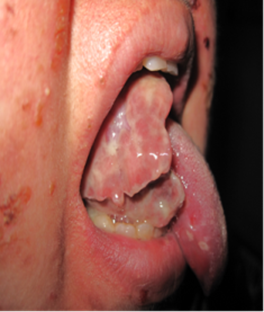
We report a 30- year-old Iranian woman presenting with a red to yellowish, well demarcated, painless exophytic and lobulated mass originating from the right hand side of the tongue (Figure 1). The lesion had started as a small tumor 4 months before. It later became extensive and friable and transformed into a rapid growing flesh to yellowish colored mass that began interfering with her speaking and eating. Additionally since almost 5 years ago she has suffered from intractable pemphigus vulgaris with oral erosions and cutaneous bullous lesions which had been treated with prednisolone and azothioprin. Otherwise physical examination and routine laboratory and HIV tests were normal. An excisional biopsy was obtained and it was diagnosed histopathologically as Kaposi's sarcoma by detecting atypical spindle cells with rare mitoses delineating blood-filled vascular slits.
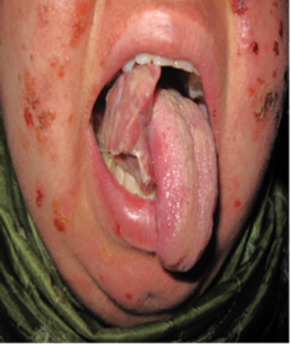
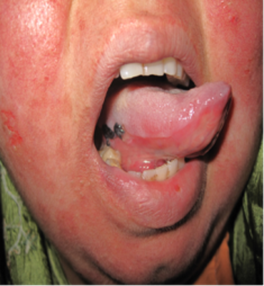
Traditionally, Kaposi sarcoma has a general distribution, often appearing first on the limbs (2). Kaposi's sarcoma is a frequently seen as an AIDS-related malignant neoplasm in the head and neck region, especially in the oral cavity, but is rarely described in HIV-negative or non-immunosuppressed individuals (3). Oral involvement occurs in approximately 11% of all cases described (4). Initial oral involvement is an even rarer occurrence, with only fourteen cases previously documented in literature for non-HIV related KS (5). A statistically increased incidence of malignancy has been observed in patients with pemphigus (6) but notably iatrogenic KS occurs infrequently in the setting of pemphigus vulgaris (7). Interestingly, while immunosuppression is highly associated with KS, it cannot be considered an etiology (5). Basically it is believed that immunosuppression creates an environment that allows an opportunistic factor to cause KS (5, 8). HHV8 is considered to be the etiological agent of all forms of Kaposi's sarcoma (9). In Iran a high prevalence of HHV8 infection has been observed in several risk groups such as haemodialysis, renal transplant and HIV-positive patients (10) but unfortunately we did not test the patient for HHV8 DNA. A number of dermatologic diseases can be present as an oral mass such as bacillary hemangioma, lymphoma and squamous cell carcinoma (11). Accordingly, Kaposi's sarcoma should be considered as a differential diagnosis of oral masses in patients especially if they are treated with immunosuppressive agents. The patient successfully underwent surgery (Figure 2) and there was no recurrence after more than 1 year of follow up.
Red to yellowish, well demarcated, painless exophytic and lobulated mass originating from right hand side of the tongue.


Immediately after surgery

Conflict of Interest
The authors have declared that no conflict of interest exists.
References
1. Avalos-Peralta P, Herrera A, Ríos-Martín JJ, Pérez-Bernal AM, Moreno-Ramírez D, Camacho F. Localized Kaposi's sarcoma in a patient with pemphigus vulgaris. J Eur Acad Dermatol Venereol. 2006;20:79-83
2. Pearce CT, Valker L.E. Multiple hemorrhagic sarcoma of the skin (Kaposi). The Ohio State Medical Journal. 1936;32:137-139
3. Bottler T, Kuttenberger J, Hardt N, Oehen HP, Baltensperger M. Non-HIV-associated Kaposi's sarcoma of the tongue. Case report and review of the literature. Int J Oral Maxillofac Surg. 2007;36:1218-20
4. Pisanty S, Garfunkel A. Kaposi's sarcoma. Journal of Oral Medicine. 1970;25:89-92
5. Jindal JR, Campbell BH, Ward TO, Almagro US.Classical Kaposi's Sarcoma Presenting in the Oral Cavity. A Case Report and Literature Review. Head Neck. 1995;17:64-8
6. Younus J, Ahmed AR. The relationship of pemphigus to neoplasia. J Am Acad Dermatol. 1990;23:498-502
7. Saggar S, Zeichner JA, Brown TT, Phelps RG, Cohen SR. Kaposi's sarcoma resolves after sirolimus therapy in a patient with pemphigus vulgaris. Arch Dermatol. 2008May;144:654-7
8. Wahman A, Melnick S.L, Rhame F.S, Potter J.D. The epidemiology of classic, african and immunosuppressed Kaposi's sarcoma. Epidemiologic Reviews. 1991;13:178-197
9. Schwartz RA, Micali G, Nasca MR, Scuderi L. Kaposi sarcoma: a continuing conundrum. J Am Acad Dermatol. 2008;59:179-206
10. Jalilvand S, Shoja Z, Mokhtari-Azad T, Nategh R, Gharehbaghian A. Seroprevalence of Human herpesvirus 8 (HHV-8) and incidence of Kaposi's sarcoma in Iran. Infect Agent Cancer. 2011;6:5
11. Mra Z, Chien J. Kaposi's sarcoma of the tongue. Otolaryngol Head Neck Surg. 2000;123:151
Author contact
![]() Corresponding author: Amir Feily, MD, Jundishapur Infectious and Tropical Diseases Research Center and Department of Dermatology, Jundishapur university of Medical Sciences, Ahvaz, Iran; Dr.feilycom
Corresponding author: Amir Feily, MD, Jundishapur Infectious and Tropical Diseases Research Center and Department of Dermatology, Jundishapur university of Medical Sciences, Ahvaz, Iran; Dr.feilycom

 Global reach, higher impact