Impact Factor ISSN: 1449-1907

 Global reach, higher impact
Int J Med Sci 2011; 8(7):547-553. doi:10.7150/ijms.8.547 This issue Cite
Case Report
Iatrogenic Mandibular Fracture Associated with Third Molar Removal
Abdulkadir Burak Cankaya1 ![]() , Mehmet Ali Erdem1, Sirmahan Cakarer1, Muhsin Cifter2, Cuneyt Korhan Oral1
, Mehmet Ali Erdem1, Sirmahan Cakarer1, Muhsin Cifter2, Cuneyt Korhan Oral1
1. Department of Oral and Maxillofacial Surgery, Faculty of Dentistry, Istanbul University, Istanbul, 34093, Turkey
2. Department of Orthodontics, Faculty of Dentistry, Istanbul University, Istanbul, 34093, Turkey
Received 2011-8-9; Accepted 2011-9-12; Published 2011-9-17
Abstract
Third molar extraction is one of the most common procedures performed in oral and maxillofacial surgery units. It is sometimes accompanied by complications such as alveolar osteitis, secondary infection, hemorrhage, dysesthesia and, most severely, iatrogenic fracture. This article describes two mandibular angle fractures that occurred in two patients during the surgical extraction of one erupted and one unerupted third molar, including a brief review of the literature.
Keywords: third molar extraction, complication, iatrogenic fracture, mandible
Introduction
The extraction of impacted mandibular third molars is a common dental procedure. The reasons for extracting these teeth include acute or chronic pericoronitis, the presence of cysts or a tumor, periodontal problems, the presence of a carious lesion, and preparation for orthodontic treatment or orthognathic surgery (1).
The management of a deeply impacted mandibular third molar presents a significant surgical challenge, and potential complications must be weighed against the potential benefits of surgical removal (2,3). Common complications of mandibular third molar surgery include alveolar osteitis (dry socket), secondary infection, nerve dysfunction, and hemorrhage. The incidence of these complications varies from 0.2% to 6% (4,5). The most severe complication of third molar surgery is mandibular fracture. Iatrogenic mandibular fracture associated with the removal of teeth, which can occur during the procedure or at a later time, is rare; reported incidences range from 0.0034% to 0.0075% (6,7).
The purpose of this paper is to discuss the risks and predisposing factors for immediate mandibular fracture and the need for surgical or nonsurgical treatment to remove impacted molars. This report is based on a thorough review of the literature and on the authors' personal experience with two iatrogenic mandibular fractures that occurred in one male and one female patient during third molar surgery.
Case 1
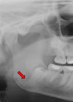
A 35-year-old female patient was admitted to the Department of Oral Surgery, Faculty of Dentistry, Istanbul University, with the complaint of mild pain in the right mandibular angle. She was systemically healthy and showed no swelling on the right side of the mandible due to infection. A panoramic radiograph revealed the presence of a mesioangular and deeply impacted mandibular right third molar surrounded by mild radiolucency. Extraction of the tooth was planned due to the pain and the radiolucency surrounding the crown and apex (Fig. 1).
Preoperative x-ray of the patient showing deeply impacted right third molar and radiolucency surrounding the tooth.
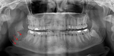
Under local anesthesia, a flap was reflected to completely reveal the buccal bony plate to the apical extent of the roots. Under constant saline cooling, a round burr was used to penetrate the cortical bony plate from the cementoenamel junction to the tooth apex. Due to the position of the third molar, the external oblique ridge was also flattened with the burr to provide sufficient visibility. During the attempt to luxate the tooth using a straight bein elevator between the second and third molars with a normal application of force, a cracking noise was heard. A panoramic radiograph was taken immediately, revealing a non-displaced, unfavorable green-stick-like fracture line extending from the base of the alveolar margin to the lower mandibular border (Fig. 2).
Perioperative x-ray of the patient showing the thin fracture line.
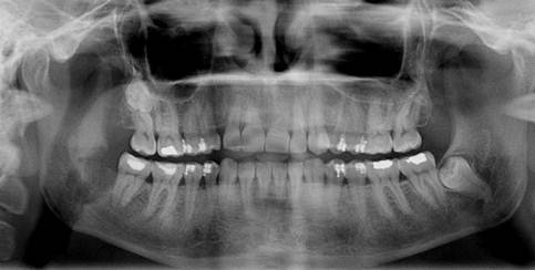
No bone fragment mobility was detected. Clinically, the patient showed no limitation or restriction of movement and could open her mouth freely. We explained the treatment options (closed reduction with intermaxillary fixation [IMF], open reduction) to the patient. Due to the good occlusion and lack of dislocation between fracture segments, we treated the fracture with 4 weeks of semi-rigid fixation by placing orthodontic brackets and elastic bands on the molars and premolars. The patient received a 5-d course of oral antibiotics, nonsteroidal anti-inflammatory analgesics, and an antimicrobial mouthwash. She was advised to follow a soft diet and was followed up. During the follow-up visits, the patient was symptom-free and no displacement of the fracture segments was noted. Bone union was observed radiologically after 1 month (Fig. 3).
Postoperative x-ray at 1 month showing the healing of the fracture line.
Case 2
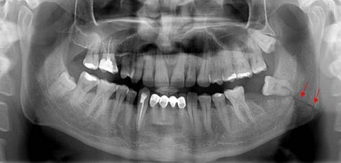
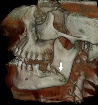
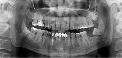
A 33-year-old male patient was referred to the Department of Oral Surgery, Faculty of Dentistry, Istanbul University, after an unsuccessful attempt to extract a partially erupted left third molar 1 week previously. The patient showed no significant disease or systemic condition. A panoramic radiograph and a volumetric tomographic scan showed an oblique and unfavorable fracture line on the left mandible that extended from the tooth roots to the mandibular angle (Figs. 4, 5).
Postoperative panoramic view of the patient showing the fracture line extending from the root to the mandibular angle.
Postoperative cone-beam computed tomography image of the patient showing the fracture line.
The patient could open his mouth without limitation and the occlusion was good. The regional lymph nodes were not palpable. The patient could not recall any pertinent medical history. No sensitivity of the lower lip, pain, swelling, facial bruising, or lingual hematoma was evident. We thus planned to take no action. The patient was advised to follow a soft diet and was followed up. During the follow-up visits, the patient was symptom-free. Bone union was observed radiologically after 1 month (Fig. 6).
Postoperative x-ray at 1 month showing the healing fracture line.
Discussion
Dentists encounter a wide range of hard-tissue injuries in practice. Dental extractions are one of the most common procedures in dentistry and may lead to several complications, including oral sinus complications, osteitis, infection, dysesthesia, pain, and bleeding (8,9). Frequently seen injuries include those associated with concomitant dentoalveolar trauma and those inadvertently caused by the dentist in practice. Factors affecting the incidence and etiology of iatrogenic mandibular fractures include the magnitude of tooth impaction, type of tooth angulation, length of roots, patient age, age and experience of the surgeon, presence of a cyst or tumor around an impacted third molar, systemic disease or medications that may impair bone strength, preoperative infections in the third molar site, and inadequate preoperative examination (10-12). A fracture occurs when the strength of the bone is overcome by the forces acting on it.
Iatrogenic fractures may occur during an operation or within 4 weeks after the procedure (classified as pathological fractures), and most are associated with third molar removal (13). The mandible is fractured 2-3 times more frequently than other facial bones because it has less bony support (14,15). The body of the mandible is naturally strengthened by a system of buttresses extending onto the rami. On the lateral surface, the strong external oblique ridge extends from the body obliquely upward to the anterior border of the ramus. Although the medial surface is thinner than the lateral surface, both are composed of dense, thick, compact cortical bone. The mylohyoid line extends diagonally downward from the area of the third molar and forward toward the genial tubercles at the midline. Because stress is localized primarily on the external oblique ridge, it is important to protect this region during surgery (16). We believe that the fracture described in Case 1 occurred primarily due to the position of the third molar, which occupied a large osseous space and thereby weakened the mandibular angle by decreasing the cross-sectional area of bone and causing the loss of supporting bone, especially in the external oblique ridge.
Open or closed reduction methods may be used for the management of mandibular fractures. In closed reduction procedures, dental wiring or bars are applied to the dental arch to achieve satisfactory occlusion. Closed reduction is indicated in nondisplaced favorable fractures. The open reduction of mandibular fractures is reserved for displaced unfavorable fractures, multiple fractures, cases in which IMF is contraindicated or impossible, and cases in which IMF is avoided to increase patient comfort. The terms “favorable” and “unfavorable” are used to describe mandibular angle fractures. The direction of the fracture line affects the resistance to muscle pull. When muscle pull resists the displacement of fragments, the fracture line is considered to be favorable; when muscle pull distracts the fragments away from one another, resulting in displacement, the fracture line is considered to be unfavorable (17). A modified IMF technique was used in Case 1 because the fracture was unfavorable. In Case 2, the fracture line passed inferiorly and posteriorly from the alveolar margin, and physical obstruction by the body of the mandible prevented the upward displacement of the posterior fragment. Although this fracture was also unfavorable, we did not use IMF to treat the patient.
Teeth along the line of a fracture were formerly considered a potential impediment to healing due to the risk of tooth death or previous infection and the possibility of infection transfer via the periodontal membrane. In current practice, a tooth in the fracture line that lacks structural damage, is not subluxated, and remains in a functional position is retained, and antibiotics are administered. A tooth that becomes infected should be extracted immediately. In Case 2, the tooth along the fracture line had no infection before the extraction attempt and was not severely damaged during extraction; we thus decided not to extract it to avoid extensive bone loss and secondary trauma.
Male patients over 40 years of age with complete dentitions are considered to be at a higher risk for mandibular fracture (18,19). However, Libersa et al. (7) found that 85% of patients with mandibular fractures were over 25 years of age and that the mean age of these patients was 40 years. In a similar study, 22/28 patients were at least 26 years of age (4). Our female patient was 35 years old, and our male patient was 33 years old; thus, both of these patients were at higher risk for mandibular fracture. Wagner (19) noted that more (70%) iatrogenic mandibular fractures occur on the left side, perhaps due to the reduced visualization of an operation site on this side provided by the surgeon's normal position. Bodner et al. (8) found no difference in the occurrence of fractures on the right and left sides. However, we believe that the higher incidence of fractures on the left side reflects the dentist's position, from which excessive and uncontrolled force may readily be applied to the left side of a patient's mandible by a right-handed surgeon. The fracture described in Case 2 was probably caused by the application of excessive force to the mandible.
The depth of impaction is related to the risk of damage to adjacent anatomical structures. Axial, coronal, and sagittal sectional computed tomography (CT) scans are the best way to determine impaction depth, the relationship of the tooth to the mandibular nerve, and buccal and lingual bone volume (20). However, the depth of impaction is not the only reason for iatrogenic fracture; this severe complication is multifactorial (8). Age-related weakening of bony elasticity makes extraction more difficult, and ankylosis can occur in older patients. Because both of our patients were middle-aged, ankylosis was not a factor in our cases.
Patients with complete dentitions have a high biting force that increases the risk of postoperative fracture. However, Al-Belasy et al. (21) claimed that mastication does not affect late mandibular fracture after the surgical removal of impacted third molars associated with no gross pathology in patients more than 25 years of age who are fully dentate or missing one or two teeth, exhibit no mandibular atrophy, and have no systemic problems that may impair bone strength. In light of these findings, we did not apply rigid fixation in Case 1, but performed semi-rigid fixation using four orthodontic brackets and elastic rondelles on both sides.
Osteoporotic women have a high risk of iatrogenic fracture due to the low resistance of the bone to standard biting forces (20,22). The decision to screen for osteoporosis remains controversial. The United States Preventive Services Task Force (USPSTF) has recommended that all women aged >65 years be screened for osteoporosis and that screening be initiated at the age of 60 years in women with additional risk factors, such as personal or family history of osteoporosis, previous fragility fracture after the age of 50 years, premature menopause, medical conditions (e.g., hyperparathyroidism, hyperthyroidism, malnutrition, rheumatoid arthritis, diabetes, or chronic liver or renal disease), or lifestyle factors (e.g., cigarette smoking, eating disorders, excessive alcohol consumption).
The extent of tooth impaction, the volume of the impacted tooth, and the relative portion of mandibular volume are also important factors. Fracture risk is higher when the relative portion of the mandible exceeds 50% (8). In Case 1, the volume of bone occupied by the third molars exceeded 50% of the mandibular angle, resulting in severe bone loss during extraction, as reported in previous studies. Some studies have found that the position of an impacted tooth was associated with the frequency of mandibular fracture, whereas others have reported no significant relationship between tooth position and fracture risk (16). The deep vertical and horizontal impaction of third molars is considered to be a risk factor for iatrogenic mandibular fracture (8), although this finding is inconsistent with our experience in Case 1.
Pre-existing pathological findings at the extraction site such as pericoronitis, periodontal pockets, and cysts may also weaken the mandible. In Case 1, radiolucency around the crown of the third molar indicated that bone loss had weakened the mandible and may have been a predisposing factor for fracture.
The professional's experience is another important factor (9), although some researchers have found that the surgeon's clinical experience does not play an important role (8). Although the operator in Case 1 was an experienced surgeon, a young and inexperienced private practitioner performed the extraction in Case 2. Although the surgeon was experienced, the misevaluation of the situation in Case 1 may also have been a factor causing the iatrogenic fracture.
CT scans and panoramic radiographs may be used to detect fractures, although some fractures are radiologically undetectable. The occurrence of a cracking noise (as in Case 1) is important in such situations because it should alert the operator to the possibility that a fracture has occurred. Dental volumetric tomographic scans should also be considered when some fracture characteristics are uncertain. In unclear situations, the surgical team must be prepared for unanticipated surgical difficulties.
Conclusion
The ability to predict the surgical difficulty of mandibular third molar extraction is essential when designing a treatment plan because it helps assess the competence of the dental practitioner for the particular operation, minimize complications, optimize patient preparation, and guide the postoperative management of inflammation and pain. Even the most targeted periapical radiograph cannot always provide sufficiently detailed visualization. Although CT scans of impacted teeth are usually not necessary, we recommend that they be taken to evaluate buccal and lingual bone volumes when a third molar is fully impacted vertically and horizontally near the mandibular angle; deficiencies in these volumes can cause unwanted complications, such as iatrogenic fractures. Preoperative risk/benefit evaluation, adequate surgical expertise, guided force application, good visualization, proper instrumentation, conservative bone removal during extraction, tooth sectioning, and attention to cracking noises are also important to avoid fractures. Although full-arch IMF is usually the preferred treatment for unfavorable mandibular fractures, we have concluded that it is unnecessary in cases showing minimal dislocation and good occlusion. The use of a modified ivy ligature technique in combination with orthodontic brackets on the maxillary and mandibular premolars provided sufficient fixation for healing. In such cases, a soft diet and antibiotic therapy should also be prescribed, and the patient should be followed up.
The mandible is the largest, heaviest, and strongest facial bone. A normal mandible provides a normal airway space and a proper facial contour. A solid, stable, mobile mandible allows normal chewing, swallowing, and speech functions. Physicians and dentists should be aware that the development of mandibular instability due to trauma may life threatening. The recent literature and practitioners' long-term experience have improved the understanding of the origin and treatment of iatrogenic mandibular fractures related to third molar surgery. As the body of literature related to iatrogenic fractures during third molar surgery expands, more techniques and evaluations will be elucidated.
Conflict of Interest
The authors have declared that no conflict of interest exists.
References
1. Chiapasco M, De Cicco L, Marrone G. Side effects and complications associated with third molar surgery. Oral Surg Oral Med Oral Pathol. 1993;76:412-420
2. Toffanin A, Zupi A, Cicognini A. Sagittal split osteotomy in removal of impacted third molar. J Oral Maxillofac Surg. 2003;61:638-640
3. Ness GM, Peterson LJ. Impacted teeth. In: (ed.) Ness GM, Peterson LJ, Miloro M. Peterson's principles of oral and maxillofacial surgery. London: BC Decker Inc. Hamilton. 2004:139-155
4. Goldberg MH, Nemarich AN, Marco WP II. Complications after mandibular third molar surgery: a statistical analysis of 500 consecutive procedures. J Am Dent Assoc. 1985;111:277-279
5. Osborn TP, Frederickson G, Small IA, Torgerson TS. A prospective study of complications related to mandibular third molar surgery. J Oral Maxillofac Surg. 1985;43:767-769
6. Nyul L. Kieferfrakturen bei zahnextrationen. Zahnarztl Welt. 1959;60:1-5
7. Libersa P, Roze D, Cachart T, Libersa JC. Immediate and late mandibular fractures after third molar removal. J Oral Maxillofac Surg. 2002;60:163-166
8. Bodner K, Brennan PA, McLeod NM. Characteristics of iatrogenic mandibular fractures associated with tooth removal: review and analysis of 189 cases. Br J Oral Maxillofac Surg. 2010 Epub ahead of print
9. Niedzielska I, Kowol I, Sroczynska-Grula A. Iatrogenic injury during extraction of lower molar teeth. Dent Med Probl. 2009;46:501-505
10. Sakr K, Farag IA, Zeitoun IM. Review of 509 mandibular fractures treated at the University Hospital, Alexandria, Egypt. Br J Oral Maxillofac Surg. 2009;44:107-111
11. Tanaka M, Tomitsuka K, Shionoya K. et al. Aetiology of maxillofacial fractures. Br J Oral Maxillofac Surg. 1994;32:19-23
12. Thorn JJ, Mogeltoft M, Hansen PK. Incidence and aetiological pattern of jaw fractures in Greenland. Int J Oral Maxillofac Surg. 1986;15:372-379
13. Krimmel M, Reinert S. Mandibular fracture after third molar removal. J Oral Maxillofac Surg. 2000;58:1110-1112
14. Subhashraj K, Ramkumar S, Ravindran C. Pattern of mandibular fractures in Chennas, India. Br J Oral Maxillofac Surg. 2008;46:126-127
15. Haug RH, Prather J, Indreasano AT. An epidemiological survey of fractures and concomitant injuries. J Oral Maxillofac Surg. 1990;48:926-932
16. Szücs A, Bujtar P, Sandor GKB, Barabas J. Finite element analysis of the human mandible to assess the effect of removing an impacted third molar. J Can Dent Assoc. 2010;76:a72
17. Neelima AM. Textbook of oral maxillofacial surgery, 2nd ed. New Delhi, India: Jaypee Brothers. 2008
18. Iizuka T, Tanner S, Berthold H. Mandibular fractures following third molar extraction. A retrospective clinical and radiological study. Int J Oral Maxillofac Surg. 1997;26:338-343
19. Wagner KW, Otten JE, Schoen R, Schmelzeisen R. Pathological mandibular fractures following third molar removal. Int J Oral Maxillofacial Surg. 2005;34:722-726
20. Chrcanovic BR, Custodio ALN. Considerations of mandibular angle fractures during and after surgery for removal of third molars: a review of the literature. Oral Maxillofac Surg. 2010;14:71-80
21. Al-Belasy FA, Tozoglu S, Ertas U. Mastication and late mandibular fracture after surgery of impacted third molars associated with no gross pathology. J Oral Maxillofac Surg. 2009;67(4):856-861
22. Contar CM, de Oliveira P, Kanegusuku K, Berticelli RD, Azevedo-Alanis LR, Machado MA. Complications in third molar removal: a retrospective study of 588 patients. Med Oral Patol Oral Cir Bucal. 2010;15:e74-78
Author contact
![]() Corresponding author: Dr. Burak ÇANKAYA, Tel: +905327654857; Fax: +902125312230; e-mail: cankayaedu.tr
Corresponding author: Dr. Burak ÇANKAYA, Tel: +905327654857; Fax: +902125312230; e-mail: cankayaedu.tr