Impact Factor ISSN: 1449-1907

 Global reach, higher impact
Int J Med Sci 2010; 7(3):101-109. doi:10.7150/ijms.7.101 This issue Cite
Research Paper
Growth of Microorganisms in Total Parenteral Nutrition Solutions Containing Lipid
Takashi Kuwahara1 ![]() , Kazuyuki Shimono1, Shinya Kaneda1, Takumi Tamura1, Masao Ichihara2, Yoshifumi Nakashima1
, Kazuyuki Shimono1, Shinya Kaneda1, Takumi Tamura1, Masao Ichihara2, Yoshifumi Nakashima1
1. Preclinical Assessment Department, Otsuka Pharmaceutical Factory, Inc., Tokushima, Japan.
2. Research and Development Center, Otsuka Pharmaceutical Factory, Inc., Tokushima, Japan.
Received 2010-2-1; Accepted 2010-5-17; Published 2010-5-18
Abstract
Background: To identify the microorganisms that can grow rapidly in total parenteral nutrition (TPN) solutions, we investigated the growth of the major causes of catheter-related blood stream infection (Staphylococcus aureus, Serratia marcescens, Bacillus cereus, and Candida albicans) in TPN solutions containing lipid. Methods: The pH value of a TPN solution containing lipid (pH 6.0, containing 20 ppm of NaHSO3) was adjusted by the addition of HCl to 5.7, 5.4, or 4.9. The pH value of another TPN solution (pH5.5, containing 400 ppm of NaHSO3) was adjusted by the addition of NaOH to 5.9, 6.3, or 6.8. A specific number of each microorganism was added to 10 mL of each test solution and incubated at room temperature. The number of microorganisms was counted as colony forming units at 0, 24, and 48 hrs later. Results: C albicans increased similarly at any pH values in the TPN solution. The bacterial species also increased rapidly at pH6.0 in the solution containing 20 ppm of NaHSO3, but growth was suppressed as the pH value was reduced, with growth halted at pH4.9. However, these bacterial species did not increase, even at pH5.9, in the other solution containing 400 ppm of NaHSO3. Conclusions: These results suggest that Candida species can grow rapidly in almost all TPN solutions regardless of the acidity, lipid, and NaHSO3; also, some bacterial species may grow in TPN solutions containing lipid unless the pH value is 5.0 or less. Therefore, each TPN solution should be examined whether or not the bacterial species can proliferate.
Keywords: CRBSI, microbial growth, TPN solution, lipid, pH, bisulfite
INTRODUCTION
Catheter-related blood stream infection (CRBSI) is one of the most common complications of intravenous catheters.1-3 To reduce or prevent CRBSI, factors that enhance or inhibit microbial growth in parenteral solutions should be investigated and identified. Total parenteral nutrition (TPN) solutions are considered to be relatively good growth media for microorganisms,1,4 whereas a number of investigators have shown that TPN solutions are poor growth media for most microorganisms that cause CRBSI, with the exception of Candida species.5-9 Likewise, we have previously confirmed that while bacterial species do not grow in TPN solutions without lipid, Candida species grow rapidly.10 Our data have shown that bacterial species cannot grow due to the acidity, but Candida species can grow regardless of the acidity.10 In contrast, most microorganisms grow rapidly in commercial lipid emulsions,11-15 and sporadic CRBSI and outbreaks have been traced to contaminated lipid emulsion.16,17 Because lipid emulsions have been shown to contribute to the rapid growth of most microorganisms that cause CRBSI, it can be assumed that TPN solutions containing lipid promote the microbial growth compared with TPN solutions without lipid.18 Consequently, the Center for Disease Control and Prevention (CDC) has recommended that when any infusion solutions containing lipid are administered, the entire delivery system such as the administration set be replaced every 24 hours.19
As the major causes of CRBSI, Staphylococcus aureus, Staphylococcus epidermidis, Serratia marcescens, Escherichia coli, Klebsiella pneumonia, Candida albicans, etc. were shown.2,3,6,11 Furthermore, blood stream infection outbreaks of Bacillus cereus via intravenous line were recently reported in Japan.20 In the present study, we investigated the growth in TPN solutions containing lipid of the same microorganisms studied previously:10 ie, Staphylococcus aureus as a delegate of gram positive cocci, Serratia marcescens as a delegate of gram negative rods, Bacillus cereus as a delegate of gram positive rods, and Candida albicans as a delegate of fungi. To clarify whether the acidity of TPN solutions suppresses microbial growth in the presence of lipid, the growth of all strains employed were investigated at various pH values in a commercial TPN solution containing lipid in the first experiment. To identify which factors enhance or inhibit the growth of each microorganism, we supplemented a TPN solution containing lipid with multivitamins, supplemented a TPN solution that did not contain lipid with lipid, and increased the bisulfite concentration in a TPN solution containing both lipid and multivitamins in the following experiments.
MATERIALS AND METHODS
Microorganisms employed
A standard American Type Culture Collection (ATCC) strain and 1 clinical isolate were used for each microorganism; the standard strain ATCC6538 and the clinical isolate N3 of Staphylococcus aureus, the standard strain ATCC13880 and the clinical isolate N4 of Serratia marcescens, the standard strain ATCC11778 and the clinical isolate H2 of Bacillus cereus, and the standard strain ATCC10231 and the clinical isolate N7 of Candida albicans.
Test solutions
A commercial TPN solution containing lipid (ML; MIXID-L, Otsuka Pharmaceutical Factory, Inc., Japan), a commercial TPN solution without lipid (AT1; AMINOTORIPA-1, Otsuka Pharmaceutical Factory, Inc.), multivitamins (MV; Otsuka MV Injection, Otsuka Pharmaceutical Factory, Inc.), a 20% lipid emulsion (IL; Intralipos 20%, Otsuka Pharmaceutical Factory, Inc.), and a physiological saline were used. The compositions of ML and AT1 are shown in Table 1.
The compositions of ML and AT1
| Solution | ML | AT1 | |
|---|---|---|---|
| (volume) | 900 mL | 850 mL | |
| Amino acids | 30.0 g | 25.0 g | |
| Glucose | 110.0 g | 79.8 g | |
| Fructose | - | 40.2 g | |
| Xylitol | - | 19.8 g | |
| Lipid | 15.6 g | - | |
| Na+ | 35 mEq | 35 mEq | |
| K+ | 27 mEq | 22 mEq | |
| Mg2+ | 5 mEq | 4 mEq | |
| Ca2+ | 8.5 mEq | 4 mEq | |
| Cl- | 44 mEq | 35 mEq | |
| SO42- | 5 mEq | 4 mEq | |
| Acetate- | 25 mEq | 44 mEq | |
| Gluconate- | 8.5 mEq | 4 mEq | |
| Citrate3- | - | 10 mEq | |
| P | 150 mg | 154 mg | |
| Zn | 10 μmol | 8 μmol | |
| NaHSO3 | 17 mg | 412 mg | |
| pH | 6.0 | 5.5 | |
| Osmotic pressure ratio to physiological saline | Approx. 4 | Approx. 5 | |
Experiment 1: ML was used as the base solution. The pH value, osmotic pressure ratio to physiological saline (OPR, the osmolality of each solution/the osmolality of physiological saline), and concentration of NaHSO3 of ML were 6.0, approximately 4, and 17 ppm (about 20 ppm), respectively. The other pH solutions of ML were adjusted to 5.7, 5.4, or 4.9 by the addition of 0.5 mol/L HCl.
Experiment 2: ML(pH6.0), ML(pH4.9), and those supplemented with MV (MLV) were used.
Experiment 3: AT1 was supplemented with MV and used as the base solution (AT1V). The pH value, OPR, and concentration of NaHSO3 of AT1V were 5.5, approximately 5, and 412 ppm (about 400 ppm), respectively. The other pH solutions of AT1V were adjusted to 5.9, 6.3, or 6.8 by the addition of 0.5 mol/L NaOH. Each pH solution of AT1V was added with 1/10 volume of IL (L+AT1V) or physiological saline (S+AT1V).
Experiment 4: ML was supplemented with MV and used as the base solution (MLV). The other pH solutions of MLV were adjusted to 5.7, 5.4, or 5.0 by the addition of 0.5 mol/L HCl. Besides, MLV(pH5.7) and MLV(pH5.4) were added with 100 ppm (MLV+100 ppm) or 200 ppm of NaHSO3 (MLV+200 ppm).
Addition of microorganism, incubation and sampling
A specific number of each test microorganism was added to each 10 mL of test solutions in sterile plastic tubes, and all tubes were allowed to stand at room temperature (23-26ºC). An aliquot of test solution was sampled at 0, 24, and 48 hours after the addition of microorganisms.
Measurement of viable microorganisms
Each aliquot of test solution sampled was inoculated in a Soybean Casein Digest (SCD) agar plate in duplicate. When necessary, the aliquot of test solution was diluted 10-fold to 107-fold with physiological saline before inoculation. After a 24-hour incubation at 37ºC, the number of colony forming units (CFU) of each microorganism was counted for each plate. The mean CFU of duplicate data was calculated for each aliquot, and number of each microorganism per mL was calculated by using the number of CFU per plate, aliquot volume, and diluting ratio. The results are shown as values of CFU/mL in semi-logarithmic graphs. As other experimental studies of microbial growth,5,12,13,21,22 the data obtained in this study were not analyzed statistically, because biological significance of these kinds of data is considered to be assessable without statistical analysis.
RESULTS
Experiment 1
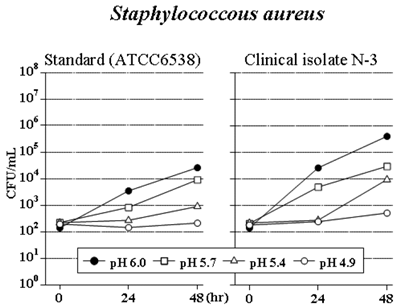
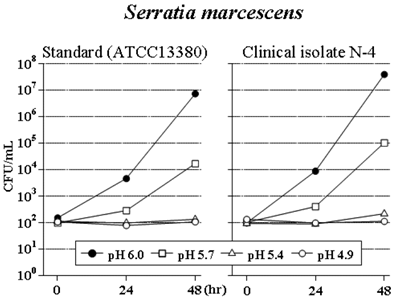
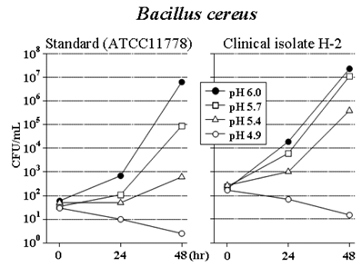
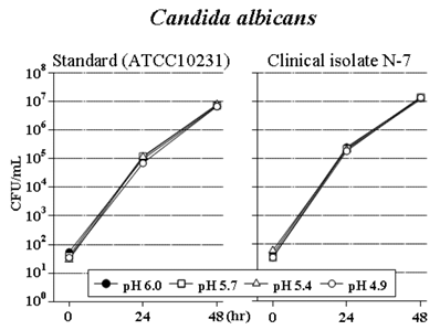
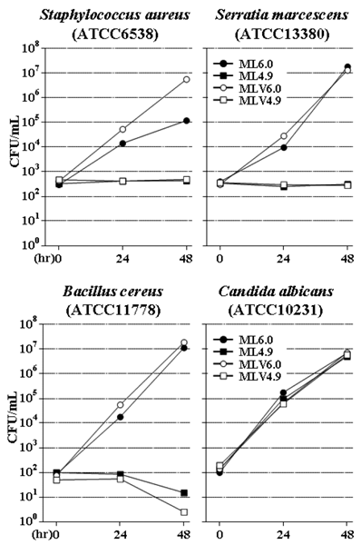
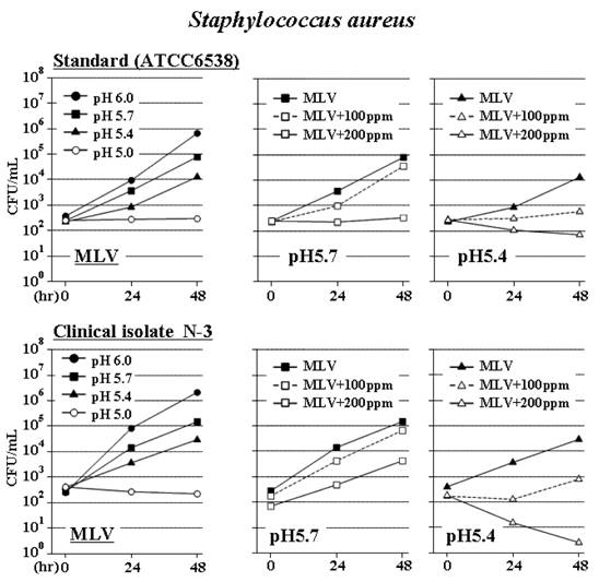
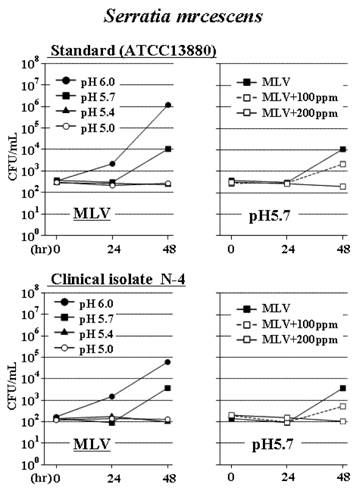
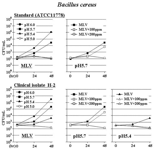
S. aureus increased in ML at the original pH of 6.0, but growth was suppressed as the pH value was reduced, coming to a halt at a pH of 4.9 (Figure 1). S. marcescens increased rapidly in ML at a pH of 6.0, also increased but with suppression at a pH of 5.7, and did not increase at pH values of 5.4 and 4.9 (Figure 2). B. cereus increased at a pH of 6.0. Its growth was suppressed as the pH value was reduced, decreasing at a pH of 4.9 (Figure 3). However, both strains of C. albicans increased rapidly and similarly at any pH value (Figure 4).
Effect of pH on the growth of Staphylococcus aureus in ML (pH6.0; NaHSO3, 20 ppm). The pH value was adjusted by addition of 0.5 mol/L HCl.
Effect of pH on the growth of Serratia marcescens in ML (pH6.0; NaHSO3, 20 ppm). The pH value was adjusted by addition of 0.5 mol/L HCl.
Effect of pH on the growth of Bacillus cereus in ML (pH6.0; NaHSO3, 20 ppm). The pH value was adjusted by addition of 0.5 mol/L HCl.
Effect of pH on the growth of Candida albicans in ML (pH6.0; NaHSO3, 20 ppm). The pH value was adjusted by addition of 0.5 mol/L HCl.
Experiment 2
Supplementation with MV to ML enhanced only the growth of S. aureus at a pH of 6.0 but did not resume the growth at a pH of 4.9 (Figure 5). Concerning S. marcescens, B. cereus and C. albicans, MV did not affect them both at pH 6.0 and at pH 4.9 (Figure 5).
Growth of Staphylococcus aureus, Serratia marcescens, Bacillus cereus, and Candida albicans at pH6.0 and pH4.9 in ML and in MLV (ML supplemented with multivitamins).
Experiment 3
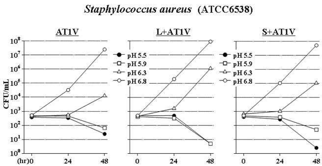
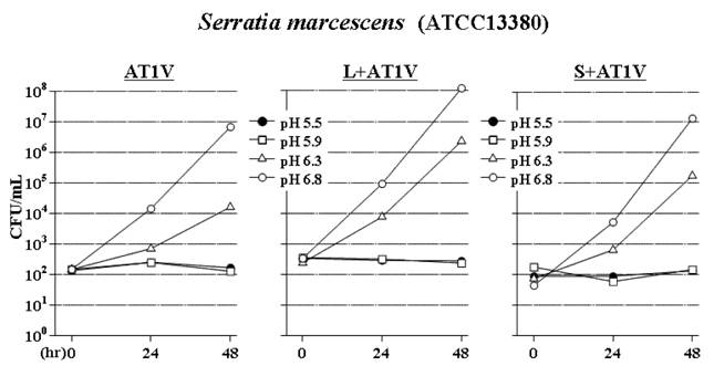
The OPR of AT1V was 4.6, and the OPR of L+AT1V (or S+AT1V) was 4.3 (or 4.2) as a result of the diluting effect from the addition of the isotonic solution.
In AT1V, the standard strains of S. aureus, S. marcescens and B. cereus did not increase at pH 5.9 as well as at the original pH of 5.5 but increased slowly at pH 6.3 and increased rapidly at pH 6.8 (Figures 6-8), similar to the results of our previous study.10
Effect of lipid on the growth of Staphylococcus aureus in AT1V (pH5.5; NaHSO3, 400 ppm). The pH value was adjusted by addition of 0.5 mol/L NaOH. To AT1V was added 1/10 volume of 20% lipid emulsion (L+AT1V) or physiological saline (S+AT1V).
Effect of lipid on the growth of Serratia marcescens in AT1V (pH5.5; NaHSO3, 400 ppm). The pH value was adjusted by addition of 0.5 mol/L NaOH. To AT1V was added 1/10 volume of 20% lipid emulsion (L+AT1V) or physiological saline (S+AT1V).
Effect of lipid on the growth of Bacillus cereus in AT1V (pH5.5; NaHSO3, 400 ppm). The pH value was adjusted by addition of 0.5 mol/L NaOH. To AT1V was added 1/10 volume of 20% lipid emulsion (L+AT1V) or physiological saline (S+AT1V).
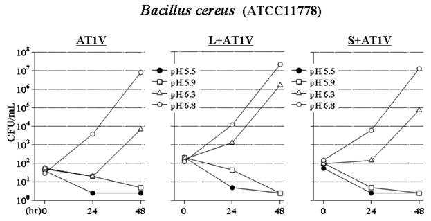
Surprisingly, none of S. aureus, S. marcescens and B. cereus increased in L+AT1V at either pH 5.5 or pH 5.9, despite the presence of lipid (Figures 6-8). These results conflicted with those of Experiment 1. At pH 6.3 and pH 6.8, however, supplementation with 1/10 volume of IL to AT1V (L+AT1V) enhanced mildly the growth of these bacterial species (Figures 6-8).
Addition of 1/10 volume of physiological saline to AT1V (S+AT1V) contributed to the reduction of OPR from 4.6 to 4.2, enhancing the growth of the bacteria less slightly than did supplementation with IL (Figures 6-8).
Experiment 4
The growths of 2 strains of S. aureus in MLV were halted only at pH 5.0 (Figure 9). The addition of 100 ppm of NaHSO3 was not enough to stop the growth of S. aureus. However, when 200 ppm of NaHSO3 was added, the growth of the standard strain was halted at pH 5.7 and the clinical isolate was halted at pH 5.4 (Figure 9).
Effect of bisulfite concentration on the growth of Staphylococcus aureus in MLV (ML supplemented with multivitamins). The pH value was adjusted by addition of 0.5 mol/L HCl. To MLV was added 100 ppm or 200 ppm of NaHSO3 (+100ppm or +200ppm).
The growth of 2 strains of S. marcescens in MLV were halted at pH 5.0 and pH 5.4 (Figure 10), corresponding with the results of Experiment 1. The addition of 200 ppm of NaHSO3 halted the growth of both strains of S. marcescens at pH 5.7 (Figure 10).
Effect of bisulfite concentration on the growth of Serratia marcescens in MLV (ML supplemented with multivitamins). The pH value was adjusted by addition of 0.5 mol/L HCl. To MLV was added 100 ppm or 200 ppm of NaHSO3 (+100ppm or +200ppm).
The growth of the standard strain of B. cereus in MLV was halted both at pH 5.0 and pH 5.4, and the growth of the clinical isolate was halted at pH 5.0 (Figure 11). The addition of 200 ppm of NaHSO3 halted the growth of both strains of B. cereus at pH 5.7, and the addition of 100 ppm of NaHSO3 halted the growth of the clinical isolate at pH 5.4 (Figure 11).
Effect of bisulfite concentration on the growth of Bacillus cereus in MLV (ML supplemented with multivitamins). The pH value was adjusted by addition of 0.5 mol/L HCl. To MLV was added 100 ppm or 200 ppm of NaHSO3 (+100ppm or +200ppm).
DISCUSSION
To reduce or prevent catheter-related blood stream infection (CRBSI), we have to understand the growth properties of the microorganisms that cause this condition. We have previously investigated the growth of the microorganisms that are known as the major causes of CRBSI (Staphylococcus aureus, Serratia marcescen, Bacillus cereus and Candida albicans) in total parenteral nutrition (TPN) solutions without lipid.10 Therefore, we investigated the growth of the same microorganisms in TPN solutions containing lipid in the present study.
In a commercial TPN solution containing lipid (ML), both standard strains and clinical isolates of all microorganisms (S. aureus, S. marcescens, B. cereus and C. albicans) increased rapidly at the original pH of 6.0, even without multivitamins. Although only C. albicans increased equally at any pH value, the growth of S. aureus, S. marcescens and B. cereus was suppressed as the pH value was reduced, with growth halted at pH4.9 (Experiment 1). However, these 3 bacterial species did not increase in another TPN solution containing lipid (L+AT1V) even at pH5.5 and pH5.9 (Experiment 3), which is the same result as obtained in the solution without lipid (AT1V or S+AT1V); this finding conflicts with the results of Experiment 1. The conflicting results from these 2 TPN solutions may be attributable to the difference in the bisulfite concentrations (ML contains NaHSO3 at a very low concentration [20 ppm], but L+AT1V contains NaHSO3 at a relatively high concentration [400 ppm]) because the bactericidal effect of bisulfite is enhanced in acidic conditions.21 Therefore, the additional experiment (Experiment 4) was performed to investigate the effect of bisulfite concentration in the TPN solution containing both lipid and multivitamins (MLV). As a result, the growth of the 3 bacterial species was suppressed or halted at the same pH (5.4 or 5.7) as the concentration of NaHSO3 increased (20 ppm, 100 ppm, and 200 ppm). These results suggest that the concentration of bisulfite in TPN solutions is an important factor for suppressing bacterial growth, especially between pH5.0 and pH6.0: the bacterial species cannot increase at pH5.9 with 400 ppm of NaHSO3, at pH5.7 or pH5.4 with 200 ppm of NaHSO3, and at pH5.0 with 20 ppm of NaHSO3.
Other findings in the present study are as follows: 1) even if lipid is contained, the acidity of TPN solution is the critical factor suppressing the bacterial growth; 2) the addition of lipid enhances mildly the growth of the bacterial species in TPN solutions but does not affect the growth substantially; 3) the addition of multivitamins further enhances the growth of S. aureus but does not affect the growth of S. marcescens, B. cereus, and C. albicans in TPN solutions containing lipid; 4) C. albicans can grow regardless of acidity, bisulfite, and lipid.
Because C. albicans could grow at pH5.5 with 400 ppm of NaHSO3 (AT1V) in our previous study,10 the effect of bisulfite concentration on the growth of C. albicans was not investigated in the present study. However, it has been reported that C. albicans could not increase in a TPN solution at pH4.4 with 500 ppm of NaHSO3, whereas C. albicans increased in the same TPN solution at pH4.4 with 40 ppm of NaHSO3 or at pH5.0 with 500 ppm of NaHSO3.23 Practically, Candida species can grow rapidly in almost all TPN solutions.
The pH values of most of the recent TPN solutions are within 5.0 and 6.0, similar to the old TPN solutions. On the other hand, the old TPN solutions contain bisulfite at relatively high concentrations, but the recent TPN solutions contain very low concentrations of bisulfite or are bisulfite-free. To investigate bacterial growth in the recent TPN solutions, referring to results from the studies that used the old TPN solutions that contained high concentration of bisulfite is not appropriate, even at the same pH range. In the recent TPN solutions containing lipid, some bacterial species may proliferate unless the pH value is 5.0 or less. Although the TPN solutions containing lipid can be theoretically improved to be bacteriostatic by reducing the pH value and/or increasing the bisulfite concentration, more studies seem needed to improve the solution because lipid emulsions become unstable as the pH value reduces or as the concentration of bisulfite increases.
In conclusion, Candida species can grow rapidly in almost all TPN solutions regardless of the acidity and the presence of lipid; also, some bacterial species may grow in TPN solutions containing lipid unless the pH value is 5.0 or less. Therefore, each TPN solution should be investigated to determine whether or not the bacterial species can proliferate.
Acknowledgements
We are very grateful to Dr. Yoshifumi Inoue, Kawasaki Hospital, Kobe, Japan, for his helpful suggestions.
Conflict of Interest
We declare that there are no conflicts of interest for all of us.
References
1. Banton J. Techniques to prevent central venous catheter infection: products, research, and recommendations. Nutr Clin Pract. 2006;21:56-61
2. Mermel LA, Farr BM, Sherertz RJ. et al. Guidelines for the management of intravascular catheter-related infection. Clin Infect Dis. 2001;32:1249-1272
3. Llop J, Badia MB, Comas D, Tubau M, Jodar R. Colonization and bacteremia risk factors in parenteral nutrition catheterization. Clin Nutr. 2001;20:527-534
4. Allwood MC. Microbiological risks in parenteral nutrition compounding. Nutrition. 1997;13:60-61
5. Didier ME, Fischer S, Maki DG. Total nutrient admixtures appear safer than lipid emulsion alone as regards microbial contamination: growth properties of microbial pathogens at room temperature. J Parenter Enteral Nutr. 1998;22:291-296
6. Rowe CE, Fukuyama TT, Martinoff JT. Growth of microorganisms in total nutrient admixtures. Drug Intell Clin Pharm. 1987;21:633-638
7. Gilbert M, Gallagher SC, Eads M, Elmore MF. Microbial growth patterns in a total parenteral nutrition formulation containing lipid emulsion. J Parenter Enteral Nutr. 1986;10:494-497
8. Melly MA, Meng HC, Schaffner W. Microbial growth in lipid emulsions used in parenteral nutrition. Arch Surg. 1975;110:1479-1481
9. Goldmann DA, Martin WT, Worthington JW. Growth of bacteria and fungi in total parenteral nutrition solutions. Am J Surg. 1973;126:314-318
10. Kuwahara T, Kaneda S, Shimono K, Inoue Y. Growth of microorganisms in total parenteral nutrition solutions without lipid. Int J Med Sci. 2010;7:43-47
11. Crocker KS, Noga R, Filibeck DJ, Krey NH, Markovic M, Steffee WP. Microbial growth comparisons of five commercial parenteral lipid emulsions. J Parenter Enteral Nutr. 1984;8:391-395
12. Jarvis WR, Highsmith AK. Bacterial growth and endotoxin production in lipid emulsion. J Clin Microbiol. 1984;19:17-20
13. Keammerer D, Mayhall CG, Hall GO, Pesko LJ, Thomas RB. Microbial growth patterns in intravenous fat emulsions. Am J Hosp Pharm. 1983;40:1650-1653
14. Kim CH, Lewis DE, Kumar A. Bacterial and fungal growth in intravenous fat emulsions. Am J Hosp Pharm. 1983;40:2159-2161
15. Deitel M, Fuksa M, Kaminsky VM, Vasic V. Growth of microorganisms in soybean oil emulsion and clinical implications. Int Surg. 1979;64:27-32
16. Jarvis WR, Highsmith AK, Allen JR, Haley RW. Polymicrobial bacteremia associated with lipid emulsion in a neonatal intensive care unit. Pediatr Infect Dis. 1983;2:203-209
17. McKee KT, Melly MA, Greene HL, Schaffner W. Gram-negative bacillary sepsis associated with use of lipid emulsion in parenteral nutrition. Am J Dis Child. 1979;133:649-650
18. Mershon J, Nogami W, Williams JM, Yoder C, Eitzen HE, Lemons JA. Bacterial/fungal growth in a combined parenteral nutrition solution. J Parenter Enteral Nutr. 1986;10:498-502
19. O'Grady NP, Alexander M, Dellinger EP. et al. Guidelines for the prevention of intravascular catheter-related infections. MMWR. 2002;51:1-34
20. Matsumoto S, Suenaga H, Naito K, Sawazaki M, Hiramatsu T, Agata N. Management of suspected nosocomial infection: an audit of 19 hospitalized patients with septicemia caused by Bacillus species. Jpn J Infect Dis. 2000;53:196-202
21. Murano A, Morinaga N, Iwamaru Y. et al. Acidic conditions enhance bactericidal effects of sodium bisulfite on Helicobacter pylori. Helicobacter. 2005;10:132-135
22. Obayashi A, Oie S, Kamiya A. Microbial viability in preparations packaged for single use. Biol Pharm Bull. 2003;26:667-670
23. Ishida K, Nakao S, Sata T. et al. Fungistatic action of the total parenteral nutrition (TPN) fluids against clinically isolated Candida albicans. Jpn J Pharm Health Care Sci. 2002;28:259-262
Author contact
![]() Corresponding author: Takashi Kuwahara, Ph.D., Preclinical Assessment Department, Otsuka Pharmaceutical Factory, Inc., 115 Tateiwa, Naruto, Tokushima 772-8601, Japan. Telephone: +81 88 685 1151 (Ext. 678) Fax: +81 88 684 0553; E-mail: kuwahat2co.jp
Corresponding author: Takashi Kuwahara, Ph.D., Preclinical Assessment Department, Otsuka Pharmaceutical Factory, Inc., 115 Tateiwa, Naruto, Tokushima 772-8601, Japan. Telephone: +81 88 685 1151 (Ext. 678) Fax: +81 88 684 0553; E-mail: kuwahat2co.jp